 WhatsApp Us (24/7)
 +91 926 888 0303 (24/7)


To Book an Appointment
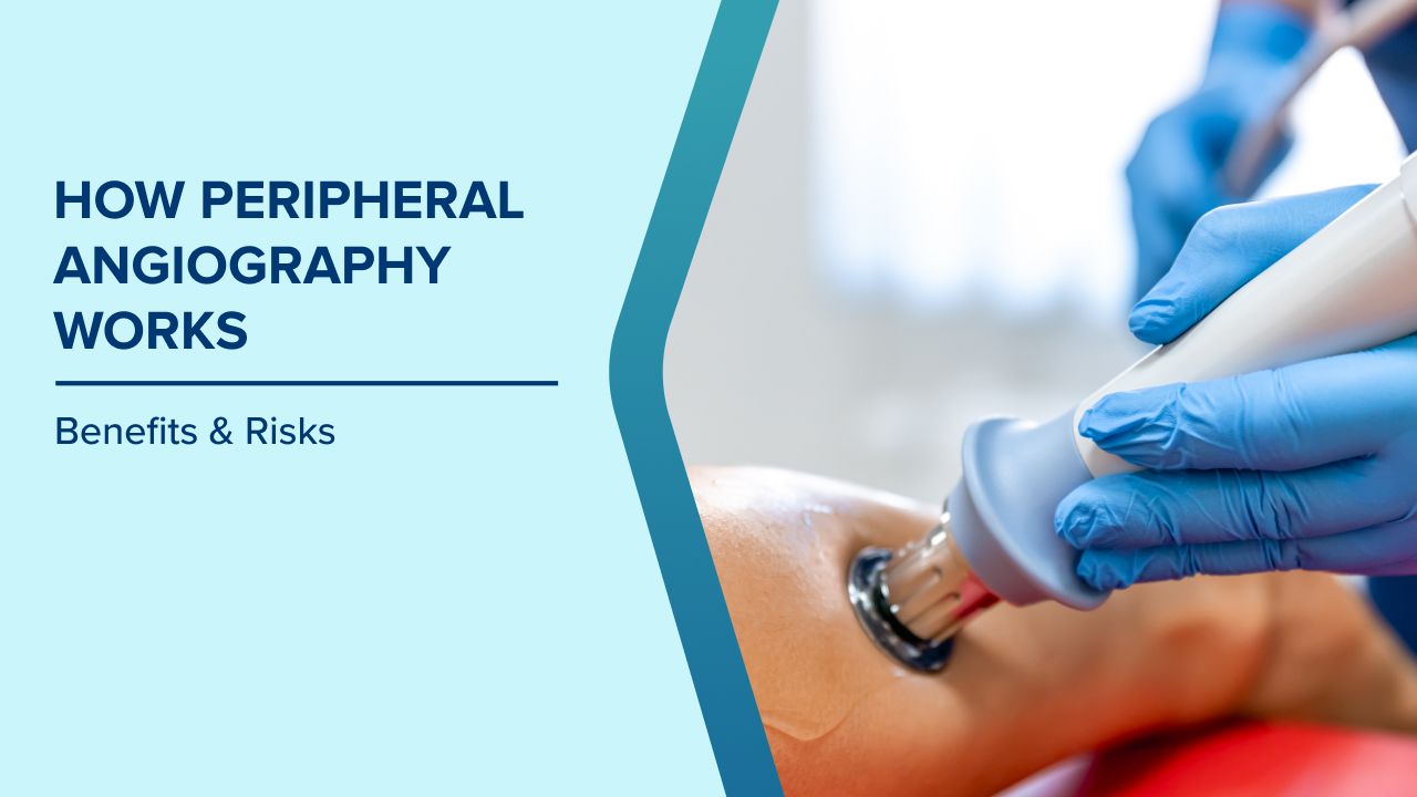
Call Us+91 926 888 0303How Peripheral Angiography Works: Benefits & Risks
By Dr. Arun Kumar Gupta in Cardiac Sciences , Cardiology , Interventional Cardiology
Apr 16 , 2026

Peripheral artery disease (PAD) is a common circulatory condition in which arteries that supply blood to the legs become narrowed or blocked. This usually happens because of plaque buildup, fatty deposits that accumulate on artery walls and restrict blood flow. One of the most reliable ways doctors diagnose these blockages is through peripheral angiography, an advanced imaging test that provides a detailed view of the arteries in the legs.
At Max Hospital, Gurugram, specialists use peripheral angiography to accurately identify arterial blockages and plan effective treatments to restore blood flow and prevent complications.
What Is Peripheral Angiography?
Peripheral angiography is a diagnostic imaging procedure that uses X-rays and a special contrast dye to visualise blood vessels in the legs, feet, arms, or other parts of the body. The dye highlights the arteries on X-ray images, allowing doctors to see areas where blood flow is restricted or blocked.
This test is commonly recommended when doctors suspect peripheral artery disease (PAD), which can reduce circulation to the lower limbs and lead to pain, non-healing wounds, or even tissue damage if untreated.
Why Doctors Recommend Peripheral Angiography
Doctors may suggest peripheral angiography if a patient experiences symptoms indicating reduced blood flow to the legs. These may include:
- Leg pain or cramping while walking (claudication)
- Cold or numb feet
- Weak pulse in the legs or feet
- Sores on the legs or feet that heal slowly
- Skin colour changes in the lower limbs
- Hair loss or shiny skin on the legs
These symptoms often signal underlying vascular issues, and angiography helps doctors locate and assess the severity of the blockage.
How Peripheral Angiography Works
Peripheral angiography is usually performed in a specialised catheterisation laboratory by experienced vascular or interventional specialists. The procedure generally involves the following steps:
- Preparation: Patients may be asked to avoid food or drink for several hours before the procedure. Doctors also review medications and check for allergies, especially to contrast dye.
- Catheter Insertion: After administering local anaesthesia, a doctor makes a small puncture, usually in the groin or the arm, to insert a thin tube, called a catheter, into an artery.
- Contrast Dye Injection: The catheter is guided to the arteries in the legs, and a contrast dye is injected. This dye makes the blood vessels visible on X-ray images.
- Imaging: X-ray images are taken as the dye flows through the arteries, helping doctors identify narrowed or blocked areas.
- Completion: Once imaging is complete, the catheter is removed and pressure is applied to prevent bleeding. Patients are monitored for a short period before returning home.
Can Treatment Be Done During the Same Procedure?
One advantage of peripheral angiography is that doctors can sometimes treat the blockage during the same session. If a narrowing is detected, procedures such as angioplasty (inflating a balloon to open the artery) or stent placement may be performed immediately to restore blood flow.
This approach reduces the need for multiple procedures and speeds up recovery.
Benefits of Peripheral Angiography
Peripheral angiography offers several advantages for diagnosing vascular conditions:
- Accurate detection of arterial blockages
- Real-time visualisation of blood flow
- Ability to plan targeted treatment
- Option for simultaneous diagnosis and intervention
Because it provides precise imaging of the arteries, it is considered one of the most effective methods for evaluating peripheral vascular disease.
Are There Any Risks?
Peripheral angiography is generally safe, but like any medical procedure, it carries minor risks. These may include:
- Bruising or bleeding at the catheter insertion site
- Allergic reaction to the contrast dye
- Infection
- Rarely, damage to the artery or blood clots
Most complications are uncommon, and doctors take precautions to minimise these risks.
Peripheral Angiography at Max Hospital, Gurugram
At Max Hospital, Gurugram, patients benefit from modern diagnostic facilities and a team of skilled vascular specialists. Peripheral angiography is performed using advanced imaging technology to ensure precise diagnosis and effective treatment planning.
Early detection of blocked leg arteries can prevent serious complications such as chronic pain, mobility issues, or limb-threatening conditions. If you experience persistent leg pain, numbness, or slow-healing wounds, consulting a vascular specialist and undergoing appropriate diagnostic tests, such as peripheral angiography, can help protect your vascular health.
Conclusion
Peripheral angiography plays a vital role in identifying blocked arteries in the legs and guiding effective treatment strategies. By providing detailed images of the blood vessels, this minimally invasive test helps doctors diagnose peripheral artery disease early and take timely action to restore healthy blood flow.
At Max Hospital, Gurugram, advanced diagnostic procedures, such as peripheral angiography, ensure patients receive accurate diagnoses and personalised care for vascular conditions.
Written and Verified by:

Related Blogs

Dr. Ritwick Raj Bhuyan In Cardiac Sciences , Cardiac Surgery (CTVS)
Nov 08 , 2020 | 2 min read

Dr. Gaurav Minocha In Cardiac Sciences
Nov 08 , 2020 | 4 min read
Blogs by Doctor

Syncope or fainting attack -do always need a treatment?
Dr. Arun Kumar Gupta In Cardiac Sciences
Oct 19 , 2015 | 2 min read

Dilated Cardiomyopathy: Improving Care and Seeking a Cure
Dr. Arun Kumar Gupta In Cardiac Sciences
Jun 28 , 2024 | 12 min read
Most read Blogs


Related Blogs
Dr. Ritwick Raj Bhuyan In Cardiac Sciences , Cardiac Surgery (CTVS)
Nov 08 , 2020 | 2 min read
Dr. Gaurav Minocha In Cardiac Sciences
Nov 08 , 2020 | 4 min read
Blogs by Doctor
Syncope or fainting attack -do always need a treatment?
Dr. Arun Kumar Gupta In Cardiac Sciences
Oct 19 , 2015 | 2 min read
Dilated Cardiomyopathy: Improving Care and Seeking a Cure
Dr. Arun Kumar Gupta In Cardiac Sciences
Jun 28 , 2024 | 12 min read
Most read Blogs