

To Book an Appointment
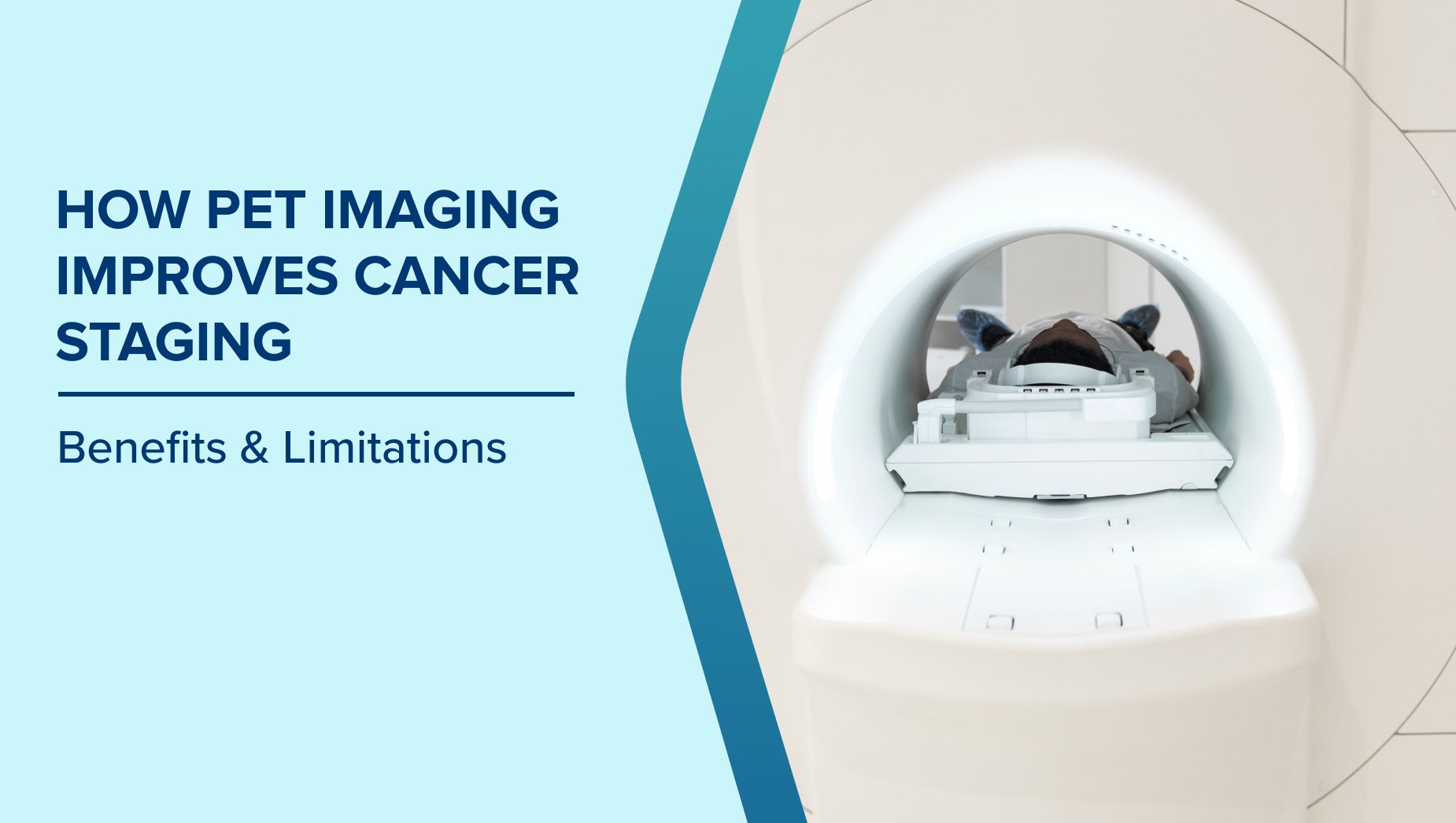
Call Us+91 926 888 0303How PET Imaging Improves Cancer Staging: Benefits & Limitations
By Dr Shashwat Verma in Nuclear Medicine
Jan 05 , 2026 | 7 min read
1
When someone is diagnosed with cancer, one of the first and most important questions is how far the disease has spread. This information decides almost everything that follows, including the type of treatment, its intensity, and the chances of recovery. Cancer staging is not just a medical step. It is the foundation on which the entire treatment plan is built.
Over the years, medical imaging has evolved to provide clearer answers with greater confidence. Among these tools, PET imaging has become one of the most powerful methods for understanding cancer behaviour inside the body. It does more than show the size of a tumour. It reveals how active the cancer is, where it may be spreading, and how the body is responding.
Why Cancer Staging Is More Than a Medical Label
Cancer staging is often described in numbers or stages, but for patients, it means something much deeper. It determines whether treatment will be surgical, medical, or a combination of approaches. It also influences how aggressive the treatment needs to be and what outcomes can realistically be expected. Accurate staging helps in the following ways:
- It guides doctors in selecting treatments that are neither excessive nor insufficient for the actual disease extent
- It avoids unnecessary procedures that may not offer a benefit
- It helps predict how the cancer is likely to behave over time
- It allows patients and families to prepare emotionally and practically for the treatment journey
When staging is incomplete or unclear, treatment decisions become more uncertain. This is where advanced imaging, such as PET imaging, plays a critical role.
What Makes PET Imaging Different From Other Scans
Many patients are familiar with CT scans or MRI scans. These tests show the structure of organs and tissues. PET imaging adds another layer of information by showing how cells function.
Cancer cells tend to use energy differently from normal cells. PET imaging detects these differences in activity, helping doctors identify areas where cancer may be present even if they look normal on other scans.
In simple terms, PET imaging answers not only where the tumour is but also how active it is.
This functional information makes PET imaging especially valuable in cancer staging, where knowing the true extent of disease can completely change treatment direction.
How PET Imaging Improves Staging Accuracy
One of the biggest challenges in cancer care is identifying a disease that is not obvious on routine imaging. Small lymph nodes or early spread may not look abnormal in size, but they may already be involved. PET imaging improves staging accuracy by:
- Detecting cancer activity in lymph nodes that appear normal in size on CT scans
- Identifying distant spread that may not cause symptoms yet
- Differentiating between scar tissue and active cancer in previously treated areas
- Clarifying whether a suspicious area truly represents cancer or a benign change
This level of detail helps ensure that staging reflects reality rather than assumptions.
The Impact of Accurate Staging on Treatment Planning
Cancer treatment works best when it is tailored to the actual stage of the disease. PET imaging often plays a decisive role in shaping the treatment plan. Accurate PET-based staging can lead to the following treatment decisions:
- Choosing surgery only when cancer is truly localised
- Avoiding surgery when cancer has already spread beyond the primary site
- Selecting targeted radiation fields instead of broad exposure
- Adjusting chemotherapy schedule based on disease burden
By guiding these choices, PET imaging helps protect patients from treatments that may not benefit them while ensuring that necessary therapy is not delayed.
Avoiding Overtreatment and Undertreatment Through PET Imaging
One of the less discussed but highly important benefits of PET imaging is its role in avoiding both overtreatment and undertreatment. Overtreatment can lead to unnecessary side effects, prolonged recovery, and long-term health issues. Undertreatment can allow cancer to progress unchecked. PET imaging supports balanced decision-making by:
- Preventing aggressive treatments when the disease is limited and manageable
- Identifying hidden spread that requires more comprehensive therapy
- Reducing trial-and-error approaches to cancer care
- Supporting evidence-based treatment choices rather than assumptions
For patients, this means receiving treatment that is appropriate, justified, and aligned with their actual condition.
PET Imaging and Personalised Cancer Care
Modern oncology increasingly focuses on personalized treatment rather than one-size-fits-all approaches. PET imaging plays an important role in this shift. By revealing how active cancer is in different parts of the body, PET imaging helps doctors personalise care in meaningful ways:
- Treatment plans can be adjusted based on how aggressive the cancer appears
- Therapy intensity can be modified to match disease behaviour
- Response to treatment can be assessed early, rather than waiting months
- Decisions about continuing or changing therapy can be made with confidence
This personalized approach improves both patient outcomes and quality of life.
The Emotional Value of Clear Staging Information
Cancer diagnosis often brings fear, confusion, and countless unanswered questions. Uncertainty about disease extent can be emotionally exhausting. PET imaging provides clarity that many patients find reassuring.
Even when results confirm advanced disease, knowing the truth allows patients and families to plan, cope, and focus on what matters most. Clear staging through PET imaging offers emotional benefits such as:
- Reducing anxiety caused by unanswered questions
- Helping patients understand why certain treatments are recommended
- Supporting realistic expectations about recovery and prognosis
- Empowering patients to participate actively in care decisions
Knowledge, even when difficult, often feels less overwhelming than uncertainty.
PET Imaging in Follow-Up and Treatment Response Assessment
Cancer care does not end with treatment initiation. Monitoring response is equally important. PET imaging is often used after treatment has started or completed to assess effectiveness. It helps distinguish between treatment-related changes and persistent disease. During follow-up, PET imaging can help by:
- Showing whether cancer activity has reduced or disappeared
- Identifying early signs of recurrence before symptoms appear
- Avoiding unnecessary additional treatments when scans show remission
- Supporting timely intervention if the disease returns
This ongoing role makes PET imaging a valuable tool beyond initial staging.
Difference Between Clinical Staging and Imaging-Based Staging
Clinical staging relies on physical examination, symptoms, and basic tests. While important, it has limitations. Imaging-based staging, particularly PET, provides a deeper and more accurate understanding of disease spread. Key differences include:
- Clinical staging may miss silent or early spread
- Imaging-based staging reveals functional cancer activity
- PET imaging often leads to stage changes that alter treatment plans
- Imaging-based staging improves confidence in treatment decisions
For many cancers, PET imaging has become an essential part of accurate staging rather than an optional test.
How PET Imaging Helps Reduce Unnecessary Procedures
Invasive procedures carry risks and emotional burden. PET imaging often helps avoid unnecessary biopsies or surgeries. By clearly identifying active disease areas, PET imaging can:
- Reduce the need for multiple diagnostic biopsies
- Prevent exploratory surgeries when cancer is already widespread
- Help target biopsies to the most relevant sites
- Minimise patient discomfort and recovery time
This practical benefit is often overlooked but deeply appreciated by patients.
Who Benefits the Most From PET Imaging in Cancer Staging
While PET imaging is not required for every cancer type or stage, it is particularly valuable in certain situations. Patients who often benefit most include those with:
- Newly diagnosed cancers where the spread is uncertain
- Lymphoma and lung cancer, where staging accuracy is critical
- Head and neck cancers with complex lymph node involvement
- Recurrence suspicion after previous treatment
- Cancers where treatment decisions depend heavily on staging
Doctors recommend PET imaging when the results are likely to change management rather than as a routine test.
Limitations and Realistic Expectations
While PET imaging is powerful, it is not perfect. Understanding its limitations helps maintain realistic expectations. Important considerations include:
- Not all cancer types show strong PET uptake
- Inflammation or infection can sometimes mimic cancer activity
- PET imaging is usually combined with other tests for accuracy
- Results must always be interpreted by experienced specialists
Patients should view PET imaging as a valuable tool rather than a standalone answer.
Preparing for a PET Imaging Study
Preparation is usually simple but important for accurate results. Patients are often advised to follow specific dietary and activity instructions before the scan. Following preparation guidelines helps ensure clear images and reliable results, reducing the need for repeat scans.
Conclusion
Cancer staging is one of the most important steps in the cancer care journey. PET imaging has transformed this process by providing clearer, more accurate, and more meaningful information than ever before. By influencing treatment choices, reducing unnecessary procedures, supporting personalised care, and offering emotional reassurance, PET imaging plays a vital role beyond diagnosis alone.
For patients and caregivers, understanding how PET imaging fits into cancer staging helps build trust in the treatment plan and confidence in the path ahead. When decisions are based on accurate information, care becomes not only more effective but also more humane.
Frequently Asked Questions
Does PET imaging expose patients to high radiation levels?
The radiation exposure is controlled and kept within safe medical limits. The benefits of accurate staging usually outweigh the minimal risk involved.
Can PET imaging replace biopsy?
PET imaging does not replace biopsy but helps guide where biopsies should be taken and whether they are necessary.
Is PET imaging painful?
The scan itself is painless. Patients need to lie still, which some may find mildly uncomfortable.
How long does it take to get results?
Results are usually available within a few days, depending on the facility and case complexity.
Can PET imaging detect cancer recurrence early?
Yes, PET imaging is often used to detect recurrence before symptoms appear, allowing earlier intervention.
Written and Verified by:

Related Blogs

Dr Shashwat Verma In Nuclear Medicine
Nov 10 , 2025 | 4 min read
Blogs by Doctor
Nuclear Medicine: Its Role in Diagnosing Heart Diseases
Dr Shashwat Verma In Nuclear Medicine
Nov 10 , 2025 | 4 min read
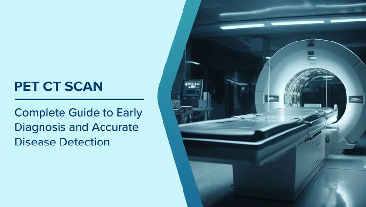
PET CT Scan: A Complete Guide to Early Diagnosis and Accurate Disease Detection
Dr Shashwat Verma In Nuclear Medicine
Nov 30 , 2025 | 8 min read
Most read Blogs


Related Blogs
Dr Shashwat Verma In Nuclear Medicine
Nov 10 , 2025 | 4 min read
Blogs by Doctor
Nuclear Medicine: Its Role in Diagnosing Heart Diseases
Dr Shashwat Verma In Nuclear Medicine
Nov 10 , 2025 | 4 min read
PET CT Scan: A Complete Guide to Early Diagnosis and Accurate Disease Detection
Dr Shashwat Verma In Nuclear Medicine
Nov 30 , 2025 | 8 min read
Most read Blogs