 WhatsApp Us (24/7)
 +91 926 888 0303 (24/7)


Abstract: The anterolateral thigh flap has been the workhorse flap for coverage of soft-tissue defects in the head and neck for decades. However, the reconstruction of multiple and complex soft-tissue defects in head and neck with multi-paddled anterolateral thigh (ALT) or chimeric flaps is still a challenge for reconstructive surgeries, particularly for total maxillectomy defects, as it requires reconstruction of nasal lining, palatal reconstruction, cheek mucosa, and even skin defect reconstruction, if any created by the oncosurgeon. Here, we report a clinical case in which a multi-paddled anterolateral thigh flap was used for a complex soft-tissue defect created after onco-resection for recurrent squamous cell carcinoma of the maxilla. In this case, total maxillectomy with hemimandibulectomy was done along with excision of the skin of the lower half of the face of that side. Most branches of the external carotid artery, along with the internal jugular vein (IJV) were ligated in previous surgery. The availability of a vascular pedicle was also a challenge. Here, reconstruction was performed using a large ALT flap that was divided into four paddles based on three perforators for reconstruction of all linings, including a de-epithelised skin paddle for orbital floor reconstruction.
Key words: Free Anterolateral Thigh (ALT) Flap, Maxillectomy, Mandibulectomy, Head and Neck OncoReconstruction.
Introduction
Complex soft-tissue defects of the head and neck that involve multiple and nonadjacent anatomic sites can arise after radical surgery for advanced-stage malignancies or multiple lesions of the upper aerodigestive tract. Immediate flap reconstruction is the gold standard for the surgical treatment of these defects after tumour ablation. However, functional and aesthetic reconstruction of complex and multiple defects in the head and neck remains a major surgical challenge, because the reconstruction of multiple and nonadjacent defects requires a flexible and feasible flap with a multiplanar configuration consisting of multiple paddles.1 For multiple and complex soft-tissue defects, reconstruction with three- dimensional microvascular flaps is often the preferred alternative. The free anterolateral thigh (ALT) flap, first described by Song et al.2 in 1984, is a versatile and reliable reconstructive option. This flap has become a workhorse for head and neck reconstruction in recent decades.3,4 The advantages of ALT flap include consistent and reliable anatomy, long vascular pedicle, feasibility to create multiple skin paddles by recruiting additional perforators, the flexibility to reconstruct composite defects by recruiting different tissue types, and low donor site morbidity. Based on multiple cutaneous perforators originating from the descending branch of the lateral circumflex femoral artery (LCFA), free ALT flap can be harvested with multiple skin paddles for multiple and complicated soft-tissue defects, which can eliminate the need for two or more separate flaps and vascular anastomosis.5-7 In this study, we present our experience with free multi-paddled ALT flap for functional and aesthetic reconstruction of complex head and neck softtissue defects involving multiple nonadjacent anatomic sites in a single operation.
Challenging reconstructive cases occur in a previously treated neck following either surgery, radiation (with or without chemotherapy), or a combination of both. Patients with these prior interventions have cervical anatomy with distorted tissue planes, inflammation, and scarred vessels, leaving the neck depleted of suitable recipient vessels. A vessel-depleted neck (VDN) occurs in around 7% of all patients receiving microvascular reconstructions in the head and neck.8
Case Report
Our patient was a 60-year-old lady who was diagnosed with recurrent cancer of the right-sided buccal mucosa. The patient was a known diabetic and hypertensive (controlled on regular medications), with wide excision and primary closure of right buccal mucosal squamous carcinoma about 5 years earlier, along with a right neck dissection.
She presented us with less than one-finger mouth opening and recurrence of carcinoma at the same location, thus a total maxillectomy with hemimandibulectomy was performed by the oncosurgeon. On the same side of the neck, all branches of the external carotid were ligated, IJV was also excised and ligated at the base of the neck.
Reconstruction was a challenge as there was a defect involving the floor of the right orbit, right lateral nasal wall, palate (two-thirds of the soft and hard palate were excised), right posterior pharyngeal wall, buccal mucosa, floor of mouth, and skin of cheek and mandibular area (Figure 1 and 2).
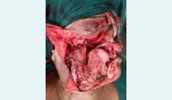
Figure 1: Front view — Post excision defect.
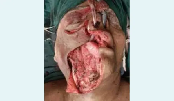
Figure 2: Lateral view (Day 5) — Reconstruction with single anterolateral thigh (ALT) flap.
On Doppler (handheld), two prominent perforators were found along the line of ALT flap, thus a flap with the full thigh length (26 cm x 9 cm wide) of skin paddle was planned, including a good bulk of vastus lateralis muscle. After the anterior skin incision, two septocutaneous and one musculocutaneous perforators were found. The flap was harvested, saving all perforators and taking a good bulk of muscle.
After getting three good perforators, the division of the flap into three parts was planned:
- Lowermost skin paddle – 13 cm x 9 cm – for external skin defect
- Skin paddle above No. 1 – 6 cm x 9 cm – for intraoral defect
- Uppermost skin paddle was used in two parts:
- 3 cm x 4 cm – terminal part de-epithelised to support orbit
- 3 cm x 5 cm – for lateral nasal wall
- After partial inset of flap in the oral cavity (Figure 3 and
- vascular anastomoses were done in the following sequence:
Flap artery – external carotid – end to side
Two flap veins – IJV – end to end with closure of middle part of the IJV. Reconstructive with free anterolateral thigh flap is shown in figure 3 (lateral view) and figure 4 (frontal view).
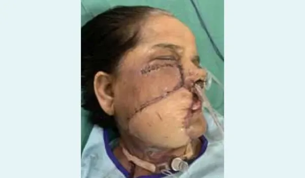
Figure 3: Lateral view — Defect after wide excision of right total maxillectomy, right hemimandibulectomy with skin of upper lip, lower lip, and cheek.
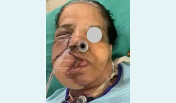
Figure 4: Front view (Day 5) — Reconstruction with free anterolateral thigh (ALT) flap.
Vascularity of the flap was excellent, as there was no marginal necrosis of the flap of even a millimetre at any margin. There was some fat necrosis of the facial skin leading to wound dehiscence, so debridement and secondary suturing were done on post-operative Day 14. The tracheostomy tube was kept for a longer period as a large part of the oral and nasal cavity was formed by the flap, leading to difficulty in breathing while attempting removal of the tracheostomy tube. The tube was removed with a gradual decrease in size of the uncuffed tube.
Discussion
In India, the majority of head and neck cancers present as locally advanced Stage III/IV disease.9 As the incidence and diagnosis of oral cancer increase, excision of the head and neck cancers will frequently yield large, complex three-dimensional defects. These defects test the limits of the reconstructive surgeons and thereby obligate imaginative planning and surgical astuteness. Since its description in 1984 by Song et al., 10 ALT flap has become the preferred flap for reconstruction in varied settings. Other good options for these extensive defects are the flaps on the scapular or parascapular axis, but these require a change in position. Deep inferior epigastric artery, vertical rectus abdominis flaps, and second free flap are also other feasible options in this situation. Chimeric flaps are also described in the literature for reconstruction of such defects, taking the antero-medial thigh (AMT ) along with the ALT flap.
In our case, we have achieved all the desired goals: providing support for the eyeball, lining of lateral nasal wall, posterior pharyngeal wall with buccal mucosa, and reconstruction of a large skin defect in a vessel-depleted post-radiation neck.
Conclusion
The ALT flap is a versatile and commonly used flap in reconstructive surgery, especially for covering defects in the head and neck, trunk, and lower extremities. ALT flap is known for its large size, reliable pedicle, and relatively low donor site morbidity. ALT flap has been the workhorse flap for coverage of soft-tissue defects in the head and neck for decades now. However, the reconstruction of multiple and complex soft-tissue defects in the head and neck with multi-paddled ALT or chimeric flaps remains a challenge for reconstructive surgeries, particularly for total maxillectomy defects. Here, we report a clinical case in which a multi-paddled ALT was used for a complex soft-tissue defect created after onco-resection for recurrent squamous cell carcinoma of the maxilla.
Anurag Pandey. Quadri Paddle Anterolateral Thigh Flap for Complex Maxillo-Mandibular-Cheek Defect
in Vessel Depleted Neck. MMJ. 2025, September. Vol 2 (3).
References
- Longo B, Nicolotti M, Ferri G, et al. Sagittal split osteotomy of the fibula for modeling the new mandibular angle. J Craniofac Surg. 2013;24(1):714.
- Song YG, Chen GZ, Song YL. The free thigh flap: a new free flap concept based on the septocutaneous artery. Br J Plast Surg. 1984;37(2):149–59.
- Koshima I, Hosoda S, Inagawa K, et al. Free combined anterolateral thigh flap and vascularized fibula for wide, through-and-through oromandibular defects. J Reconstr Microsurg. 1998;14(08):529–34.
- Shieh SJ, Chiu HY, Yu JC, et al. Free anterolateral thigh flap for reconstruction of head and neck defects following cancer ablation. Plast Reconstr Surg. 2000;105(7):2349–57.
- Park CW, Miles BA. The expanding role of the anterolateral thigh free flap in head and neck reconstruction. Curr Opin Otolaryngol Head Neck Surg. 2011;19(4):263–8.
- Huang WC, Chen HC, Jain V, et al. Reconstruction of throughand-through cheek defects involving the oral commissure, using chimeric flaps from the thigh lateral femoral circumflex system. Plast Reconstr Surg. 2002;109(2):433–41.
- Lai CL, Ou KW, Chiu WK, et al. Reconstruction of the complete loss of upper and lower lips with a chimeric anterolateral thigh flap: a case report. Microsurgery. 2012;32(1):60–3.
- Jacobson AS, Eloy JA, Park E, et al. Vessel-depleted neck: Techniques for achieving microvascular reconstruction. Head Neck. 2008;30(2):201–7.
- Jiang C, Guo F, Li N, et al. Multipaddled anterolateral thigh chimeric flap for reconstruction of complex defects in head and neck. PLoS One. 2014;9(9):e106326.
- Shah SB, Sharma S, D’Cruz AK. Head and neck oncology: The Indian scenario. South Asian J Cancer. 2016;5(03):104–5.