 WhatsApp Us (24/7)
 +91 926 888 0303 (24/7)


Abstract: A 26-year-old male reported with high-grade fever accompanied by chills and rigours persisting for four days, followed by vomiting and dull, aching pain in the right hypochondrium. Laboratory investigations revealed leucocytosis with raised procalcitonin levels. An abdominal ultrasound (USG) showed no liver parenchymal abnormalities. As the patient’s symptoms persisted despite antibiotic therapy, a comprehensive pyrexia of unknown origin (PUO) workup was undertaken. Serological tests for Toxoplasma, Epstein–Barr virus (EBV), anti-capsid antibodies, tropical fever panel, cytomegalovirus (CMV) immunoglobulin G (IgG) and immunoglobulin M (IgM), as well as blood cultures (aerobic and anaerobic), proteinase-3 (PR-3, cytoplasmic antineutrophil cytoplasmic antibody [c-ANCA]), and myeloperoxidase (MPO, perinuclear antineutrophil cytoplasmic antibody [p-ANCA]) were all negative. Anti-nuclear antibody (ANA) testing showed cytoplasmic speckled positivity. A positron emission tomography–computed tomography (PET–CT) scan revealed a liver abscess (amoebic) with marked hepatomegaly and an abscess in segment VIII of the liver. Intravenous (IV) metronidazole 750 mg three times daily and oral hydroxychloroquine 200 mg once daily were immediately started. CT-guided drainage of the abscess yielded around 210 mL of anchovy-sauce-like pus, which tested positive for Entamoeba histolytica (EH). The patient improved clinically, remained haemodynamically and vitally stable and was subsequently discharged.
Key words: Right Hypochondrial Pain, USG Abdomen, Anchovy Sauce, Intravenous Metrogyl, CT-Guided Drainage of Abscess.
Introduction
Amoebic liver abscess (ALA) is a parasitic infection resulting from Entamoeba histolytica (EH), an intestinal protozoan. Transmission typically occurs through the consumption of food or water contaminated with the cyst form of EH, which is the infective stage. These cysts withstand gastric acidity and travel to the distal ileum, where they excyst to release trophozoites — the active, feeding form of the parasite. In over 90% of infected individuals, trophozoites remain confined to the intestinal lumen, feeding on bacteria and mucosal cells without causing clinical symptoms. However, in less than 1% of cases, the trophozoites invade the intestinal mucosa and spread to the liver via the portal circulation, leading to the formation of liver abscesses. Among the various clinical presentations of amoebiasis, ALA is the most prevalent and carries the highest risk of mortality. It continues to be the leading cause of liver abscess in developing and resource-limited settings.1
Although asymptomatic EH infections occur at similar rates in both genders, invasive amoebiasis, particularly ALA, is significantly more common in males, who are four to nine times more likely to develop the condition than females. This gender disparity has been documented even among travellers from non-endemic regions. The heightened susceptibility in males is believed to be associated with hormonal influences — particularly testosterone — as well as higher rates of alcohol consumption. Once EH trophozoites invade the colonic mucosa, they can reach the liver through the portal venous system. The right lobe of the liver is most frequently affected, likely because it receives the majority of portal blood via the superior mesenteric vein, which drains the caecum — the primary site of trophozoite invasion.2
Each year, EH is responsible for an estimated 40,000 to 100,000 deaths worldwide, making it the second leading cause of mortality among parasitic infections, surpassed only by malaria. Although invasive amoebiasis develops in less than 10% of infected individuals, once the parasite overcomes the host’s innate immune defences, it can lead to serious complications such as amoebic colitis and liver abscesses. At the molecular level, EH employs a key virulence factor known as the Gal/GalNAc lectin to facilitate tissue invasion. This lectin plays a dual role: it enables the parasite to adhere to and invade host tissues and simultaneously triggers an antibody response that can inhibit further adherence, offering some degree of protection against recurrent or severe disease.3
Clinically, amoebiasis presents in two primary forms: intestinal and extraintestinal. The intestinal form, which is the most common, arises when EH invades the colonic mucosa — typically in the caecum or ascending colon. Clinical manifestations range from mild abdominal discomfort to severe ulcerative colitis, characterised by bloody and mucus-laden stools, also known as amoebic dysentery. On the other hand, extraintestinal amoebiasis occurs when the parasite spreads beyond the gastrointestinal tract, affecting other organs. Among these, hepatic amoebiasis (liver abscess) is the most frequent, followed by pulmonary involvement, making the liver and lungs the two most commonly affected sites in extraintestinal disease.4
Case Report
A 26-year-old male patient presented with complaints of high-grade fever with chills and rigours persisting for four days, followed by vomiting containing food particles (non-bilious, non-blood stained) and dull aching, continuous, abdominal pain in the right hypochondrial region. The patient had a recent history of outside food consumption. There were no complaints of loose stools, haematochezia, or malaena. There was no significant past medical history, drug allergies, chronic illnesses or chronic drug use.
On presentation, the patient’s vitals were as follows: he was febrile with a temperature of 101.9°F, blood pressure 110/70 mmHg, pulse rate 114 beats per minute (bpm), and SpO₂ 98% on room air. Physical examination revealed intercostal tenderness in the right hypochondrial region. Bowel sounds were normal, and the rest of the systemic examination was unremarkable. The patient was empirically started on intravenous (IV) levofloxacin 500 mg once daily and ceftriaxone 1 g twice daily, along with other supportive measures.
Initial laboratory investigations indicated an infectious aetiology, as shown by leucocytosis (white blood count [WBC]-13,150) and raised procalcitonin (1.6). Imaging studies, including abdominal ultrasonography (USG), suggested partial gallbladder distension but no liver parenchymal involvement. Chest X-ray was normal. The prothrombin time/international normalised ratio (PT/ INR) was 14.6/1.34. Urine routine was normal. Laboratory investigations are shown in Table 1.
| Day of Admission | Hb (g/dL) | WBC (/μL) | Platelets (/μL) | Na (mEq/L) | K/Cl (mEq/L) | Creatinine (mg/dL) | PCT (ng/mL) | SGOT/SGPT (U/L) |
|---|---|---|---|---|---|---|---|---|
| Day 1 | 12.2 | 13150 | 237000 | 133 | 4.0/97 | 0.8 | 1.6 | |
| Day 2 | 12.1 | 15330 | 259000 | |||||
| Day 3 | 12.3 | 16820 | 270000 | 0.9 | 1.87 | 33.8/42.7 | ||
| Day 4 | 12.4 | 18080 | 309000 | 136 | 4.0/100 | 0.85 | ||
| Day 5 | 12.6 | 23390 | 354000 | 1.64 | ||||
| Day 7 | 11.4 | 25790 | 377000 | |||||
| Day 9 | 11.2 | 21680 | 447000 | 136 | 3.33/101 | 0.63 | ||
| Day 10 | 11.2 | 15580 | 581000 | 133.4 | 3.4/97.4 |
Table 1: Laboratory investigations.
Abbreviations: Cl: Chloride; Hb: Haemoglobin; K: Potassium; Na: Sodium; PCT: Procalcitonin; SGOT: Serum Glutamic-Oxaloacetic Transaminase; SGPT: Serum Glutamic-Pyruvic Transaminase; WBC: White Blood Cell.
Considering persistent fever and abdominal symptoms, and as the patient did not respond to antibiotics, further investigations for pyrexia of unknown origin (PUO) were conducted. Serologies for Toxoplasma, Epstein–Barr virus (EBV), anti-capsid antibodies, and a tropical fever panel were all negative. Further investigations revealed cytomegalovirus (CMV) immunoglobulin G (IgG) at 11 (negative) and immunoglobulin M (IgM) non-reactive; blood cultures (aerobic and anaerobic) showed no growth, proteinase-3 (PR-3, cytoplasmic antineutrophil cytoplasmic antibody [c-ANCA]) and myeloperoxidase (MPO, perinuclear antineutrophil cytoplasmic antibody [p-ANCA]) were negative, antinuclear antibody (ANA) showed cytoplasmic speckled positivity; ferritin was elevated at 739 ng/mL; lactate dehydrogenase (LDH) was 185 U/L, and triglycerides were 113 mg/dL.
A positron emission tomography–computed tomography (PET–CT) scan was subsequently performed to determine the fever aetiology, which revealed findings consistent with a liver abscess (amoebic) (Figure 1 and 2). There was marked hepatomegaly and an abscess in segment VIII of the liver. The cupola of the right diaphragm was elevated, secondary to hepatomegaly. The liver measured about 17.0 cm in the long axis and extended below the right costal margin. A large, peripherally enhancing lesion in segment VIII, extended to the hepatic capsule, measuring 8.0 x 7.6 cm in maximal axial dimension, with a cephalocaudal extent of about 7.3 cm and an approximate volume of 220 cm3 . The wall thickness ranged from 5 to 9 mm. It was surrounded by a hypoattenuating zone of hepatic oedema, exhibiting metabolic activity with maximum standardised uptake value (SUVMax) of 7.0.
Periportal hypoattenuation was suggestive of periportal oedema, with no dilatation of the intrahepatic biliary radicals. The intrahepatic vasculature was normal.
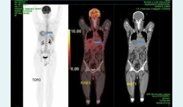
Figure 1: Positron emission tomography–computed tomography (PET–CT) imaging with blue arrows showing the amoebic liver abscess.
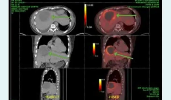
Figure 2: Positron emission tomography–computed tomography (PET–CT) imaging with blue arrows showing the amoebic liver abscess.
Discussion
The term amoebic liver abscess (ALA) is somewhat misleading, as cavity formation and liquefaction do not result from suppuration but rather from a distinctive type of necrosis. The necrotic tissue appears as though chemically dissolved, leading to the early belief that EH secretes a toxin capable of lysing hepatocytes — hence the species name histolytica. However, it is now understood that proteolytic enzymes released primarily by inflammatory cells, play the central role in hepatic tissue destruction.
The gross morphology of ALA varies according to disease stage and severity. In the early phase, lesions consists of a central area of liquefied necrotic tissue, often described as chocolate-coloured sterile pus. This liquefied material within the abscess contains red blood cells (RBCs), leucocytes, and disintegrated hepatocytes, producing the classic reddish-brown appearance often likened to chocolate sauce or anchovy sauce. This necrotic core is surrounded by a zone of partially liquefied and solid tissue, giving the abscess wall a ragged or shaggy appearance. Notably, a mature fibrous capsule is absent at this stage. The surrounding hepatic tissue is typically congested, compressed, and oedematous, and mass effect may cause pressure on adjacent liver parenchyma or the hepatic capsule. In fatal cases, venous thrombosis and ischaemic infarction are frequently observed.
Clinically, ALA manifests in three forms:
- Subacute mild ALA (~80%) is the most common form. It develops over 2–4 weeks with low-grade symptoms like fever, right upper quadrant pain, and hepatomegaly. Symptoms usually resolve rapidly with medical treatment. A characteristic finding on physical examination is localised tenderness over the right intercostal spaces. Organ dysfunction is minimal or absent.
- Acute aggressive ALA (~13%) is severe and rapidly progressive, presenting with high-grade fever, chills, and pronounced right upper quadrant tenderness due to exquisitely tender hepatomegaly. One of the hallmark features of this form is spontaneous abscess rupture, which may be the initial presentation in up to 57% of cases. Free intraperitoneal rupture frequently results in generalised peritonitis, necessitating hospitalisation, abscess drainage, and carries high mortality. Due to its severity, hospitalisation is required in up to 90% of cases, and around 13% may necessitate intensive care unit (ICU) admission. Misdiagnosis is common, as clinical features overlap with acute cholecystitis, appendicitis, or bowel perforation. Unlike in subacute disease, medical therapy alone is often insufficient. In most cases, abscess drainage — either via needle aspiration or catheter insertion — is required to achieve clinical improvement.
- Chronic indolent ALA (~10%–20%) is prolonged and milder, presenting with dull pain, systemic symptoms like weight loss, and complications like secondary infection. A history of previous medical therapy or even prior needle aspiration is frequently reported. Imaging plays a key role in evaluation.
Imaging plays a critical role in differentiating these clinical forms. Contrast-enhanced CT is particularly valuable, as it differentiates viable hepatic tissue from necrotic areas, allowing better distinction between chronic necrotic masses and active, aggressive abscesses. Acute ALA (Type 1) typically appears as ragged-edged lesions; subacute ALA (Type II) shows rim-enhancing walls with a thin rim of perilesional oedema, producing a “halo sign”; chronic ALA (Type III) demonstrates thick, non-enhancing fibrotic walls.1
ALA has a highly variable presentation, causing diagnostic difficulties. Uncommon manifestations occur due to abscess rupture into neighbouring cavities — pleura, pericardium, peritoneum — or compression of tubular ALA has a highly variable presentation, causing diagnostic difficulties. Uncommon manifestations occur due to abscess rupture into neighbouring cavities — pleura, pericardium, peritoneum — or compression of tubular structures in the hepatic hilum, or distant embolic dissemination.5 The diagnosis of ALA is confirmed by microscopic examination of aspirated pus, which characteristically resembles anchovy sauce. Aspiration is particularly indicated when the abscess exceeds 10 cm, shows a thin rim of liver parenchyma suggesting impending rupture, or demonstrates rapid enlargement.6
The laboratory diagnosis of amoebiasis relies on a combination of parasitological, serological, and molecular methods. Parasitological techniques, such as microscopy and culture, remain foundational, with microscopic detection of EH in stool, tissue, or body fluid samples still considered the diagnostic gold standard. However, microscopy is limited by low sensitivity and specificity, especially in distinguishing EH from non-pathogenic species. Serological tests such as enzyme-linked immunosorbent assay (ELISA), indirect haemagglutination assay (IHA), and latex agglutination are widely used for diagnosing extraintestinal amoebiasis, where stool microscopy often fails. In recent decades, the polymerase chain reaction (PCR) technique has emerged as a highly sensitive and specific tool for detecting EH deoxyribonucleic acid (DNA). In addition, imaging modalities such as USG, CT, magnetic resonance imaging (MRI), and radiography have become valuable adjuncts for diagnosing hepatic and other extraintestinal forms of amoebiasis, aiding in localisation, characterisation, and monitoring of abscesses and other lesions.4
Metronidazole remains the most effective agent, with cure rates approaching 90%.1 It is typically administered IV at 750 mg three times daily for 7–10 days. In severe cases, other agents such as emetine, dehydroemetine, and chloroquine may be considered. The risk of complications increases with larger abscess size and multiplicity of abscesses. Autopsy studies reveal that up to 60% of fatal cases involve multiple abscesses, often measuring 10 to 15 cm. Comparative data from Southeast Asia report a higher incidence of large (> 5–10 cm) and multiple abscesses in approximately 50% of cases, highlighting geographic variation.1
Conclusion:
Liver abscess is usually diagnosed by USG abdomen as a hypoechoic lesion; however, in its early stages, it may appear isoechoic and thus be missed. Hence, in early stages if sonography is inconclusive, particularly in patients with PUO, CT or PET imaging should be performed to facilitate early diagnosis.
Nitin Rathod, Andrea Janice Fonseca, Rajat Shende, Harsh Jani. Revealing the Hidden Pathogen: The Role
of Positron Emission Tomography in Diagnosing Amoebic Liver Abscess in an Immunocompetent Patient.
MMJ. 2025, September. Vol 2 (3).
References
- Priyadarshi RN, Kumar R, Anand U. Amebic liver abscess: Clinico-radiological findings and interventional management. World J Radiol. 2022;14(8):272–85.
- Kumar R, Patel R, Priyadarshi RN, et al. Amebic liver abscess: An update. World J Hepatol. 2024;16(3):316–30.
- Usuda D, Tsuge S, Sakurai R, et al. Amebic liver abscess by Entamoeba histolytica. World J Clin Cases. 2022;10(36):13157– 66.
- Nasrallah J, Akhoundi M, Haouchine D, et al. Updates on the worldwide burden of amoebiasis: A case series and literature review. J Infect Public Health. 2022;15(10):1134–41.
- Mukhopadhyay M, Saha AK, Sarkar A, et al. Amoebic liver abscess: presentation and complications. Indian J Surg. 2010;72(1):37–41.
- Bansal Y, Maurya V, Tak V, et al. Clinical and laboratory profile of patients with amoebic liver abscess. Trop Parasitol. 2022;12(2):113–8.