 WhatsApp Us (24/7)
 +91 926 888 0303 (24/7)


Abstract: Magnetic resonance imaging (MRI) plays a pivotal role in antenatal diagnosis of placenta percreta, particularly when ultrasound findings are equivocal. Timely intervention improves maternal and foetal outcomes. We present a case of a 35-year-old gravida 2, para 1, live 1 (G2P1L1) woman with a history of prior lower segment caesarean section (LSCS) at 31 weeks of gestation with leaking per vaginum. This was a rare but life-threatening case of placenta percreta diagnosed antenatally by MRI in a patient with preterm premature rupture of membranes. MRI revealed features of placenta percreta, including loss of the uteroplacental interface, placental bulge, and bladder invasion. Caesarean delivery and hysterectomy were performed with intraoperative management of massive haemorrhage and bladder invasion. With this case study, we aim to emphasise the value of prompt imaging in maternal-foetal outcomes.
Key words: Placenta Percreta, Magnetic resonance imaging (MRI), Placenta Accreta Spectrum (PAS), Bladder Invasion, Obstetric Haemorrhage, Hysterectomy.
Introduction
Placenta accreta spectrum (PAS) includes placenta accreta, increta, and percreta — defined by progressive degrees of abnormal placental adherence and invasion into the myometrium and adjacent pelvic organs. With the global rise in caesarean deliveries, PAS incidence has surged from 1 in 2500 to nearly 1 in 533 pregnancies.1 Early and accurate diagnosis is crucial for reducing maternal morbidity and mortality associated with massive haemorrhage and organ injury during delivery. Ultrasound remains the initial imaging modality, but magnetic resonance imaging (MRI) is increasingly recognised for its superior soft tissue contrast and ability to delineate the depth of invasion.2
Case Report
A 35-year-old gravida 2, para 1, live 1 (G2P1L1) woman with a prior lower segment caesarean section (LSCS) presented at 31 weeks of gestation with complaints of leaking per vaginum since morning. The patient informed us that her last menstrual period was on 25th October 2024. She had been diagnosed with gestational diabetes mellitus a month prior and was on metformin 500 mg daily.
Foetal movements were normal, and baseline investigations, including complete blood count (CBC), coagulation profile, renal and liver function tests, were within normal limits.
Ultrasound investigation
Antenatal ultrasound showed an anterior placenta with grade II–III maturity. Multiple placental lakes were visualised. The myometrial-placental interface was poorly visualised in the lower uterine segment, raising concern for PAS. Colour Doppler studies revealed normal resistive indices. Mild polyhydramnios was also present. Based on these findings, MRI was advised to further evaluate suspected placental invasion (Figure 1).
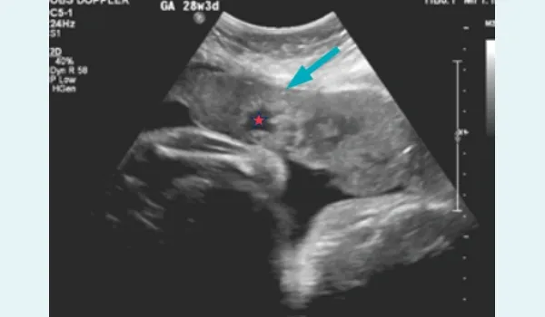
Figure 1: Antenatal ultrasound showing an anterior placenta with placental lake indicated by a red star and loss of the myometrium and placental junctional complex indicated by the blue arrow.
MRI investigation
MRI performed on a 3T scanner revealed a single live intrauterine gestation in cephalic presentation. The anterior placenta appeared heterogeneous in signal intensity, with loss of the uteroplacental interface and a placental bulge measuring 12.9 cm (SI) × 12 cm (TR). Numerous placental lacunae and prominent intra-placental flow voids were seen. Importantly, there was a loss of fat plane between the placenta and urinary bladder, with mild irregularity at the interface, strongly suggesting bladder invasion, consistent with placenta percreta (Figures 2–6).
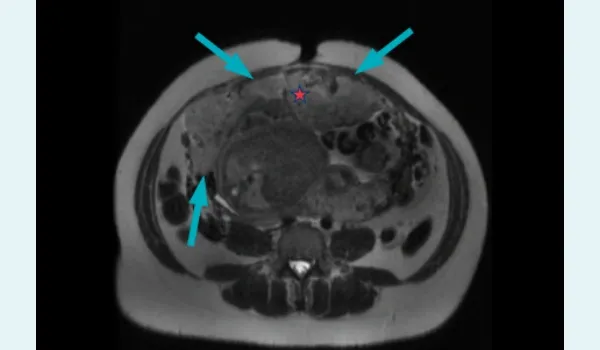
Figure 2: Axial T2-weighted magnetic resonance imaging (MRI) showing placental bulge with heterogeneous signal intensity indicated with a red star and loss of uteroplacental interface indicated by blue arrows.
MRI investigation
MRI performed on a 3T scanner revealed a single live intrauterine gestation in cephalic presentation. The anterior placenta appeared heterogeneous in signal intensity, with loss of the uteroplacental interface and a placental bulge measuring 12.9 cm (SI) × 12 cm (TR). Numerous placental lacunae and prominent intra-placental flow voids were seen. Importantly, there was a loss of fat plane between the placenta and urinary bladder, with mild irregularity at the interface, strongly suggesting bladder invasion, consistent with placenta percreta (Figures 2–6).
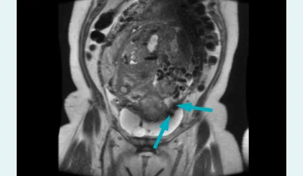
Figure 3: Coronal T2 magnetic resonance imaging (MRI) showing bladder wall invasion with prominent blood vessels indicated by blue arrows.
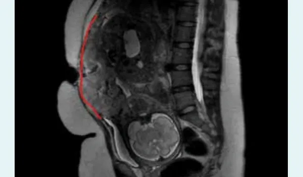
Figure 4: Sagittal magnetic resonance imaging (MRI) showing absence of myometrium along the entirety of the anterior placental margin, represented by a red line.
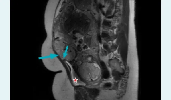
Figure 5: Sagittal magnetic resonance imaging (MRI) showing placental extension beyond the uterine serosa with obliteration of the fat plane between placenta and bladder (indicated by red star) as indicated by blue arrows.
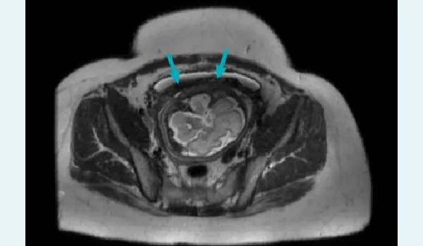
Figure 6: Axial T2-weighted magnetic resonance imaging (MRI) showing loss of the bladder interface with bladder wall invasion (blue arrows).
Surgical and postoperative management
The patient underwent emergency caesarean section followed by an obstetric hysterectomy. Dense invasion of the posterior bladder wall by the placenta was confirmed intraoperatively (Figure 7). Multidisciplinary involvement of cardiovascular and urology teams was necessary. Bilateral internal iliac artery ligation was performed to control haemorrhage. The invaded bladder segment was partially resected and repaired. Estimated blood loss was 7.5 litres; the patient received multiple blood products. The neonate was stabilised in the neonatal intensive care unit (NICU).
Postoperatively, the patient required intensive care monitoring but had an uneventful recovery and was discharged on Day 10.
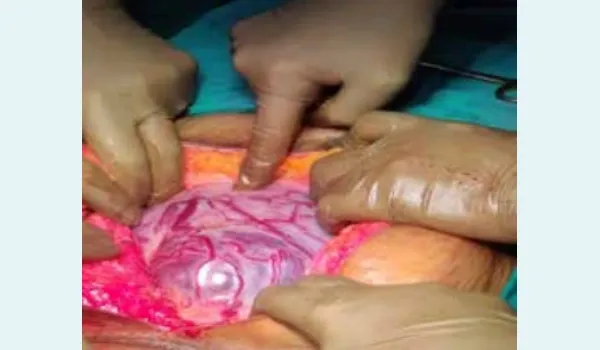
Figure 7: Intraoperative image showing completely thinned out myometrium with the underlying placenta, along with bulging vessels.
Surgical and postoperative management
The patient underwent emergency caesarean section followed by an obstetric hysterectomy. Dense invasion of the posterior bladder wall by the placenta was confirmed intraoperatively (Figure 7). Multidisciplinary involvement of cardiovascular and urology teams was necessary. Bilateral internal iliac artery ligation was performed to control haemorrhage. The invaded bladder segment was partially resected and repaired. Estimated blood loss was 7.5 litres; the patient received multiple blood products. The neonate was stabilised in the neonatal intensive care unit (NICU).
Postoperatively, the patient required intensive care monitoring but had an uneventful recovery and was discharged on Day 10.
Discussion
Placenta percreta is the most severe form of PAS and can result in catastrophic haemorrhage, bladder injury, and maternal death if not diagnosed preoperatively.3 Risk factors include prior caesarean section, placenta previa, advanced maternal age, and multiparity. In this case, the presence of a scarred uterus from prior LSCS and an anterior placenta raised the index of suspicion.
Role of imaging
Ultrasound is the first-line modality for PAS screening. The key features include:
- Loss of clear zone between placenta and myometrium
- Multiple placental lacunae ("swiss-cheese" appearance)
- Myometrial thinning (< 1 mm)
- Bladder wall interruption on colour Doppler4
However, ultrasound has limitations, especially in posterior placentas or when the lower uterine segment (LUS) is obscured. In such cases, MRI serves as a powerful adjunct. It offers:
- High soft tissue resolution
- Multiplanar evaluation
- Detailed depiction of the depth and lateral spread of invasion
- Better visualisation of posterior placentas, bladder interface, and extrauterine extension.5,6
In this patient, MRI demonstrated with clarity:
- Uteroplacental interface loss
- Placental bulge and heterogeneity
- Flow voids (indicative of increased vascularity)
- Loss of bladder fat plane with irregularity — predictive of bladder invasion, later confirmed surgically
MRI findings in PAS have shown high specificity (82%–100%) and positive predictive value (90%–100%), especially for placenta percreta and bladder involvement.5
Multidisciplinary planning
MRI allows surgeons to plan the extent of surgery, anticipate intraoperative challenges, and arrange blood products and multidisciplinary teams. The preoperative diagnosis enabled prompt involvement of urologists and vascular surgeons in this case, crucial in managing massive bleeding and bladder resection.
Acknowledgements
We thank the Departments of Obstetrics, Anaesthesia, and Urology for intraoperative support and the NICU staff for neonatal care.
CONCLUSION:
This case underscores the critical role of MRI in the evaluation of PAS, particularly placenta percreta. MRI not only enhances diagnostic accuracy but also facilitates optimal surgical planning, reduces intraoperative surprises, and improves maternal-foetal outcomes. Integration of MRI into protocols for suspected PAS is strongly recommended, especially in high-risk patients.
Nikita Singh, Mitusha Verma, Deepak Patkar. Through the Veil: Magnetic Resonance Imaging Mapping
of Placenta Percreta with Bladder Invasion in a Scarred Uterus. MMJ. 2025, September. Vol 2 (3).
References
- Wu S, Kocherginsky M, Hibbard JU. Abnormal placentation: twenty-year analysis. Am J Obstet Gynecol. 2005;192(5):1458– 61.
- Maldjian C, Adam R, Pelosi M, et al. MRI appearance of placenta accreta. J Comput Assist Tomogr. 1999;23(5):780–5.
- Silver RM, Landon MB, Rouse DJ, et al. Maternal morbidity associated with multiple repeat cesarean deliveries. Obstet Gynecol. 2006;107(6):1226–32.
- D'Antonio F, Iacovella C, Palacios-Jaraquemada J, et al. Prenatal identification of invasive placentation using ultrasound: systematic review and meta-analysis. Ultrasound Obstet Gynecol. 2013;42(5):509–17.
- Kilcoyne A, Shenoy-Bhangle AS, Roberts DJ, et al. MRI of placenta accreta spectrum: a stepwise approach for diagnosis and reporting. Radiographics. 2017;37(3):1045–61.