 WhatsApp Us (24/7)
 +91 926 888 0303 (24/7)


Abstract: The nose is not only an important part of human appearance but also performs important functions, including olfaction, humidification, and breathing. The history of nasal reconstruction dates back to 600 BC (Sushruta, India), and the Indian method using a forehead flap remains to this day the criterion standard for large defects. The art of nasal reconstruction has travelled across many countries and continents and benefited from their surgical contributions. An understanding of nasal subunits, arterial anatomy, and knowledge of skin zones is critical in planning any reconstruction. A stepwise logical planning process helps in deciding the right choices for reconstruction. It is vital to recreate all three layers of the nose, namely skin, skeletal support, and nasal lining. Local nose and facial flaps form the backbone of nasal reconstruction. Skeletal support is vital to providing shape and stability to any nasal construct. Nasal lining restoration is important for maintaining the patency of the airways. Patience, persistence, and the use of reliable techniques can make nasal reconstruction a predictable and rewarding experience for every plastic surgeon
Key words: Nasal Reconstruction, Forehead Flap, Bilobed Flap, Dorsal Nasal Flap, Nasal Lining Flaps, Nasal Subunits, Nasal Skin Zones, Nasal Anatomy.
Introduction
The nose is hugely relevant to us in both form and function. Physiologically it helps in olfaction, along with the humidification and filtration of inhaled air by forming an entrance to the respiratory passage
It is of immense emotional value to humans as well. It is a known fact that although we are constantly looking at our nose, our brain always chooses to ignore it. So, you may think that if human beings were not able to see themselves in the mirror, they would never have become conscious of the way their nose looks! This, however, would not have stopped others from seeing it.
It might be rare for anyone to get compliments for a beautifully proportionate nose, but it is almost impossible for an onlooker to keep their focus away from a disfigured or abnormal nose. Such a nose can make the owner suffer from self-consciousness about appearance and damage their confidence and self-esteem.1
Tissue loss of soft or skeletal elements of the nose can result from trauma, animal bites, human bites, infective necrosis, or carcinoma. The objective of this article is to simplify the reconstructive approaches to such nasal defects and discuss the nuances of various surgical techniques to achieve a ‘near-normal’ nose.
History of Nasal Reconstruction
While the eyes are often regarded as a defining feature of the face, the nose remains equally important as the most prominent and projecting structure.
It is thus no wonder that the nose has been traditionally equated with one’s honour. Across cultures, history is full of descriptions of nasal amputation being used to disgrace or punish offenders. In India, this led to the development of the Indian method of nasal reconstruction using the forehead flap, believed to be the first in the world. Even today, this remains the foremost method of nasal reconstruction for tissue loss.
It is, in general, attributed to the revered ancient Indian surgeon Sushruta (600 BC). The method described in his treatise ‘Sushruta Samhita’ used a cheek flap, modified perhaps by an unknown disciple to the forehead flap.
In 1793, two British surgeons — Thomas Cruso and James Findlay — witnessed restorative surgery of the nose of a bullock-cart driver named Cowasjee and his four associates by the Indian potter-surgeons using the ancient ‘Indian forehead flap’ in Poona (Pune). They first published this in ‘The Madras Gazette’ in 1793, and later the same description was published in ‘The Gentleman’s Magazine’, describing the technique step by step. This caught the attention of British army surgeon J.C. Carpue, who — after practicing on cadavers first — used it on live subjects in 1889.
Important milestones of nasal reconstruction can be listed in the following interesting chronological order. 2,4
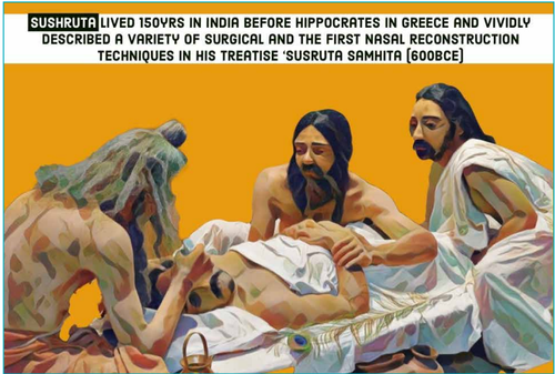
Figure 1: Artistic rendition of Sushruta performing nasal reconstruction surgery in 600 BCE (digital art by the authors based on images in the public domain).
600 BC
Indian Method: The first documented evidence of nasal reconstruction comes from India in the Ayurvedic surgical treatise ‘Sushruta Samhita’ (Sanskrit: Suśrutasaṃhitā), wherein cheek or forehead skin was used for nasal reconstruction. The author Sushruta is rightly called the ‘Father of Plastic Surgery’. He belonged to the potter clan of Varanasi, on the banks of the holy river Ganges. He taught surgical skills to his students on various simulation models like vegetables (such as watermelon, gourd, cucumber) for incisions (Figure 1).
1442 AD
Italian Modification of Indian method: T It is widely acknowledged that plastic surgery in Europe as practiced in the medieval ages, was a direct descendant of classical Indian techniques. Italian surgeon Branca de'Branca introduced the Indian method (which came from India, probably via Arabia) of nasal reconstruction in Italy in 1442.
He later modified this with delayed flaps from the arm and passed on this knowledge to his son, Antonio. This was the Italian method
Italian Method: 100 years later, Tagliacozzi modified and popularised the Italian method by transferring an arm flap to the nose in 6 stages in 4 weeks (Figure 2A and B)
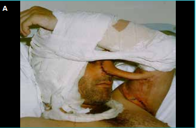
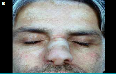
Figure 2: A. The second stage of nasal reconstruction with the ‘Tagliacozzi’ Italian method is shown in this figure. In the first stage, a bi-pedicled vertical flap was raised from the medial arm, and the proximal end was detached after 6 weeks, then attached to the nasal defect for reconstruction, B. The Tagliacozzi flap was detached after another 6 weeks in the third stage to achieve the final shape of the nose.
1793 AD
‘The Madras Gazette’ published the Indian forehead technique after witnessing the procedure in Pune, India. A year later, the same technique was published in ‘The Gentleman's Magazine’ of London by a British engraver Barak Longmate (‘B.L.’) while he was visiting India. He had observed such reconstruction on a bullockcart driver named Cowasjee. This famous letter inspired the English surgeon Joseph Carpue (1764–1846) to practice the Indian method of nasal reconstruction, first on cadavers and later on two officers of the British Army.
It is a lesser-known fact that 100 years before this famous letter, the Venetian adventurer Niccolao Manucci (1639–1717) had already witnessed and documented the Indian method of nasal reconstruction in his memoirs describing the history of the Mughal Empire (Storia do Mogor) in 1690 AD.
1818 AD
German Method: This was described by German surgeon Karl von Gräfe, wherein a free skin graft from the arm was used instead of a delayed flap.
1845 AD
German surgeon Johann Friedrich Dieffenbach described rhinoplasty in ‘Operative Chirurgie’. He also described secondary revision to enhance outcomes.
1887 AD
John Roe, an American otolaryngologist, first described aesthetic rhinoplasty techniques.
1889 AD
Indian surgeon Tribhovandas Motichand Shah published his record-breaking 300 cases of forehead flap for nasal reconstruction in ‘The Indian Medical Gazette’. This was a year before the British Surgeon J.C. Carpue had performed his first live forehead flap.
1913 AD
Harold Delf Gillies, during World War I, recommended the forehead flap for nasal reconstruction and described it in his book ‘Plastic Surgery of the Face’.
1946 AD
Kazanjian popularised the median forehead flap in the United States. Several modifications were contributed by many to prevent external nasal deformities due to contracture. Attention was also given to the intranasal lining and its significance.
1986 AD
Burget and Menick refined the art of nasal reconstruction with the paramedian forehead flap and introduced the concept of nasal lining flaps as the ‘fourth dimension’. Their seminal articles on this subject should be read by every enthusiastic nasal reconstruction surgeon.
computer-aided manufacturing (CAM) technique have become an aid for planning and are replacing the clay models that were used previously. Bioengineered tissues are being experimented upon and tested for minimal donor site morbidity
Relevant Clinical Anatomy
Subunits
The nose has a complex topographic shape that can be easily divided into 9 subunits based on the contour lines and skin type. The aesthetic nasal subunits were first illustrated by Gonzáles-Ulloa in 1957, but the ‘subunit principle’ for nasal reconstruction was first described by Burget and Menick in 1985.5 Its subunits are divided into three paired and three unpaired structures. The unpaired subunits are the dorsum, columella, and nasal tip. The nasal sidewall, soft triangle, and ala comprise the paired subunits. The 9 aesthetic nasal subunits are shown in Figure 3.
Future of nasal reconstruction
Present-day three-dimensional (3D) reconstruction models with computer-aided design (CAD)/
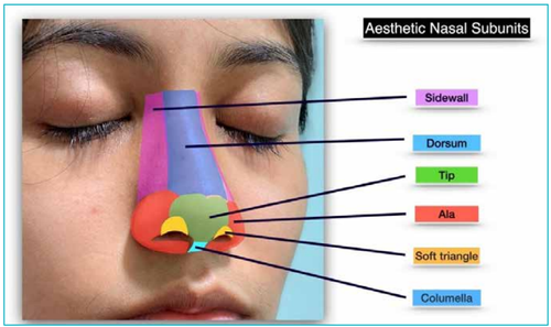
Figure 3: Nine topographic aesthetic subunits of the nose are based on contour lines and skin zones of the nose.
Skin zones
The quality and texture of skin can be used to describe three distinct zones of the surface of the nose. This is useful in planning reconstruction as skin pliability is a deciding factor in the design of local flaps. Figure 4 illustrates the three skin zones of the nose and Table 1 describes the three zones based on skin type and their relevance.
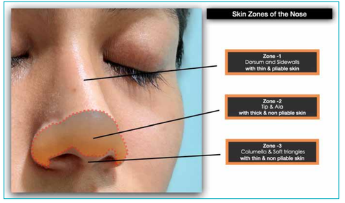
Figure 4:Three skin zones of the nose based on thickness and skin pliability
| Zone-wise Description |
|---|
| Zone 1 — Dorsum & Sidewall |
| Mobile pinchable smooth skin |
| Full thickness skin grafts or Single lobe flap can be effective |
| Zone 2 — Tip & Ala |
| Thick, sebaceous, non-pliable skin. |
| Usually needs bilobed or complex design local flap. |
| Zone 3 — Columella & Soft triangles |
| Thin but non-pliable skin |
| Difficult to reconstruct. |
| Avoid undermining and reconstruct with the tip as a subunit or use small local flaps or composite grafts from the anterior helical rim of the ear. |
Table 1: Characteristics and relevance of the three skin zones of the nose.
The choice of reconstruction thus depends on the nature of the tissue to be replaced. The structural support is given by bone cranially and cartilage caudally. The upper lateral part is supported by the nasal bone and the frontal process of the maxilla, followed by upper and lower lateral cartilage inferiorly. The midline is held by a bony and cartilaginous septum constituted by the perpendicular plate of the ethmoid and vomer, and quadrangular cartilage, respectively. The ala and tip are supported by lower lateral cartilages, which are folded on themselves twice at the medial and the lateral genu, thus dividing it into lateral, middle, and medial crus. The lining is formed by the mucosa.
Blood and nerve supply of the nose
TThe blood supply and its anatomy help in identifying the potential donor sites to raise local flaps to cover various nasal defects. The venous drainage is largely through the anterior facial and ophthalmic veins for the external nose. The septum has a rich plexus of blood vessels formed by branches of the ophthalmic artery (anterior and posterior ethmoidal arteries), maxillary artery (sphenopalatine branches), and facial artery (superior labial artery). The arterial supply of the nose is illustrated in Figure 5.
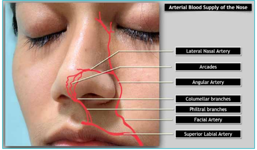
Figure 5: Arterial blood supply of the nose
The nerve supply of the nose is via the nasociliary and infratrochlear branches of the ophthalmic nerve and the infraorbital branches of the maxillary nerve. The septum is innervated by the nasociliary branch of the ophthalmic nerve, the alveolar branch of the maxillary nerve, the nerve of the pterygoid canal, and the nasopalatine, anterior palatine, and nasal branches of the sphenopalatine ganglion
The Stepwise Approach to Nasal Reconstruction
When confronted with the reconstruction of a complex 3D facial feature like the nose, it is best to use a logical planning process, as illustrated in Table 2. The whole process is simplified into four essential steps:
Step 1:
Visualise the 3D defect of the nose and account for the missing layers, namely skin, skeletal support, and mucosal lining. This is important as often different means of reconstruction have to be employed to reconstruct
Step 2:
Map the defect in terms of the missing subunits. The subunits have been described and illustrated earlier. According to the subunit principle proposed by Burget and Menick for defects with more than 50% of a subunit missing, it was advised to replace the whole subunit by converting the partial defect into a complete subunit defect, provided a suitable method of reconstruction is available.5 It is not always advisable to apply this principle of subunit reconstruction fully, and maximum native skin preservation may be advisable in many cases. Exceptions to the classical subunit principle may include the elderly (to keep it simple), children (due to a lack of excess lax skin), very large defects (due to paucity of available tissues), and medically unfit patients (to keep the surgery short and simple). The surgeon should never use a cookiecutter approach with any of these guidelines and instead use their judgement based on personal knowledge and experience to customise the most suitable approach. They should also not hesitate to perform staged refinements in flap contour and bulk, as the excellent vascularity of the area allows for repeated elevation and thinning of the flaps. Restoration of nasal contour without sacrificing the whole subunit can yield good results, and subsequent scar treatments with dermabrasion and fractional lasers can further help in the restoration of contour and blending of scar lines.6
Step 3:
The timing of nasal reconstruction is very relevant and is dependent on local disease control. Reconstruction should be:ontour and blending of scar lines.6
- Immediate if the wound is free of infection and disease (e.g., malignancy)
- Delayed in cases of infection, animal or human bite, incomplete or unconfirmed oncological margins, or poor medical condition, etc.
Step 4:
The final step is the execution step, wherein the selection of the most suitable techniques to reconstruct the nose will logically flow from the information gathered in the first three steps.7,8 Assess the local available sites for potential flaps and choose the appropriate donor site depending on the availability of adjacent tissues with a good match. Sometimes the presence of old scars may not allow the raising of a forehead flap due to previous surgeries or injury. For small defects less than 1.5 cm, one usually resorts to local nasal skin flaps only. There may be a combination of techniques involved in the reconstruction of various layers of structures like the lining, cartilaginous or bony framework, and external skin defect, depending on the subunit involved. A defect less than 1.5 cm without cartilage deficiency is considered small. Autologous materials are best for nasal reconstruction whenever possible, as artificial implants and allografts have higher risks of infection.
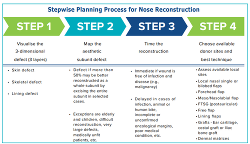
Table 2: Stepwise planning of nasal reconstruction.
Abbreviation: Full-Thickness Skin Graft.
Choice of Reconstructive Techniques and Available Options
It is important to remember that the ideal template for reconstruction is the opposite side, which can be mirrored in cases of unilateral defects with the help of simple paper or foil patterns. Nowadays, surgeons are using modern 3D planning and printing technologies, especially in larger defects where the opposite side is not available for mirroring.9 Artificial intelligence tools can help in reconstructing a virtual nose, and 3D models may be generated by CAD/CAM technique and printed for intraoperative planning. 3D surface scanning is also helpful in designing a prospective nasal structure suitable for the face and for designing prosthetic models. A 2D template may be generated from this scan, which can be used to design the flap with a calculated slight overcorrection to account for the ‘fourth dimension’ of healing, contraction, and flap perfusion.
Various common options available for the nasal reconstruction are discussed below in order of increasing complexity:
1. Tissue-engineered dermal matrices
Different artificial dermal substitutes are available in the market, and they can be used conveniently as an off-the-shelf option for small defects up to 1 cm in diameter with good results. If the defect is deep, they can be stacked with a delay of 7–10 days once the first matrix has vascularised. The biggest advantage is the lack of donor site morbidity. They can also be used for pre-lamination of forehead flaps for nasal lining as dermal matrices mucosalise very fast once inside the nose. 10
2. Healing with secondary intention
Wounds that are less than 1 cm in diameter and less than 0.5 cm in depth, located in non-sebaceous areas and natural concavities like alar grooves, heal best by secondary intention. They can be improved further by modalities like laser, platelet-rich plasma, or subcision, etc., a few weeks post-healing. 11
3. Primary closure
Midline wounds less than 1 cm with good skin laxity, such as in the elderly, may be closed with simple elliptical excision. Vertical orientation is preferred for small elliptical excisions and primary closures in the tip and supra-tip areas to avoid alar distortions. The surgeon should not limit the length of the ellipse to shorten the scar, as this may result in the formation of permanent, aesthetically undesirable dog-ear deformities. The dorsal aesthetic lines should be restored whenever possible. The nasal side walls form a normal cleavage line between the cheek and the nose, and linear primary closures in this area yield good results for small defects. Due to the zone of adherence, the base of the nasal side wall can take the tension of the mobile cheek flap with the help of marginal quilting dermal sutures. However, to avoid any unwanted ectropion of the lower lid, the linear closure should be obliquely placed with an upward slant, aiming towards the nasal side.
4. Full-thickness skin graft (FTSG) and split skin grafts
Full-thickness skin grafts can be good choices for simple skin defects of Zone 1 nasal dorsum, lateral walls, and especially for the inner canthal areas, where the flaps would be unacceptably thick. They are not usually a good match for thick Zone 2 areas as they cannot match the thick sebaceous skin, but may still be a simple option in elderly, unfit patients.
Preauricular, post-auricular, supraclavicular area, forehead hairline, and medial arm sites have all been used due to their hairless, smooth texture. FTSGs go through a prolonged phase of healing, can suffer partial take, and often undergo epithelial loss before healing. An FTSG should not be put on denuded cartilage but may take if the perichondrium or periosteum is well preserved. The final colour of these can be variable, especially in coloured skin, as they can suffer from post-inflammatory hyperpigmentation (PIH) and contracture.
Split skin grafts can also be used for nasal skin defects but often leave a depressed contour and are prone to contractures, which may deform the nose in the long term. FTSGs are preferred over split skin grafts for these reasons.
5. Composite grafts
Zone 3 defects of the columella and nasal rim in young patients, particularly children, can be wellsuited to reconstruction by composite grafts. These can be harvested from the anterior helical rim of the ear lobule as part FTSG and part composite graft with the helical cartilage. They can be used for Zone 3 defects for reconstruction of the nostril rim and columellar defects, usually up to 1 cm in width.12 The graft take is enhanced if a large portion of their ‘skin alone’ part is in contact with the nasal bed. They often undergo a prolonged healing phase and turn into a black eschar before separating in a few weeks, leaving a well-healed tissue underneath
Attention to accurate edge-to-edge approximation, good skin–bed contact and avoiding disturbance to the composite graft with a light bolster dressing can increase their take. The success rate is variable, with a higher risk of failure in smokers, the elderly, and in those with uncontrolled medical conditions or larg- sized defects.
6. Single lobe flaps
Single lobe flaps are best suited for small skin defects less than 1.5 cm. These are simple subcutaneous random Banner-style or rhomboid flaps. They are best suited for the pliable, thin, superior nose Zone 1 defects. 13
7. Dorsal nasal flap
The dorsal nasal flap can slide the entire nasal dorsum from the glabella down to the tip like a big vertical pillar with incisions along the dorsal wall aesthetic lines with V-Y design. It can fill in the defects of the tip and dorsal subunits (Figure 6 A-C). It includes the nasalis muscle and is elevated in the loose areolar supraperiosteal plane. Various modifications, like the laterally based hatchet glabellar V-Y design can incorporate a Z-plasty superiorly to avoid dog-ear formation. The islanded, skin-only V-Y design is based on the nasalis muscle with bilevel undermining. This flap is capable of easily resurfacing defects upto 2 cm. The main disadvantage is extensive dissection and the resulting long scars. 14,15
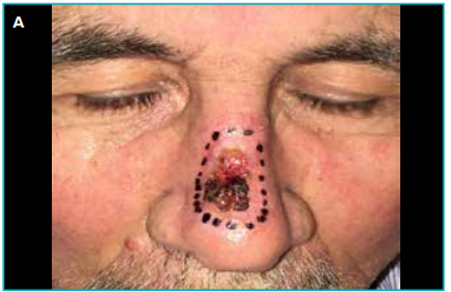
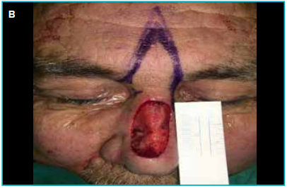
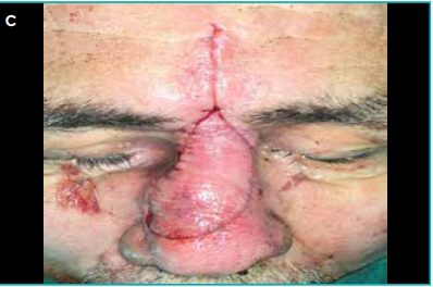
Figure 6: A. Large basal cell carcinoma of the nasal dorsum, B. Wide local excision defect of approximately 3.5 x 2 cm, C. Reconstruction with a sliding V-Y islanded dorsal nasal glabellar flap.
8. Bilobed flap
The bilobed flap is a double transposition flap and an excellent option for Zone 2 thick skin defects of the tip and ala (inferior nose), especially for defects located at least 0.5 cm away from the nostril rim. As the nasal skin in this zone is stiff, inflexible, and adherent, the ‘bilobed principle’ is applied to transfer skin from the thin, pliable superior nasal area by means of two adjacent flaps.
After the defect is triangulated by excising Burow’s triangle, the first lobe flap is transposed from the adjacent thick skin of almost the same size and a contiguous second flap is used from the pinchable flexible superior nose skin, usually of a slightly longer length (to avoid dog-ears) but half the width to fill the space left by the first lobe flap. This efficiently transfers the skin laxity from the superior to the inferior nose in an effective manner. The flap should include the underlying muscle for a robust blood supply.
Most surgeons use the geometric design described by Zitelli, wherein each adjacent flap rotates in an arc-like fashion by 45°, bringing the final rotation to 90–100°. These are useful for defects up to 1.5 to 2 cm (Figure 7 A and B). Small cartilage defects may not need any separate cartilage grafts, as the flap is quite thick. 16 Medially based design is used for alar defects, and laterally based design is used for tip defects. The second flap donor site should be closed first, and any dog-ear should be excised. This makes the further inset of the two flaps essentially tension-free. Accurate layered closure is important to avoid future ‘pin-cushioning’ of the scar. The main disadvantages are extensive dissection and scars that can pincushion or become hypertrophic.
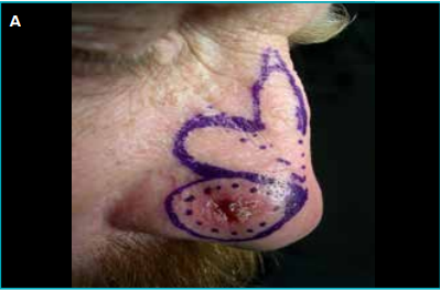
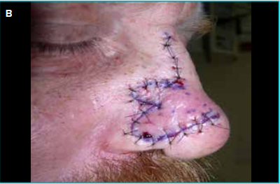
Figure 7:
A. Right alar basal cell carcinoma planned for a wide local excision and reconstruction with a medially based bilobed flap using Zitelli’s design, B. Final appearance of bilobed flap reconstruction.
9. Nasolabial and melolabial flap
The adjacent area of the cheek or the nasolabial area provides natural skin laxity along the resting lines of tension to raise a melolabial or nasolabial flap, respectively.17,18 Both are based on perforators emanating from the terminal branches of the facial artery. They can be used for large adjacent ala and sidewall nasal defects of up to 2 to 3 cm.
For adjacent defects, both flaps can be used as a pedicled transposition or an islanded flap (Figure 8 A–E). If the flap is bulky, a second stage for thinning and alar groove or lateral wall contouring can be performed after 3 weeks. Cartilage grafts can be placed underneath in the very first stage.
Larger defects of the ala and tip or columella can also be reconstructed with two-stage pedicled nasolabial flaps. The main facial artery, which runs deep to the muscle, needs to be included in large-sized flaps; for smaller defects, however, islanded perforator-based flaps minimise donor site morbidity.
The width of the flap can vary from 2 to 4 cm, depending on cheek skin laxity, and is an excellent choice in elderly patients, for sidewall, ala, and composite inferior nose side defects due to its simplicity and reliability. In younger patients, the nasolabial flap is often limited to a maximum 2 cm width due to tight skin and a higher incidence of hypertrophic scars.
10. Forehead flap
The forehead flap is the best reconstructive option in large, complex defects involving the nose.19-22 It offers an excellent tissue match with acceptable donor site morbidity. It may be harvested based on the supratrochlear vessels as the paramedian flap or based on canthal and paracentral vessels as the midline flap. Apart from these two designs, many variations have been described, such as oblique, sickle, and converse scalping flaps. However, the vertical paramedian forehead flap has continued to enjoy immense popularity for hundreds of years due to its reliable vascularity, great skin texture match, large tissue availability, and good flap reach with minimal donor site morbidity.
Paramedian forehead flap is a workhorse flap for nasal reconstruction and is mostly performed in two stages, wherein the pedicle base and inset is performed in 3 weeks’ time. The pedicle must always be at least 1.5 cm wide and may be rotated to the ipsilateral or contralateral side. If required, the flap can be extended to include the hair-bearing area, especially in short foreheads. Such patients benefit from hair removal with diode laser sessions later. The flap’s reach can be extended by incising the medial margin of the flap below the brow hairline. Another method to increase the reach is to score the galea or frontalis without damaging the pedicle. This may give an extension of about 1 cm.
The subunits requiring reconstruction should be marked first. A foil or paper template of the defect can be made and transposed 180° to design the flap. The supratrochlear vessels are marked with the help of a handheld Doppler probe and are anatomically located just lateral to the frown crease. The exact pattern of the defect is marked on the forehead, which may include the hairline depending on the length of the flap required. The flap is elevated in the loose areolar plane from cranial to caudal direction towards the pedicle. Although the superior tip of the flap can be elevated in the subcutaneous plane to
reduce the bulk, more than two-thirds of the inferior flap must include the frontalis muscle, and the lower 2 cm must also include the periosteum to prevent inadvertent division of the tightly coursing pedicle vessels, which lie closely against the periosteum at the superior orbital rim. Skeletal support in the form of costal or auricular cartilage grafts can be implanted at the very first stage in most cases. It is often combined with turnover flaps or nasolabial flaps for the inner lining of large defects (Figure 9A–K).
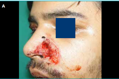
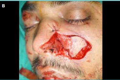
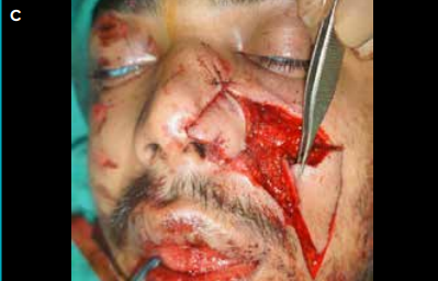
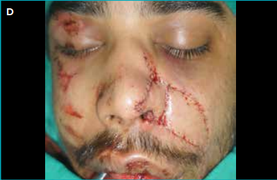
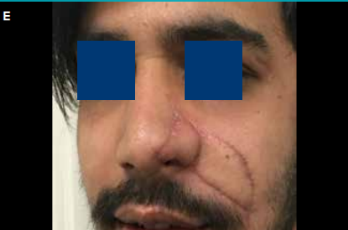
Figure 8 :
A. Traumatic loss of the left ala and cheek tissue, B. Adjacent mesolabial flap being raised as an islanded design (after verifying the arterial supply from the angular artery superiorly using a Doppler probe), C. V-Y islanded nasolabial flap raised to fill the donor defect of the mesolabial flap, D. Mesolabial flap inset to the left alar defect and nasolabial V-Y flap advanced to close the donor defect simultaneously. The two flaps successfully reconstructed a complex ala–cheek defect in this case, E. 2 months postoperative result. The patient was advised to undergo fractional laser resurfacing for further scar modulation.
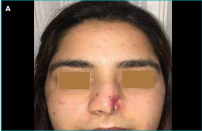
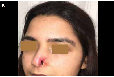
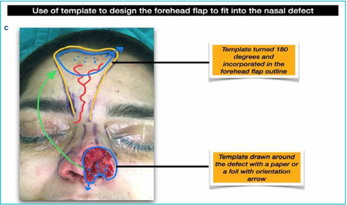
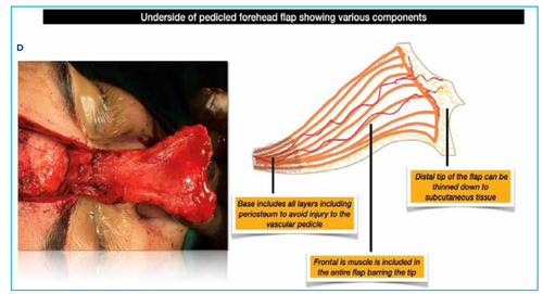
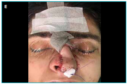
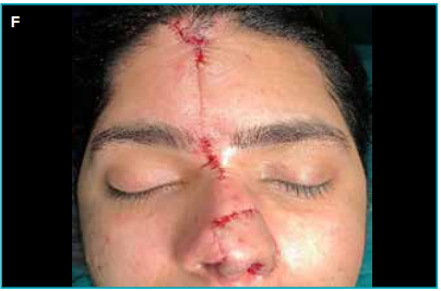
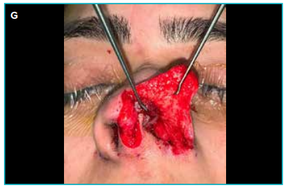
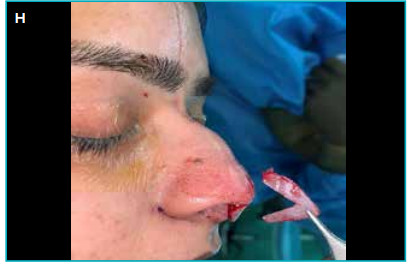
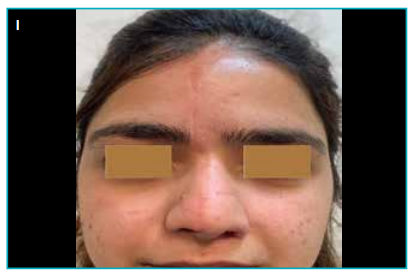
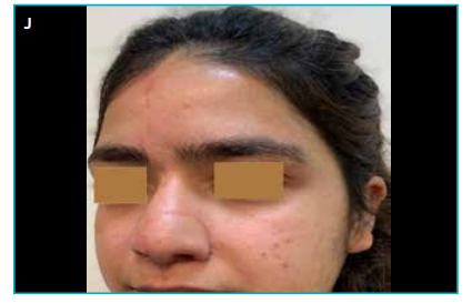
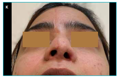
Figure 9:
A and B. Showing a composite dorsum, left ala, tip, and columella defect secondary to a dog bite, C. Paramedian forehead flap drawn on the contralateral pedicle based on the defect template. Dorsal turnover flaps are used for alar lining, D. Underside of raised forehead flap showing inclusion of various deep components in different parts of the flap, E. Flap inset to reconstruct the nasal defect. The pedicle is protected with a nonadherent foam dressing, F. Stage 2 of flap division and inset at 3 weeks after the first stage, G and H. Stage 3 after another 3 months, showing further nasal lining addition with a full-thickness postauricular skin graft and septal extension and tip cartilage graft, I, J and K. Final result of nasal reconstruction with the forehead flap after one year.
Donor forehead closure may be achieved primarily in smaller cases; in larger defects, partial areas may be left for healing by secondary intention. This is preferred to split skin graft as secondary healing scars are excellent in the forehead area and the scalp, and do not suffer any contracture in the long term. The majority of surgeons prefer the 3-week period for the
second stage of pedicle division, but the duration can be shortened to 2 weeks if good vascularity of the distal flap can be validated with indocyanine green angiography, whilst temporary pedicle occlusion is performed with a gentle vascular clamp or rubber tourniquet (Figure 10A and B).
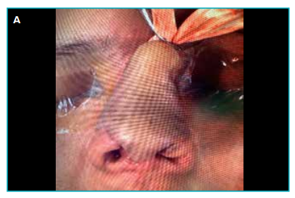
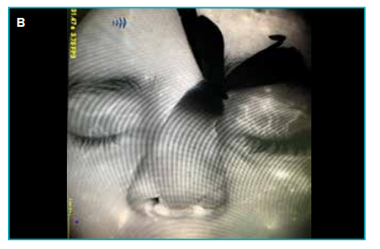
Figure 10: A. Glove tourniquet is tied at the forehead flap pedicle base on the forehead side. This flap is 2 weeks post-first stage, and indocyanine green (ICG) angiography will be performed to see if the distal part of the attached flap is well perfused from the nasal side, B. Bright white fluorescence of ICG angiography clearly demonstrates excellent flap vascularity despite the pedicle being occluded with a tourniquet. This indicates that the flap has started to derive full vascularity from the nasal side, and the pedicle can be safely divided at the base. In smokers, elderly patients, or those with vasculopathies, no flap thinning should be done, and the duration of the second stage division should be increased to 4–5 weeks for safety. The flap can also be performed in three stages with an intermediate stage at 2–3 weeks for cartilage grafting, flap thinning, or contouring.
When faced with partially scarred foreheads or small foreheads, tissue expansion can be used before elevating the flap to increase its size. However, this is generally not a preferred option as it thins out the subcutaneous tissue and may result in later contraction on the nose, thus compromising the result. Forehead flap also lends itself to prelamination with a skin graft or a dermal matrix for the inner lining.
11. Radial artery forearm (RAFA) free flap
When no local options are available for nasal reconstruction due to failed previous reconstructions or scarred local sites, RAFA free flap is quite the saviour.23 It can reconstruct all subunits of the nose. It is preferred over other free flaps because of its long pedicle and thin, pliable tissue that can easily
be moulded to the desired shape and size. However, the colour match is not as good as the forehead flap, especially for coloured skin. It can also be used for nasal lining in large central defects and may be combined with a forehead flap for external cover. Cartilage or bone grafts should be sandwiched between two layers when necessary. It has the added advantage of bringing vascularity to severely scarred areas, besides promoting healing. In cases of large septal defects, a dorsal cantilever graft can be used for support without any formal attempt at reconstructing the septum, as the large septal fistula provides enough room for adequate airflow without stenosis.
The flap may also be prefabricated or prelaminated with cartilage graft on the forearm and later transferred to the recipient site.24 The skin paddles may be longitudinally arranged like a string of pearls and placed according to the defect to restore the lining, floor, and columella separately. Attention must be paid to flap design and orientation of the pedicle to avoid kinking and vascular compromise to the skin paddle (Figure 11A–C).
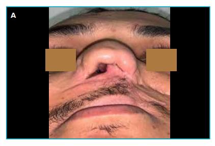
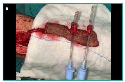
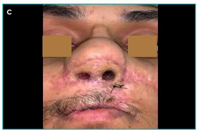
Figure 11A. Patient with loss of the entire tip and nostrils from a bomb blast injury. The forehead flap was previously used unsuccessfully to reconstruct elsewhere, B. Radial artery forearm (RAFA) free flap raised and crafted whilst still attached in the forearm to make the nostril lining and the nasal tip complex. The construct was sutured over appropriately sized paediatric endotracheal tubes, which were also used as internal splints. The flap was anastamosed to the facial vessels, C. Excellent reconstruction of composite nostril tip complex along with internal lining with the RAFA free flap. Patient is advised to wear nasal splints for 3 months to prevent contracture. Patient will undergo further revision for refining the eternal nasal profile.
Structural Support Reconstruction
Stable nasal architecture, shape, function, and sturdiness of the reconstruction rest firmly on skeletal integrity. Split cranial grafts or rib grafts can be used for large nasal bone defects. Most nasal reconstructions require only the cartilage framework, for which septal, ear or costal cartilage grafts can be used.
Septal cartilage is easily available and provides good support, but its use may be limited in cases of large structural requirements. Ear cartilage is another site providing excellent tip and alar grafts. It is, however, rarely used for constructing a linear columella or dorsal graft owing to its curved nature and limited quantity. The ear cartilage is harvested from the conchal bowl using an anterior, or preferably a posterior technique in patients with keloid tendencies
Costal rib cartilage is an excellent and almost unlimited source of cartilage grafts for a depleted nose. The sixth, seventh, and eighth rib cartilages are reliable donor sites when a large amount of structural reconstruction is necessary. Floating ribs can be used as a cantilever graft. The rib grafts often warp in the future, causing delayed deformation and deviation of the reconstructed nose. To prevent this, the cartilage is carved based on Gibson’s principle of balance — equal carving on the sides of the core to prevent warping. Keeping the half-carved cartilage immersed in a bowl of sterile saline solution for half an hour can also make the future warping obvious and thus allow for pre-emptive corrections. Another method to minimise warping is to harvest half bone and half cartilage. A costochondral graft can be used as an L-shaped construct with a columellar strut for stable projection. The graft should be fixed securely to the remnant structures in the midline to prevent deviation. Additional K-wire fixation for 10 days can also be done. Anatomically shaped costal cartilage grafts can be used to reconstruct the septum, lateral nasal walls, tip, and alar regions just like in a standard open rhinoplasty approach (Figure 12) .19,25M.
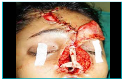
Figure 12: Rib cartilage grafts used as a dorsal cantilever graft with a columellar strut and bilateral cartilage grafts. External nasal skin cover is restored with a paramedian forehead flap seen as being harvested (it is vital to fix all the grafts with the remaining base in a stable way at multiple points).
Split calvarial, ischial crest bone, and rib grafts can be used as cantilever grafts, which are fixed to the nasal radix with a screw but may suffer from absorption and dislodgement. Lack of columellar fixation is a weak link in the entire cantilever construct.
Artificial materials such as silicone and porous polyethylene implants are usually not preferred for complex nasal reconstruction as they are associated with high risks of extrusion and infection.
Nasal Lining
Nasal lining is important for maintaining a patent airway. Small defects can be reconstructed with a bi-pedicled nasal lining flap or even an inferior turbinate mucosal flap. Larger defects may be covered by a septal mucosal pivot flap based on the septal branch of the superior labial artery. The midvault lining may be obtained from a contralateral septal hinge mucosal flap based on the superior ethmoidal vessels, passed through an incision in the ipsilateral septum dorsally .8
Turnover skin flaps from the defect rim, folded forehead flap, nasolabial flap, or, in severe deficiency, a microvascular RAFA free flap can all be used as needed for the nasal lining.
It is also possible to use FTSG or a dermal matrix sutured to the underside of the forehead or a nasolabial flap.
Post-Operative Rehabilitation
Prevention of nostril stenosis, airway collapse, and skin contracture is essential in all major reconstructions. Sequential conforming silicone nasal retainers may need to be worn for a few months to prevent collapse and fibrosis (Figure 13).
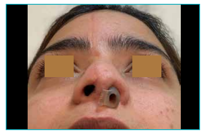
Figure 13:Nostril retainer is advised to be worn for at least 3 months after nostril reconstruction, and retainers are worn in incremental sizes till the opening has been over-dilated by 10%–15%. In this particular case left side nostril retainer-dilator can be seen in place
In extended forehead flaps that transfer hair-bearing skin to the nose, laser hair removal is hugely beneficial.
Fractional resurfacing lasers — such as carbon dioxide lasers — are helpful in improving scar appearance and texture. Triamcinolone intralesional injections may be needed to treat scar hypertrophy
In patients for whom surgical reconstruction is not possible due to poor medical conditions or personal preference, an external removable or osseointegrated nasal prosthetic can be an option. They often become loose and need replacement. The side seams need regular cleaning and may develop crusting and infection. 26
Face transplant may be indicated in patients with severe facial deformity when autologous reconstruction and prosthetics are not possible. The entire face or midface may be transplanted, especially in patients with large neurofibromas and extensive facial trauma. The challenge is to obtain an ideal donor. There is, of course, the risk of rejection before and after the procedure, and lifelong immunosuppression is required. Composite tissue allotransplantation is a debatable procedure, as failure due to rejection leaves very little choice and often leaves the patient in a condition worse than their original state. In any case, given the lack of donors for such a procedure, it will always remain within the domain of advanced research institutes with unlimited resources. 27
Conclusion
The nose is a unique part of human appearance and identity. The reconstruction of a natural-looking and harmonious nose not only restores form and function, but also reinstates human dignity. An artistic blend of techniques, along with stepwise logical planning and layer-by-layer restoration of skin, skeletal support, and nasal lining, is the key to a good result. Local flaps, especially the ‘old-is-gold’ forehead flap, still retain their winner status in this arena. The aim is to make a good-looking nose that rests above a beautiful smile!
Take Home Messages
- The nose has a complex topographic shape that can be easily divided into 9 subunits based on the contour lines and skin type. In defects with more than 50% of a subunit missing, it is often best to replace the whole subunit by converting the partial defect into a complete subunit defect, provided a suitable method of reconstruction is available.
- Quality and texture of skin can be used to describe 3 distinct zones of the nasal surface. This is useful in planning reconstruction as skin pliability is a deciding factor in the design of local flaps.
- When confronted with the reconstruction of a complex three-dimensional facial feature like the nose, it is best to use a logical stepwise planning process focusing on restoration of all three anatomical layers.
- Bilobed flap is an excellent option for Zone 2 thick skin defects of the tip and ala (inferior nose).
- The paramedian forehead flap is the workhorse flap for large, complex defects involving the nose. It provides an excellent tissue match for nasal skin. It lends itself beautifully to moulding in multiple stages and the addition of skeletal grafts and other inner flaps. Scars on the donor forehead also settle well.
- Nasal lining flaps and skeletal support are vital to give stability and predictability to any nasal reconstruction.
Sunil Choudhary, Soumya Khanna, Raghav Mantri, Prateek Arora. Art and Artistry of Three-Dimensional
Nasal Reconstruction. MMJ. 2025, September. Vol 2 (3).
Multiple Choice Questions (MCQs)
1. The ‘Italian modification’ of Indian nasal reconstruction secret was guarded and modified by:
A. Tagliacozzi
B. Alessandro Benedetti
C. Pfalzpaint
D. Antonio Branca
2. Which is the most important layer when performing a total nasal reconstruction?
A. Skin
B. Structural support
C. Lining
D. Cartilage
3. A bilobed nasal flap is best suited for:
A. Defects of the tip and ala with thick skin
B. Defects of Zone 3 nasal skin
C. Defects of the dorsum and side walls with thin skin
D. Nasal lining
4. Which of the following statements is incorrect regarding the nasolabial flap?
A. It is the best flap for ala reconstruction
B. It can be used as a nasal lining flap
C. Its blood supply is from the superficial temporal artery
D. It can be used as a single-stage reconstruction
5. A patient who is a smoker underwent nasal reconstruction with a forehead flap. The base of the flap needs to be divided for inset at three weeks, but the surgeon is concerned about the flap survival as the patient has a history of smoking. What is the best imaging modality to assess the vascularity prior to dividing the flap?
A. Computed tomography (CT) angiography
B. Magnetic resonance (MR) angiography
C. Indocyanine green (ICG) angiography
D. Duplex Doppler
6. According to Burget and Menick, what is the best time to provide skeletal nasal support in nasal reconstruction?
A. Prior to skin reconstruction as prelamination
B. At the time of reconstruction, at the first stage itself
C. 3 to 4 weeks after skin flap reconstruction
D. Any time after the flap seems to have healed
7. Which is the best form of nasal lining in total nasal reconstruction following gunshot injury with a palatal and nasal defect and intact maxillary arch?
A. Prelamination with skin grafts and staged forehead flap reconstruction
B. Single radial forearm flap with separate skin paddles for lining with external cover
C. Radial forearm flap for lining and forehead flap for external skin cover
D. Mid-face transplant
8. Which of the following statements regarding tissue expansion is true?
A. There is a rebound contracture of the flap
B. Tissue expansion is commonly done for nasal reconstruction
C. There is no need to excise the capsule
D. The flap can be easily prelaminated
9. Which of the following is not a modification of the forehead flap?
A. Scalping flap (converse)
B. Gull Wing flap
C. Washio flap
D. Juri flap
10. Burget’s principles of nasal reconstruction include all except:
A. Restore the normal shape, contour, and texture of tissue
B. Three-dimensional planning of tissues using computer-aided design (CAD)/computer-aided manufacturing (CAM) technique
C. Replace like with like tissues
D. Replace the entire subunit whenever practical
MCQ Answers
1. (D) Antonio Branca. The Italian family Branca de’Branca guarded the secret of nasal reconstruction with the Indian method. Later, Antonio Branca modified the reconstruction using a forearm flap in the fourteenth century. Alessandro Benedetti first reported the Italian method 100 years before Tagliacozzi. In the midfifteenth century, a similar technique was described by Pfalzpaint (a German surgeon).
2. (C) Lining. It determines the final outcome. Without a lining, the reconstruction will collapse. It should be vascular, supple, and thin to support the cartilage and or bone.
3. (A) Defects of the tip and ala less than 1.5 cm can be easily covered with a bilobed flap as the skin of this area is thick and immobile. The bilobed flap effectively transfers the skin laxity from the superior to the inferior nose in an efficient manner.
4. (C) Nasolabial flap is supplied by branches of the facial artery.
5. (C) Indocyanine green angiography (ICG angiography) is a simple and accurate imaging modality to assess the vascularity of the forehead flap before dividing the pedicle (a temporary glove tourniquet is tied to the pedicle). It is capable of showing dynamic cutaneous vascularity on the table intraoperatively. If the distal part of the flap fails to light up with the fluorescence, then it is not ready for pedicle division, and a further period of 1–2 weeks should be given before attempting division of the flap.
6. (C) According to Burget and Menick, 3–4 weeks post first stage nasal reconstruction with forehead flap is the preferred time to add skeletal support in the form of cartilage or bone grafts. They would divide the pedicle subsequently at the third stage 3–4 weeks later.
7. (C) In a severe gunshot injury, there is extensive damage resulting in scarred residual nasal and oral mucosa with a large palatal and midface defect. The palatal defect and nasal lining can both be reconstructed with a double paddle radial forearm flap. The best skin match for the external cover can be provided by the forehead flap.
8. (A) Rebound contraction of the flap is an unfavourable outcome seen with expanded forehead flaps, mainly used if the defect skin size is huge and the donor’s forehead is small. This leads to contraction of the lining. Expanded flaps are better suited when nasal lining reconstruction is not required. The capsule is usually excised during transfer. Skeletal support is also always inserted at the first stage to limit flap skin contracture.
9. (D) Juri flap is a temporal scalp flap based on temporal artery branches, mainly used for the treatment of male pattern baldness. The other options are forehead flap modifications. The scalping flap has a skin paddle designed on the lateral forehead. It can be used to reconstruct large nasal defects. It requires a bicoronal incision and leaves a large secondary scalp defect to be covered with a skin graft. Washio flap utilises post auricular and mastoid skin based on the lateral forehead and temporal area and is supplied by the superficial temporal artery and retroauricular artery. The Gull Wing flap was designed by Millard using a median forehead flap. It is used for columella reconstruction.
10. (B) Three-dimensional planning, as described by Burget and Menick, involves using a foil or an empty suture packet as a source for configuration.
References
- Faris C, Heiser A, Quatela O, et al. Health utility of rhinectomy, surgical nasal reconstruction, and prosthetic rehabilitation. Laryngoscope. 2020;130(7):1674–9.
- Whitaker IS, Karoo RO, Spyrou G, et al. The birth of plastic surgery: the story of nasal reconstruction from the Edwin Smith Papyrus to the twenty-first century. Plast Reconstr Surg. 2007;120(1):327–36.
- Sykes PJ, Santoni-Rugiu P, Mazzola RF. Nicolò Manuzzi (1639-1717) and the first report of the Indian Rhinoplasty. J Plast Reconstr Aesthet Surg. 2010;63(2):247–50.
- Shaye DA. The history of nasal reconstruction. Curr Opin Otolaryngol Head Neck Surg. 2021;29(4):259–64.
- Burget GC, Menick FJ. The subunit principle in nasal reconstruction. Plast Reconstr Surg. 1985;76(2):239–47.
- Rohrich RJ, Griffin JR, Ansari M, et al. Nasal reconstruction—beyond aesthetic subunits: a 15-year review of 1334 cases. Plast Reconstr Surg. 2004;114(6):1405–16; discussion 1417–9.
- Michelotti B, Mackay D. Nasal reconstruction. Clin Anat. 2012;25(1):8698.
- Menick FJ. Aesthetic nasal reconstruction. In: Neligan P, Rodriguez ED, eds. Plastic Surgery. 3rd Edition. London: Elsevier Saunders; 2013. p. 134–86.
- Bauermeister AJ, Zuriarrain A, Newman MI. Three-Dimensional Printing in Plastic and Reconstructive Surgery: A Systematic Review. Ann Plast Surg. 2016;77(5):569–76.
- Shridharani SM, Tufaro AP. A systematic review of acellular dermal matrices in head and neck reconstruction. Plast Reconstr Surg. 2012;130(5 Suppl 2):35S–43S.
- Cook JL. The examination of several common misconceptions in nasal reconstruction. Semin Cutan Med Surg. 2003;22(4):281–94.
- Chen C, Patel R, Chi J. Comprehensive Algorithm for Nasal Ala Reconstruction: Utility of the Auricular Composite Graft. Surg J (N Y). 2018;4(2):e55–e61.
- Masson JK, Mendelson BC. The banner flap. Am J Surg. 1977;134(3):419–23.
- Bitgood MJ, Hybarger CP. Expanded applications of the dorsal nasal flap. Arch Facial Plast Surg. 2007;9(5):344–51.
- Nahai FR, Papadopoulos DJ, Papadopoulos JP, et al. Nasal Reconstruction Using a Myocutaneous Island Pedicle Flap Based on the Nasalis Muscle With Bilevel Undermining: Review of 57 Cases. Ann Plast Surg. 2021;86(2):171–4.
- Zitelli JA. Design aspect of the bilobed flap. Arch Facial Plast Surg. 2008;10(3):186.
- Patel AA, Cheng A. The Nasolabial Flap. Atlas Oral Maxillofac Surg Clin North Am. 2020;28(1):7–12.
- Carucci JA. Melolabial flap repair in nasal reconstruction. Dermatol Clin. 2005;23(1):65–71,vi.
- Menick FJ. Nasal reconstruction. Plast Reconstr Surg. 2010;125(4):138e–150e.
- Phillips TJ. Total nasal reconstruction: a review of the past and present, with a peak into the future. Curr Opin Otolaryngol Head Neck Surg. 2019;27(5):420–5.
- Lo Torto F, Redi U, Cigna E, et al. Nasal Reconstruction With Two Stages Versus Three Stages Forehead Flap: What is Better for Patients With High Vascular Risk? J Craniofac Surg. 2020;31(1):e57–e60.
- Shokri T, Kadakia S, Saman M, et al. The Paramedian Forehead Flap for Nasal Reconstruction: From Antiquity to Present. J Craniofac Surg. 2019;30(2):330–3.
- Gasteratos K, Spyropoulou GA, Chaiyasate K. Microvascular Reconstruction of Complex Nasal Defects: Case Reports and Review of the Literature. Plast Reconstr Surg Glob Open. 2020;8(7):e3003.
- Cavadas PC, Torres A. Total Nasal Reconstruction With Prefabricated and Prelaminated Free Flap. Ann Plast Surg. 2019;83(6):e35–e38.
- Owusu J, Nesbitt B, Boahene K. Management of Complicated Nasal Defects. Facial Plast Surg. 2020;36(2):158–65.
- Dings JPJ, Vijverberg MA, Hol MKS, et al. Autologous versus prosthetic nasal and auricular reconstruction - patient, professional and layperson perceptions. Int J Oral Maxillofac Surg. 2020;49(10):1271–8.
- Kiwanuka H, Bueno EM, Diaz-Siso JR, et al. Evolution of ethical debate on face transplantation. Plast Reconstr Surg. 2013;132(6):1558–68.