 WhatsApp Us (24/7)
 +91 926 888 0303 (24/7)


Abstract: Classical Hodgkin lymphoma (cHL) typically presents with localised lymphadenopathy with contiguous spread. Rarely, it can mimic infections or disseminated malignancies, leading to diagnostic delays. We report a case of a 25-year-old female with extensive systemic involvement and massive ascites resembling disseminated tuberculosis or metastatic disease. Diagnosis was confirmed via lymph node biopsy and immunohistochemistry. The patient responded well to combined immunotherapy and chemotherapy using nivolumab with doxorubicin, vinblastine, and dacarbazine (AVD). This case underscores the importance of considering cHL in the differential diagnosis of systemic symptoms in young adults and highlights the evolving role of immunotherapy in frontline treatment.
Key words: Hodgkin, Lymphadenopathy, Immunotherapy.
Introduction
Classical Hodgkin lymphoma (cHL) is a B-cell lymphoid malignancy characterised by the presence of Hodgkin and Reed-Sternberg (HRS) cells within a reactive cellular milieu. It shows a bimodal age distribution and is most commonly observed in adolescents and young adults. While most cases present with painless peripheral lymphadenopathy, atypical, disseminated presentations complicating diagnosis are rare but clinically significant.1,2
Case Report
A 25-year-old female student presented with intermittent fever and unintentional weight loss over six months, progressing to abdominal distension and cervical/axillary swelling in the last two months. The initial complete blood count (CBC) was within normal limits. Empirical antibiotic therapy provided only transient relief, and symptoms recurred, prompting further evaluation.
Investigations
Her haemogram revealed haemoglobin 7.8 g/dL, white cell count: 55,400/µL with neutrophilic leucocytosis (Figure 1), platelets 203,000/µL. Peripheral blood smear demonstrated macrocytic anaemia, and elevated lactate dehydrogenase (LDH) 314.7 U/L. She also had prolonged prothrombin time and activated partial thromboplastin time (APTT). Abdominal ultrasound (USG) revealed hepatosplenomegaly and abdominal lymphadenopathy. Whole-body positron emission tomography (PET)–computed tomography (CT) scan revealed fluorodeoxyglucose (FDG)-avid lymphadenopathy in the cervical, mediastinal, abdominal, and pelvic regions; FDG-avid lesions in the liver, spleen, and right lung nodule; bilateral pleural effusions; and gross ascites (Figure 2).
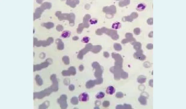
Figure 1: Peripheral blood smear showing neutrophilic leucocytosis with left shift and toxic granulation, indicative of systemic inflammatory response.
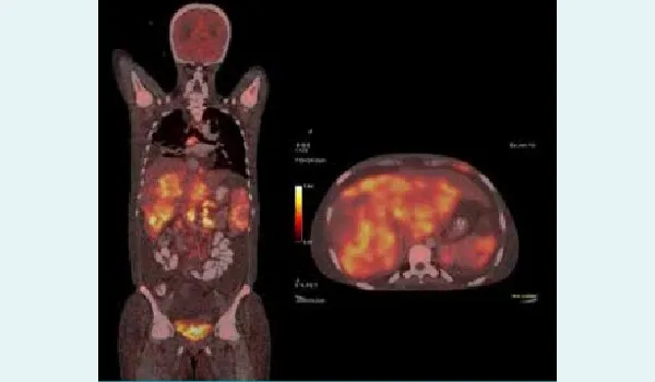
Figure 2: Baseline positron emission tomography (PET)– computed tomography (CT) scan showing widespread fluorodeoxyglucose (FDG)-avid lymphadenopathy and visceral involvement.
The differential diagnoses included disseminated tuberculosis, haematolymphoid malignancy (lymphoma, leukaemia), fungal infection, solid tumours (germ cell tumour, carcinoma of unknown primary), or rare conditions such as haemophagocytic lymphohistiocytosis (HLH) or Castleman disease. Excisional biopsy of supraclavicular lymph node with immunohistochemistry revealed HRS cells positive for CD30 and CD15, weakly positive for PAX5, MUM1 positive, and negative for CD3, CD20, Epstein–Barr virus (EBV). The final diagnosis was nodular sclerosing cHL. Bone marrow aspiration and biopsy showed reactive changes with haemophagocytosis. Echocardiography demonstrated an ejection fraction of 40%–45% with mild pericardial effusion (Figures 3–5).
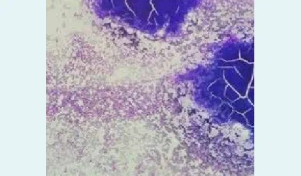
Figure 3: Bone marrow aspiration smear (low power view) showing increased cellularity with areas of haemophagocytosis.
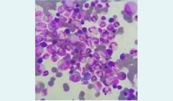
Figure 4: Bone marrow aspiration smear (high power view) demonstrating reactive features and scattered mononuclear cells.
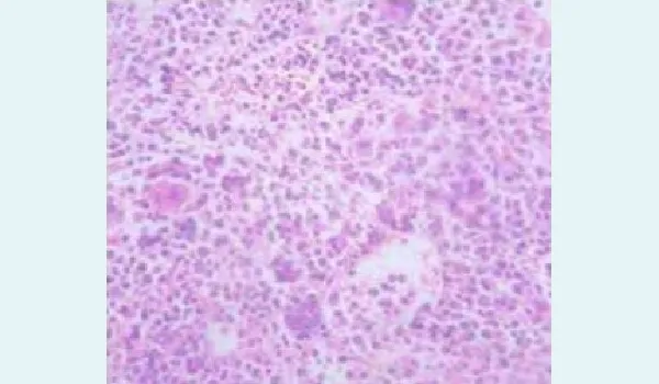
Figure 5: Bone marrow biopsy (Haematoxylin and Eosin stain) displaying increased cellularity with reactive changes and haemophagocytosis.
Treatment
The patient was started on immunochemotherapy using nivolumab with doxorubicin, vinblastine, and dacarbazine (AVD). Interim PET–CT, after 3B cycles, showed metabolic resolution of lymphadenopathy, liver and spleen lesions, and reduction of pleural effusion and ascites (Figure 6)
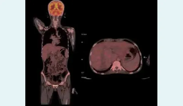
Figure 6: Interim positron emission tomography (PET)– computed tomography (CT) after 3 cycles of nivolumab with doxorubicin, vinblastine, and dacarbazine (AVD) showing Deauville Score 3 and significant metabolic response.
Discussion
cHL is characterised by its unique tumour microenvironment, where few HRS cells orchestrate a rich inflammatory infiltrate.1-4 This case highlights the disease's potential for widespread organ involvement and clinical masquerade. The patient, presenting with persistent abdominal distension, was initially treated for disseminated tuberculosis. Histologically, the nodular sclerosis subtype — characterised by lacunar variant HRS cells and collagen bands — is most prevalent in young females.2,3,5 CD30 and CD15 positivity, along with weak PAX5 expression and loss of B-cell markers, are diagnostic immunophenotypic hallmarks.3
Checkpoint blockade using programmed cell death protein 1 (PD-1) inhibitors like nivolumab has revolutionised the management of relapsed/refractory cHL, and recent trials now demonstrate efficacy in first-line settings.1,4,6 This case supports these findings, showing early metabolic response and symptom relief. The patient has completed six cycles of chemo-immunotherapy and returned to her usual routine.
Conclusion:
cHL can rarely present with features mimicking disseminated infections or malignancies. High clinical suspicion, histological confirmation, and evolving therapies like immune checkpoint inhibitors are critical to achieve optimal outcomes.
Viral Jain, Sanjeev Kumar Sharma, Anamika Bakliwal, Neelkumar Patel. Atypical Presentation of Classical
Hodgkin Lymphoma Mimicking Disseminated Infection or Metastatic Disease in a Young Female. MMJ.
2025, September. Vol 2 (3).
References
- Khan M, Hagemeister F, Wang M, et al. A review of pathobiology and therapies for classic Hodgkin lymphoma. Blood Rev. 2022;55:100949.
- Gupta S, Craig JW. Classic Hodgkin lymphoma in young people. Semin Diagn Pathol. 2023;40(5):379–91.
- El Hussein S, O'Malley DP. Classic Hodgkin lymphoma: An illustrative review of diagnostic limitations and immunomorphological challenges. Hum Pathol. 2025;156:105678.
- Alibrahim MN, Gloghini A, Carbone A. Classic Hodgkin lymphoma: Pathobiological features that impact emerging therapies. Blood Rev. 2025;71:101271.
- Brice P, de Kerviler E, Friedberg JW. Classical Hodgkin lymphoma. Lancet. 2021;398(10311):1518–27.
- Ansell SM. Immunotherapy in Hodgkin Lymphoma: The Road Ahead. Trends Immunol. 2019;40(5):379–87.