 WhatsApp Us (24/7)
 +91 926 888 0303 (24/7)


Abstract: Chikungunya is a mosquito-borne viral disease caused by the chikungunya virus, typically presenting with a sudden onset of fever and severe joint pain. While the disease generally follows a benign course, it can lead to severe complications like myocarditis, encephalitis, and Guillain–Barré syndrome (GBS). We report the case of a 53-year-old male with a history of hypertension and diabetes mellitus, who was recently diagnosed with chikungunya infection. He presented with shortness of breath and fever and was subsequently diagnosed with chikungunya-associated myocarditis. During his hospital stay, he developed GBS. He was managed with corticosteroids, diuretics, intravenous immunoglobulin (IVIG), and supportive care. This case highlights the rare but severe neurological and cardiac complications that may arise in the course of chikungunya infection.
Key words: Chikungunya, Myocarditis, Guillain–Barré Syndrome (GBS), Viral Complications, Case Report.
Introduction
Chikungunya is a mosquito-borne viral disease caused by the chikungunya virus (CHIKV), a ribonucleic acid (RNA) virus belonging to the Alphavirus genus of the Togaviridae family. It is primarily transmitted by Aedes aegypti and Aedes albopictus mosquitoes, which can also transmit dengue and Zika viruses.1
The incubation period ranges from 2 to 12 days, with symptom onset typically occurring between 4 and 8 days post-infection. The disease is characterised by an abrupt onset of fever and severe joint pain, which may persist for weeks to months.1
Although most cases are self-limiting, chikungunya can lead to serious complications involving the eyes, heart, and nervous system. Neurological manifestations include Guillain–Barré syndrome (GBS) acute disseminated encephalomyelitis (ADEM), and oculomotor nerve palsy. Cardiac involvement, such as myocarditis, has also been reported.2-4
This case report presents a rare and severe manifestation of chikungunya infection involving both myocarditis and GBS in a single patient.3,4
Case Report
A 53-year-old male with a history of hypertension, diabetes mellitus, and obstructive sleep apnoea presented to the Emergency Department of Max Super Speciality Hospital, Nagpur, with complaints of progressive shortness of breath and fever for one day. He tested positive for CHIKV via reverse transcriptase– polymerase chain reaction (RT–PCR) (Figure 1).
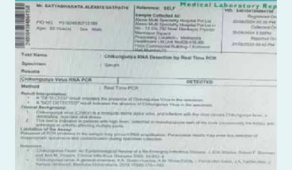
Figure 1: Chikungunya virus ribonucleic acid (RNA) reverse transcriptase–polymerase chain reaction (RT–PCR) report showing virus detected in serum.
On examination, the patient was tachycardic with a heart rate of 116 beats per minute (bpm), tachypnoeic with a respiratory rate of 36 breaths per minute, and dyspnoeic with an oxygen saturation of 92% on room air. Blood pressure was 140/80 mmHg. Laboratory investigations revealed a haemoglobin level of 12.6 g/dL, total leucocyte count of 10,300/mm³, and platelet count of 1.66 lakhs/mm³. Inflammatory markers were significantly elevated, with a C-reactive protein (CRP) level of 178.2 mg/L and ferritin at 1045.8 ng/mL. Cardiac markers showed elevated levels, with high-sensitivity troponin I (Trop-I hs) at 314.22 pg/mL and N-terminal pro–B-type natriuretic peptide (NT-proBNP) at 1602 pg/mL. D-dimer was markedly elevated at 2380 ng/mL. Serum creatinine was 1.4 mg/dL, while liver enzymes were within normal limits, with serum glutamic-oxaloacetic transaminase (SGOT)/Serum glutamic pyruvic transaminase (SGPT) levels of 32/11 U/L.
Pulmonary embolism was suspected due to elevated D-dimer and tachycardia; however, computed tomography (CT) pulmonary angiography ruled out embolism. The patient was started on antiplatelets, anticoagulants, statins, diuretics, bronchodilators, and nasal oxygen. Despite treatment, respiratory distress persisted. CT of the chest and abdomen revealed trace bilateral pleural effusion and multiple large groundglass opacities (Figure 2).
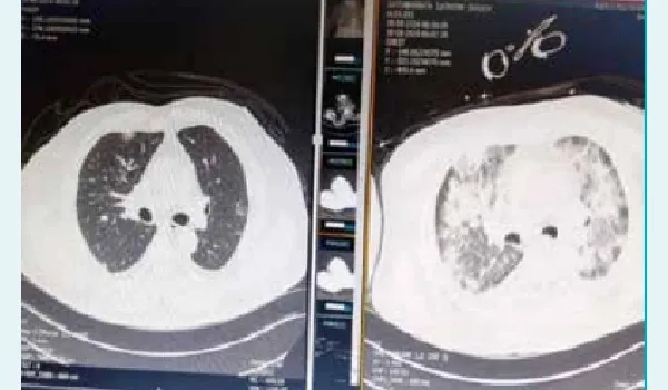
Figure 1: Computed tomography (CT thorax) showing groundglass opacities and trace bilateral pleural effusion (right side) compared to previous scan (left side).
Differential diagnosis included
- Pulmonary oedema secondary to acute coronary syndrome
- Viral pneumonia
A flu panel was sent, and oseltamivir was initiated. The patient developed atrial fibrillation with a pulse rate of 150 bpm, followed by desaturation. He was started on amiodarone and non-invasive ventilation. Injectable steroids were added due to worsening respiratory distress. Viral serology was negative.
Mechanical ventilation was initiated. Post-intubation CT of the thorax showed multiple large bilateral groundglass opacities and pleural effusion, suggestive of pulmonary oedema. Viral myocarditis was suspected, and serum ferritin was elevated at 1045.8 ng/mL. Intravenous methylprednisolone and a diuretic infusion were started. The patient improved and was weaned off the ventilator.
Following transfer to the ward, he developed lower limb weakness and was unable to lift his legs. Deep tendon reflexes were absent. Electroencephalography (EEG) and nerve conduction studies confirmed acute demyelinating inflammatory polyradiculopathy, consistent with GBS. He was treated with intravenous immunoglobulin (IVIG) for 5 days with clinical improvement. The remainder of his hospital stay was uneventful except for intermittent low-grade fever.
Discussion
CHIKV is known for its classical presentation of fever and arthralgia, but rare complications such as myocarditis and GBS can occur.1,2 Myocarditis may result from direct viral invasion or immune-mediated injury, while GBS is thought to be triggered by postinfectious autoimmune mechanisms.3,4
This case is unique due to the simultaneous occurrence of two serious complications — myocarditis and GBS — in a single patient.3,4 Early recognition and multidisciplinary management were crucial for recovery. The use of corticosteroids, IVIG, and supportive care proved effective.3,5
Conclusion:
This case highlights the importance of considering rare complications in chikungunya infection. Clinicians should maintain a high index of suspicion for myocarditis and GBS in patients presenting with respiratory distress and neurological symptoms.2-5 Timely diagnosis and appropriate treatment can significantly improve outcomes.
Nitin Wadaskar, Hitisha Agrawal. A Rare Triad: Chikungunya-Associated Myocarditis and Guillain–Barré
Syndrome — A Case Report. MMJ. 2025, September. Vol 2 (3).
References
- Moizéis RN, Fernandes TA, Guedes PM, et al. Chikungunya fever: a threat to global public health. Pathog Glob Health. 2018;112(4):182–94.
- Obeyesekere I, Hermon Y. Arbovirus heart disease: Myocarditis and cardiomyopathy following dengue and chikungunya fever—A follow-up study. Am Heart J. 1973;85(2):186–94.
- Cotella JI, Sauce AL, Saldarriaga CI, et al. Chikungunya and the Heart. Cardiology. 2021;146(3):324–34.
- Pattanaik A, Marate S, Mani RS, Pai AR, et al. Guillain-barré syndrome (GBS) with antecedent chikungunya infection: a case report and literature review. Neurol Res Pract. 2024;6(1):1-1.
- Lebrun G, Chadda K, Reboux AH, et al. Guillain-Barré syndrome after chikungunya infection. Emerging Infectious Diseases. 2009;15(3):495–7.