 WhatsApp Us (24/7)
 +91 926 888 0303 (24/7)


Abstract: Cardiac tamponade is a life-threatening condition that requires urgent diagnosis and intervention. Its occurrence in patients with complex pulmonary histories, such as bronchial carcinoid and previous tuberculosis (TB) infection, is rare and clinically challenging. We report the case of a 76-year-old male with a prior history of endoscopically treated bronchial carcinoid and treated pulmonary TB (2016), who presented with acute breathlessness, restlessness, chest discomfort, and gastrointestinal symptoms. He had a background of chronic productive cough and recurrent lower respiratory tract infections. On admission, he was tachycardic, tachypnoeic, hypotensive, and hypoxic. Clinical examination and 2D echocardiography confirmed massive pericardial effusion causing cardiac tamponade. Emergency pericardiocentesis drained 300 mL of turbid yellow pericardial fluid, which showed lymphocytic predominance. Imaging revealed bilateral pleural effusion, atelectasis, and left lung collapse. The patient was managed with intravenous antibiotics (meropenem and Targocid), vasopressors, bronchodilators, supportive care, and empiric antitubercular therapy. His condition improved significantly, and follow-up echocardiography showed only mild residual effusion without tamponade. He was discharged in stable condition with a comprehensive treatment plan and scheduled follow-up.
Key words: Cardiac Tamponade, Pericardial Effusion, Pericardiocentesis, Bronchial Carcinoid, Tuberculosis Infection, Bronchial Carcinoid, Elderly Patient.
Introduction
Lower respiratory tract infections (LRTIs) are among the most common and potentially serious infections affecting the lungs and airways. When combined with rare complications such as cardiac tamponade, a lifethreatening condition where fluid builds up around the heart, the risks increase significantly (Ref 1). This report analyses a complex medical case involving a patient with a bronchial carcinoid tumour and a history of tuberculosis (TB) infection, who developed both a LRTI and cardiac tamponade. By examining the interactions between these conditions, this report aims to shed light on how overlapping respiratory and cardiac issues can complicate diagnosis and treatment, especially in individuals with a history of lung disease. The goal is to provide insight into the medical challenges of such cases in an understandable way.
Case Report
A 76-year-old male with a known history of bronchial carcinoid status post-endoscopic removal and treated TB infection (2016) presented with an acute onset of breathlessness, restlessness, chest discomfort, and gastrointestinal symptoms, including nausea, vomiting, and abdominal pain. He had a background of recurrent LRTIs and chronic productive cough. On admission, he exhibited tachycardia, tachypnoea, hypotension, and desaturation. Clinical evaluation and 2D echocardiography revealed a massive pericardial effusion causing cardiac tamponade (Figure 1). Approximately 300 mL of turbid yellow-coloured pericardial fluid was drained via pericardiocentesis after insertion of a sheath (Figure 2). The fluid analysis showed lymphocytic predominance. Chest imaging further revealed bilateral pleural effusion, atelectasis, and left lung collapse. Initial management included intravenous (IV) vasopressors, meropenem, Targocid, supportive care, and bronchodilator therapy. The patient was also started on antitubercular therapy, considering his history and clinical findings. Serial monitoring showed clinical improvement and stabilisation of vital signs. A follow-up 2D echo showed only mild residual pericardial effusion without signs of tamponade (Figure 3). Computed tomography (CT) of the chest was carried out which revealed bilateral pleural effusion and left lung collapse (Figure 4). The patient was discharged in a vitally and haemodynamically stable condition with a comprehensive medication plan and advised follow-up after one week.
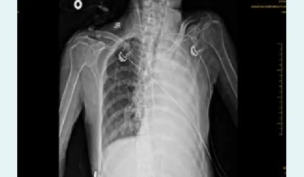
Figure 1: Chest X-ray on admission showing bilateral pleural effusion with left lung collapse.
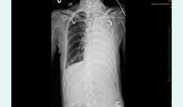
Figure 2: Chest X-ray after pericardiocentesis demonstrating resolution of cardiac silhouette enlargement.
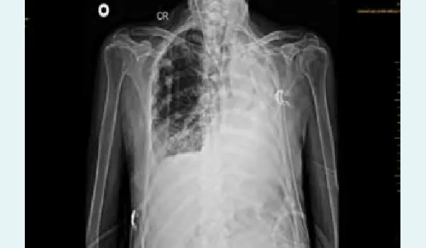
Figure 3: Chest X-ray at discharge showing significant improvement in lung expansion and reduced pleural effusion.
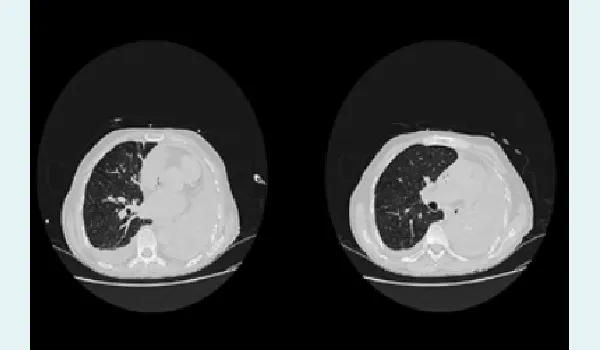
Figure 4C: PComputed tomography (CT) chest revealing bilateral pleural effusion, atelectasis, and left lung collapse.
Discussion
Pericardial effusion can lead to cardiac tamponade, a life-threatening condition in which fluid accumulation within the pericardial sac exerts pressure on the heart, impairing ventricular filling, especially during diastole, and reducing cardiac output.2-,4 This can result in shock or cardiac arrest if not promptly addressed. Clinical signs of tamponade include hypotension, muffled heart sounds, and jugular venous distension (Beck’s triad), and require urgent diagnosis via echocardiography and intervention such as pericardiocentesis.2,4 In this case, the patient initially presented with features of an LRTI, which can, especially when bacterial or viral, extend to the pericardium either through direct spread or haematogenous dissemination, leading to pericarditis and subsequent effusion. The systemic inflammatory response associated with infection may further contribute to fluid accumulation.5 This case highlights how an LRTI can mask evolving pericardial complications and underscores the need for heightened vigilance in deteriorating patients, particularly elderly individuals with multiple comorbidities.5,6 Cardiac tamponade, although uncommon, can develop rapidly in such vulnerable populations and necessitates timely, life-saving intervention.5 In this instance, pericardiocentesis was crucial, and a multidisciplinary approach involving cardiology and pulmonology played a key role in ensuring the patient’s optimal recovery.
Conclusion:
This case highlights the importance of prompt recognition and management of pericardial effusion leading to cardiac tamponade, particularly in elderly patients with a complex medical history including prior pulmonary infections and malignancy. Timely intervention with pericardiocentesis and appropriate antibiotic and supportive therapy resulted in significant clinical improvement. Comprehensive evaluation and a multidisciplinary approach played a crucial role in the successful stabilisation and recovery of the patient. Regular follow-up and adherence to prescribed treatment are essential to prevent recurrence and monitor for potential complications.
Hemalata Arora, Pritisha Pillai. Case Report: Lower Respiratory Tract Infection with Cardiac Tamponade in a
Patient with Bronchial Carcinoid and History of Tuberculosis Infection. MMJ. 2025, September. Vol 2 (3).
References
- Adler Y, Charron P, Imazio M, et al. 2015 ESC guidelines for the diagnosis and management of pericardial diseases. Eur Heart J. 2015;36(42):2921–64.
- Spodick DH. Acute cardiac tamponade. N Engl J Med. 2003;349(7):684–90.
- Azim A, Rao PB, Srivastav P, et al. Cardiac tamponade mimicking septic shock diagnosed by early echocardiography. J Emerg Trauma Shock. 2010;3(3):306.
- Caplin ME, Baudin E, Ferolla P, et al. Pulmonary neuroendocrine (carcinoid) tumors: European Neuroendocrine Tumor Society expert consensus and recommendations for best practice for typical and atypical pulmonary carcinoids. Ann Oncol. 2015;26(8):1604–20.
- World Health Organization. Treatment of tuberculosis: guidelines. 4th Edition. Geneva: WHO Press; 2017.
- Light RW. Pleural diseases. 6th Edition. Philadelphia: Lippincott Williams & Wilkins; 2013.