 WhatsApp Us (24/7)
 +91 926 888 0303 (24/7)


Abstract: Laparoscopic surgery for inguinal hernia repair has become the gold standard in the treatment of inguinal hernias in patients who are fit to undergo general anaesthesia. The total extra-peritoneal (TEP) technique is a preferred approach; however, it may be difficult for beginners. In this paper, we describe the “tri-compartment, V-Technique” for simplifying the understanding of extra-peritoneal space into three distinct compartments in a systematic manner. This technique involves precise and accurate partitioning of the medial, lateral, and central compartments, ensuring a clear view and effective mesh placement with minimal blood loss which can lead to improvised patient response and fewer complications.
Key words: V-Technique, Tri-Compartment, Laparoscopy, Totally Extra-Peritoneal, Inguinal Hernia.
Introduction
Ger et al. were the first to report laparoscopic surgery for inguinal hernia repair.1 McKernan was the first to report a complete extra-peritoneal laparoscopic repair method, called total extra-peritoneal (TEP) hernia repair.2 In TEP inguinal hernia repair, the principal concept is similar to that promoted by Stoppa, in which a large mesh is placed in the pre-peritoneal space to cover all potential hernia defects. This distributes intra-abdominal pressure over a wide area and requires little fixation. The operative steps for performing TEP and transabdominal pre-peritoneal (TAPP) repairs are very similar.3
Simplifying TEP with the V-technique essentially involves compartmentalising the pre-peritoneal space into three compartments, as shown in Figure 1, through laparoscopic dissection under clear vision. This approach provides a bloodless field and helps prevent vascular or nerve injuries.4
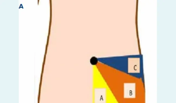
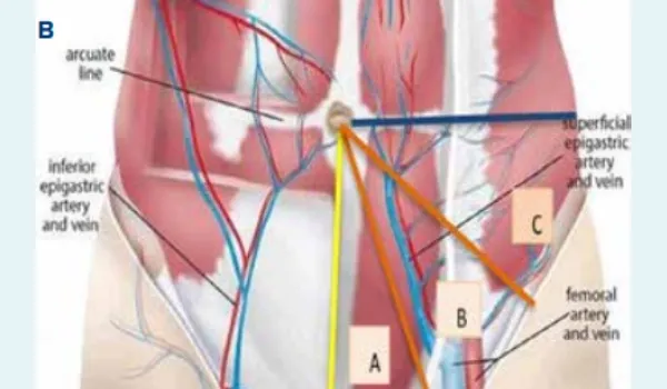
Figure 1A and B: Representation of the tri-compartment dissection for total extra-peritoneal inguinal hernia repair: A. Medial dissection, B. Central dissection, C. Lateral dissection in the pre-peritoneal space.
Generally, balloons or blunt instruments are used to create the pre-peritoneal space, a process that is usually not performed under direct vision. This often causes bleeding, which can obscure the anatomy of the pre-peritoneal space and increase the risk of complications.5
Technique: The tri-compartment V-technique includes the following steps:
A standard infra umbilical transverse incision of 2 cm, dissecting the adipose tissue, a transverse of 1.5 cm anterior rectus sheath incision is made and a rectus abdominis muscle is retracted laterally to expose the posterior sheath, and then, the Hassan’s trocar is placed in that space, after which a telescope (10 mm, 0 degree) is introduced.6 Then, insufflation is done after insertion of the camera under vision so that proper extra-peritoneal space is created, as shown in Figure 2.
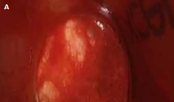
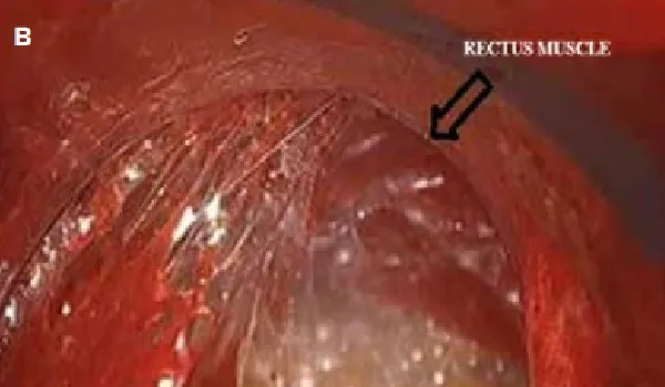
Figure 2: A. Pre-insufflated view, B. Post-insufflated view.
Firstly, surgical dissection in a medial compartment is done, using the telescope. The dissection is initiated posterior to the rectus muscle, cephalo-caudally up to the pubic crest, and then into the Retzius’ space as shown in Figure 3.
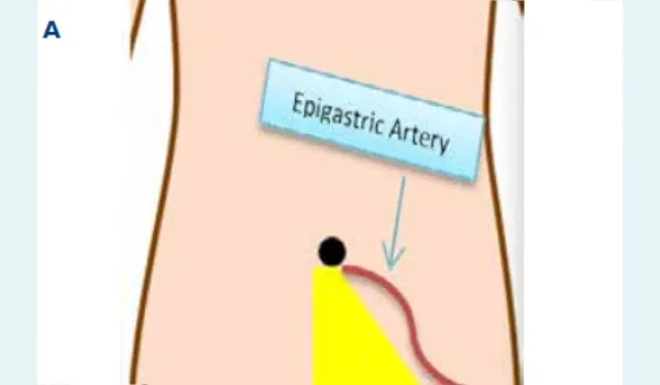
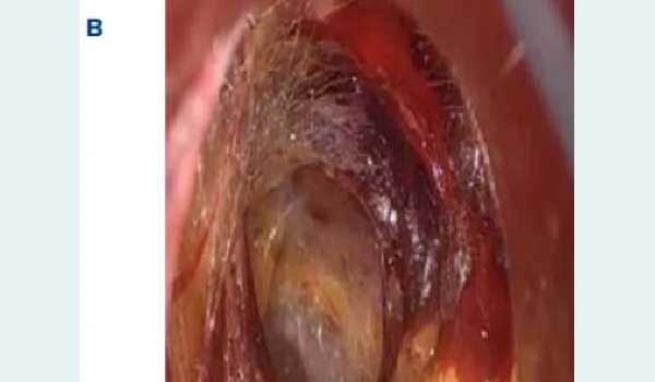
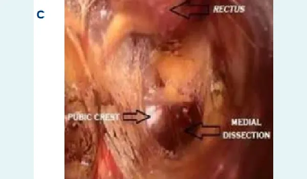
Figure 3: A. Showing the Retzius’ space and representation of medial dissection, B. Initial view of medial compartment, C. Dissected medial compartment.
After completing medial dissection secondly, the lateral compartment dissection (Bogros’ space) is done. This is achieved by dissecting laterally preserving the inferior epigastric vessels (IEV). The IEV should always be under direct vision to avoid any injury to them, once the IEV is preserved, lateral compartment space is created and the dissection completed up to the peritoneal reflection8 as shown in Figure 4. Posteriorly, the Psoas muscle should be visible and the lateral nerves must be preserved.
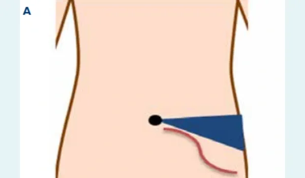
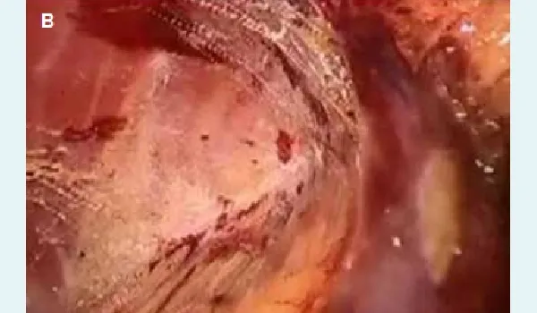
Figure 4: A. Showing the Bogros’ space, B. Representation of lateral dissection
Lastly, the central compartment dissection (iliac vessel, the hernia sac and cord structure) as shown in Figure 5 is completed with gentle blunt dissection of the adipose tissue which is closely adherent to the inferior epigastric vessels away from the peritoneum posteriorly, the epigastric vessels are left undisturbed along with the anterior abdominal wall.9
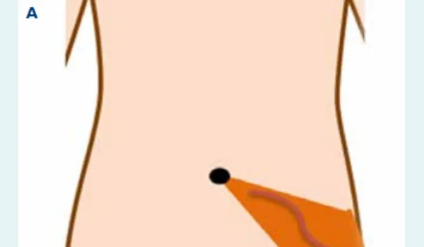
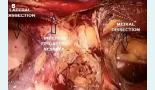
Figure 5: A. Showing the epigastric vessels’ space, B: Representation of central dissection.
Then, the inguinal hernia surgery is completed in a conventional manner, reducing and dissecting the sac in front of the cord structures. Finally, a standard polypropylene mesh is placed comfortably as sufficient space is created and there is less chance of folding.
Discussion
Laparoscopy for inguinal hernia repair is now considered a gold-standard technique; however, it has a steep learning curve. The laparoscopic inguinal anatomy can be difficult for new surgeons to grasp, as the pre-peritoneal space offers few landmarks and is both narrow and enclosed. The technique described by the author aids in understanding the extraperitoneal inguinal anatomy by compartmentalising it into three areas and addressing each compartment systematically. This makes it easier to understand and perform the extra-peritoneal dissection, which is the most important aspect of the surgery. This technique is comparatively simple to perform and is cost-effective, making it an easy technique for experienced surgeons and those who are new to laparoscopic technique.10 This is performed entirely under the direct vision of the surgeon, which minimises the risk of errors. The anterior peritoneal space can be accessed by various methods such as mirror push method, ballooning, finger separation method and retropulsion.11 However, TEP repair results in complete separation of the anterior peritoneal space through the extra-peritoneal cavity without entering into the abdominal cavity. When both TEP and TAPP are compared, they show similar outcomes in terms of recurrence rates, hospital stay, and complications.12
Conclusion:
Laparoscopic extra-peritoneal technique using the V-technique, and addressing the inferior epigastric vessels as well as the major iliac vessels, can further minimise blood loss compared with other conventional techniques. This results in quicker recovery and reduces the risk of complications.13 It also clearly defines the pre-peritoneal space, which helps the surgeon to locate significant areas like the "triangle of doom" and "triangle of pain".1 A surgeon should create enough space for proper mesh placement, which is crucial for hernia repair.15 The V-technique allows easy understanding of inguinal anatomy which results in better visualisation of extra peritoneal anatomical structures and having better control to reduce any complications.
Vikas Panwar, Sourav Panda. Simplifying Laparoscopic Totally Extraperitoneal Inguinal Hernia Repair
with V-Technique. MMJ. 2025, September. Vol 2 (3).
References
- Ger R, Monroe K, Duvivier R, et al. Management of indirect inguinal hernias by laparoscopic closure of the neck of the sac. Am J Surg. 1990;159:370–3.
- McKernan JB, Laws HL. Laparoscopic repair of inguinal hernias using a totally extraperitoneal prosthetic approach. Surg Endosc. 1993;7:26–8.
- Takata MC, Duh QY. Laparoscopic inguinal hernia repair. Surg Clin North Am. 2008;88:157–78.
- Lorenz A, Augustin C, Konschake M, et al. The Preperitoneal Space in Hernia Repair. Front Surg. 2022;9:869731.
- Chu HC, Hu SW, Wu WL, et al. Comparison of balloon dissection and telescopic dissection of the preperitoneal space in laparoscopic totally extraperitoneal hernia repair: a systematic review and meta-analysis. Langenbecks Arch Surg. 2023;408(1):15.
- Ferzli GS, Fingerhut A. Trocar placement for laparoscopic abdominal procedures: a simple standardized method. J Am Coll Surg. 2004;198(1):163–73.
- Retzius A. Some Remarks on the Proper Design of the Semilunar Lines of Douglas. With Three Illustrations. Edinb Med J. 1858;3(10):865–67.
- Bond S, Ferder M, Grossman L, et al. The effect of epigastric vessel ligation on the vascular territories of the pig rectus muscle. Plastic and reconstructive surgery. 1996;97(3):616–20.
- Alabi A, Haladu N, Scott NW, et al. Mesh fixation techniques for inguinal hernia repair: an overview of systematic reviews of randomised controlled trials. Hernia. 2022;26:973–87.
- Cao C, Shi X, Jin W, et al. Clinical data analysis for treatment of adult inguinal hernia by TAPP or TEP. Front Surg. 2022;9:900843.
- Akin E, Bas E, Firat N, et al. Comparison of balloon trocar versus telescopic dissection method for TEP inguinal hernia repair. J Coll Physicians Surg Pak. 2021;30:623–6.
- Yildiz A. Laparoscopic transabdominal preperitoneal and totally extraperitoneal in inguinal hernia surgery: comparison of intraoperative and postoperative early complications of two techniques. J Minim Invasive Surg. 2022;25:18–23.
- Goksoy B, Yilmaz G, Azamat IF, et al. Laparoscopic inguinal hernia repair-TAPP versus TEP: results of 301 consecutive patients. Surg Technol Int. 2021;39:191–5.
- Lomnicki J, Leszko A, Kuliś D, et al. Current treatment of the inguinal hernia - the role of the totally extraperitoneal (TEP) hernia repair. Folia Med Cracov. 2018;58:103–14.
- Zhou XL, Luo JH, Huang H, et al. Totally Extraperitoneal Herniorrhaphy (TEP): lessons learned from anatomical observations. Minim Invasive Surg. 2021;2021:5524986.