 WhatsApp Us (24/7)
 +91 926 888 0303 (24/7)


Abstract: A 46-year-old perimenopausal woman, with a history of previous three caesarean sections, presented with progressive abdominal distension, heavy and prolonged menstrual bleeding, and fatigue over several months, accompanied by poor appetite and lower abdominal discomfort. She experienced two episodes of severe vaginal bleeding with syncope, resulting in very severe anaemia and thrombocytopenia, necessitating multiple transfusions of packed red blood cells (PRBCs) and fresh frozen plasma (FFP). Clinical examination revealed pallor, tachycardia, and a massive firm, irregular abdominopelvic mass with hepatosplenomegaly. Laboratory investigations confirmed anaemia, thrombocytopenia, and hyperbilirubinemia. Imaging identified a massive uterine fibroid (measuring up to 32.2 × 28.4 cm) with degenerative changes and heterogenous fluorodeoxyglucose (FDG) uptake, associated with ureteric compression, hepatosplenomegaly, and mild ascites. Multidisciplinary evaluation, including bone marrow biopsy excluded marrow suppression. The patient underwent total abdominal hysterectomy with bilateral salpingo-oophorectomy (TAH-BSO) and liver biopsy. Intraoperatively, a large, lobulated uterine mass with multiple fibroids and myxoid degeneration was confirmed, weighing 8.3 kg; intraoperative frozen section and final histopathology confirmed benign leiomyomas. Liver biopsy and ascitic fluid cytology were negative for malignancy. The postoperative course was uneventful, with close monitoring and supportive transfusions. This case highlights a rare presentation of giant uterine leiomyomas causing life-threatening anaemia and coagulopathy, mimicking malignancy and necessitating a multidisciplinary approach with definitive surgical management
Key words: Perimenopausal, Thrombocytopenia, Fibroid, Hepatosplenomegaly
Introduction
Uterine fibroids are the most prevalent benign tumours in women of reproductive age, often asymptomatic or presenting with menorrhagia and pelvic pressure.1 They arise from the smooth muscle cells of the uterus and are usually diagnosed by ultrasound or pelvic examination. Rarely, massive degenerative myomas may mimic malignant neoplasms both clinically and radiologically, posing significant diagnostic challenges.1,2 Their unusual presentation can include cystic, myxoid, or red degeneration, leading to large, heterogeneous pelvic masses with features suggestive of sarcomas or ovarian malignancies.
This case report highlights a rare presentation of a degenerative fibroid, clinically resembling a malignant pelvic mass. Diagnostic complexity was further compounded by severe anaemia, thrombocytopenia, and indirect hyperbilirubinemia, occurring in the background of hepatosplenomegaly, suggestive of hypersplenism. Such haematologic abnormalities — especially in the presence of splenomegaly — raised suspicion for systemic disorders such as lymphoproliferative disease or chronic liver pathology, requiring a broad differential diagnosis. Such a constellation of findings is unusual in benign gynaecologic conditions and raises concern for underlying haematologic or systemic pathology, necessitating a multidisciplinary diagnostic approach.
Through this case, we emphasise the importance of thorough clinical evaluation, appropriate imaging, and histopathological correlation to accurately distinguish benign from malignant pelvic masses, particularly when atypical systemic manifestations coexist.
Case Report
A 46-year-old perimenopausal woman presented with progressive abdominal distension, heavy and prolonged menstrual bleeding, and fatigue over several months. She also complained of poor appetite and lower abdominal discomfort, with no significant weight loss. On 20th May 2025, she experienced heavy vaginal bleeding followed by syncope, and was admitted to a local hospital in Bareilly, from 20th to 25th May. Her haemoglobin was 4.8 g/dL and platelet count 60,000/µL, requiring transfusion of two units of packed red blood cells (PRBCs). She was discharged in a stable condition on 25th May.
On 26th May she again experienced profuse vaginal bleeding with extreme weakness and reported to our hospital. On examination, she appeared pale, with a pulse rate of 116 beats per minute (bpm) and blood pressure of 90/60 mmHg. Abdominal examination revealed a firm, irregular, non-tender abdominopelvic mass extending up to the xiphisternum. Pelvic examination confirmed a large irregular uterine mass, and hepatosplenomegaly was also noted. Obstetric history included three caesarean sections, the last 12 years ago. There was no history of chronic medical illness, malignancy, liver disease, or haemolytic disorders.
Initial laboratory investigations showed haemoglobin 8.8 g/dL, platelet count 61,000/µL, total bilirubin 2.77 mg/dL and direct bilirubin 1.54 mg/dL. Magnetic resonance imaging (MRI) of the whole abdomen demonstrated a large uterine fibroid measuring 21.5 × 32.2 × 28.4 cm with degenerative changes, extending into the subphrenic region. Hepatosplenomegaly and mild ascites were also noted. Bilateral ovaries were not clearly visualised.
A positron emission tomography–computed tomography (PET–CT) scan revealed a large soft-tissue lesion with necrotic areas and heterogeneous flourodeoxyglucose (FDG) uptake measuring approximately 23 × 28 × 29 cm, occupying the abdomen and pelvic cavity, with ureteric compression. A multidisciplinary team including haematology and surgical gastroenterology was consulted. Bone marrow biopsy demonstrated trilineage haematopoiesis, ruling out marrow suppression.
Subsequently, the patient underwent a total abdominal hysterectomy with bilateral salpingo-oophorectomy (TAH– BSO) and liver biopsy on 30th May 2025. Intraoperatively, mild ascites was present, and fluid was aspirated for cytological evaluation. A large, irregular, lobulated mass consistent with a 36-weeks pregnant uterus was visualised. Multiple fibroids with cystic and degenerative changes were noted, distorting the uterus (Figure 1). The adnexa appeared grossly normal but were elongated and distorted due to the size of the fibroids.
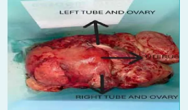
Figure 1: Multiple fibroids distorting the uterus.
Intraoperative frozen section analysis of a suspicious fibroid confirmed a benign leiomyoma, prompting completion of the planned TAH–BSO. A urologist assisted with bladder dissection and DJ stenting because of severe adhesions from three previous caesarean sections and distorted pelvic anatomy due to large multiple fibroids. The surgical specimen weighed 8.3 kg (Figure 2).
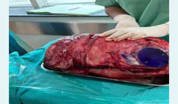
Figure 2: Multiple fibroids forming a huge mass involving the uterus, fallopian tubes, ovaries and cervix.
The patient was transferred to the intensive care unit (ICU) postoperatively for 3 days. Histopathological examination confirmed multiple benign leiomyomas with myxoid degeneration. Liver biopsy and ascitic fluid cytology were negative for malignancy. Her postoperative course was largely uneventful, with close monitoring of haematological parameters and liver function.
During her hospital stay, she received 11 units of leucodepleted PRBCs (LDPRBCs ), 12 units of fresh frozen plasma (FFP), 10 units random donor platelets (RDP), and 10 units of single donor platelets (SDP). Following stabilisation, she was transferred to the ward.
The patient was discharged on 7th June 2025 in a stable condition, with haemoglobin of 9 g/dL and platelet counts recovered to over 300,000/µL. The patient has been on regular follow up for the last one month and is doing well.
Discussion
Multiple uterine fibroids uterus exhibiting cystic and degenerative changes can mimic malignancy, and can be associated with multifactorial anaemia, hyperbilirubinemia (suggestive of haemolysis), thrombocytopenia, and hepatosplenomegaly (indicative of underlying hypersplenism).2-4
Uterine fibroids are the most common benign pelvic tumours in women. Only a very small percentage of fibroids undergo malignant change or red degeneration. With the widespread use of ultrasound in clinical practice, uterine fibroids are generally diagnosed early and are usually not missed. Hence, the development of an unusually large myoma is rare.6
Preoperative imaging is essential to determine the extent of the fibroid and to assess the likelihood of malignancy, particularly in cases of infiltrative growth.6
The differential diagnosis in this case included uterine leiomyosarcoma, ovarian neoplasm, lymphoproliferative disorder, haematological malignancy, and chronic liver disease with secondary hypersplenism.4,5
The prognosis was favourable, as histopathology confirmed benign myomas with myxoid degeneration. The haematologic abnormalities improved after surgery with resolution of splenic sequestration. This confirms that the mass effect of the fibroid on surrounding organs likely contributed to hypersplenism and reactive cytopenias, which normalised following tumour removal.
Conclusion:
This case highlights that large uterine fibroids with cystic and degenerative changes, can closely mimic malignancy, both clinically and radiologically. When associated with systemic features such as anaemia, thrombocytopenia, hyperbilirubinemia, and hepatosplenomegaly, the diagnostic complexity increases, raising suspicion for malignancy or haematologic disorders. It is crucial to maintain a broad differential diagnosis including benign aetiologies. A multidisciplinary approach, incorporating imaging, laboratory evaluation, and definitive histopathology, remains essential to accurately diagnose and appropriately manage such rare and complex cases.
Anuradha Kapur, Alka Gujral, Vijay Kumar, Ujjwala. Large Uterine Mass with Multiple Myomas Mimicking
malignancy: A Rare Case Report. MMJ. 2025, September. Vol 2 (3).
References
- Baird DD, Dunson DB, Hill MC, et al. High cumulative incidence of uterine leiomyoma in black and white women: ultrasound evidence. Am J Obstet Gynecol. 2003;188(1):100–7.
- Fasih N, Prasad Shanbhogue AK, Macdonald DB, et al. Uterine leiomyomas: understanding the imaging appearance of benign uterine tumors with atypical features. Radiographics. 2008;28(4):1005–26.
- Hodge JC, Morton CC. Genetic heterogeneity among uterine leiomyomata: insights into malignant progression. Hum Mol Genet. 2007;16(Spec No. 2):R203.
- Nishida M, Watanabe K, Suginami H, et al. Clinical manifestations of atypical leiomyoma of the uterus. Gynecol Obstet Invest. 1999;48(1):43–6.
- Viva W, Juhi D, Kristin A, et al. Massive uterine fibroid: a diagnostic dilemma: a case report and review of the literature. J Med Case Rep. 2021;15(1):344.
- Duhan N, Bansal S. Uterine sarcomas: current perspectives. Int J Womens Health. 2014;6:295–304.