 WhatsApp Us (24/7)
 +91 926 888 0303 (24/7)


Abstract: Anterior giant skull base meningiomas are technically challenging tumours to treat because of their deep vascular supply that can preclude early devascularisation during resection. Preoperative embolisation of these arterial feeders is thought to decrease blood loss and facilitate resection, especially in these large-sized lesions. It is essential that both the endovascular and skull base neurosurgeons understand the potential benefits of preoperative embolisation. We present a case which highlights the applicability and benefits of pre operative embolisation in the surgical resection of a giant skull base meningioma.
Key words: Meningioma, Embolisation, Skull Base.
Introduction
Skull base meningiomas can be highly vascular tumours, and their vascularity may complicate or limit resection because of intraoperative blood loss.1,2 Surgical devascularisation can be challenging in practice, particularly for tumours arising along the skull base in locations that prevent access to the vascular pedicle until later stages of the resection.3,4 Embolisation of meningiomas may be performed preoperatively when surgical devascularisation is expected to be challenging. Preoperative embolisation of meningiomas has been advocated to facilitate intraoperative resection by decreasing surgical blood loss through occlusion of feeding arteries, as well as by shrinking, softening, and sometimes inducing necrosis in the tumour in ways that sometimes facilitate mechanical removal and reduced operative duration.3
Case Report
A 45-year-old female patient presented with complaints of headache and personality changes for the past two months. She also had difficulty in perceiving smell. On examination, she had anosmia, 6/6 vision in both eyes, and fundus examination revealed bilateral papilloedema. The rest of the clinical examination was unremarkable. Magnetic resonance imaging (MRI) revealed a large, contrast-enhancing lesion arising from the anterior skull base involving the crista galli, planum sphenoidale and tuberculum sellae, suggestive of a giant anterior skull base meningioma (Figure 1A–C).
In view of the large size of the lesion, the decision to perform a pre-operative digital subtraction angiography (DSA) with tumour embolisation was taken. Diagnostic angiographic runs, from the right external carotid artery, revealed feeders from the accessory middle meningeal artery (MMA) (distal branch of internal maxillary artery) giving rise to a characteristic tumour blush (Figure 2A–B). A similar tumour blush was noted from the left accessory MMA.
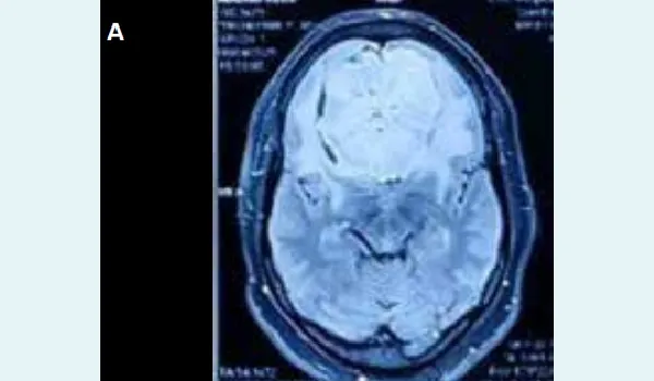
Figure 1A: Preoperative magnetic resonance imaging (MRI) axial view showing a giant, contrast-enhancing anterior skull base lesion involving the crista galli, planum sphenoidale, and tuberculum sellae, suggestive of a meningioma.
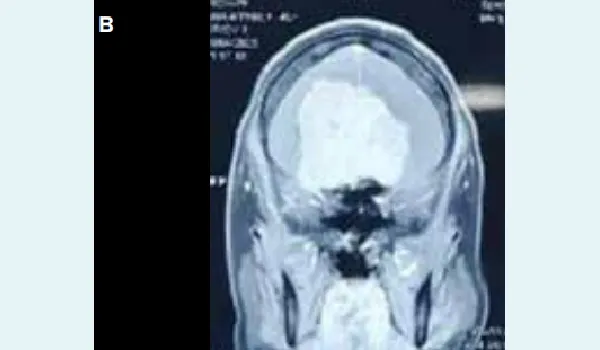
Figure 1B: Preoperative magnetic resonance imaging (MRI) coronal view demonstrating the extent of the anterior skull base meningioma with significant mass effect.
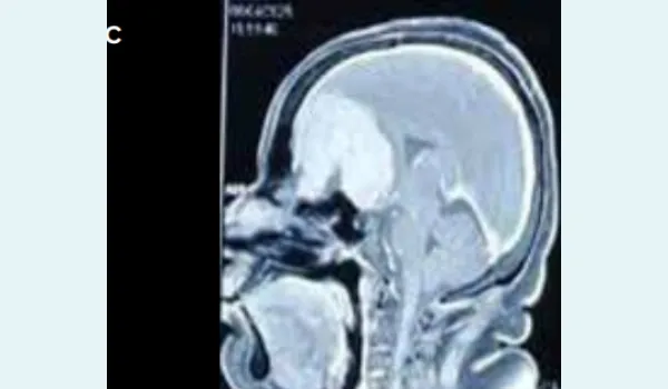
Figure 1C: Preoperative magnetic resonance imaging (MRI) sagittal view depicting the large anterior skull base tumour with superior extension.
Superselective cannulation of the accessory MMA was done using a microcatheter (on both right and left sides), and feeders were embolised with lipiodol and nectacryl glue (n Butyl cyanoacrylate) (Figure 2A and B).
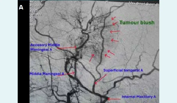
Figure 2A: Selective digital subtraction angiography (DSA) showing tumour blush from branches off the external carotid artery.
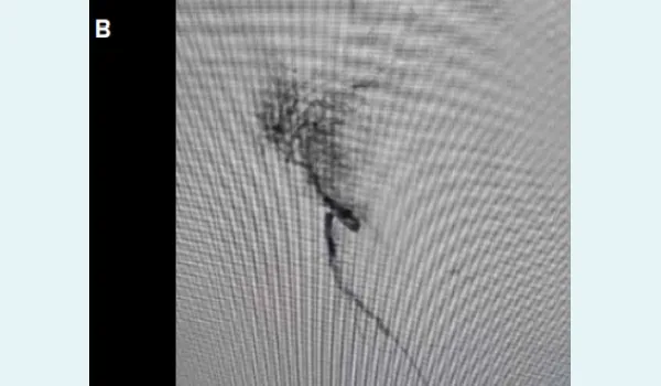
Figure 2B: Superselective shoot from the accessory meningeal artery through a microcatheter.
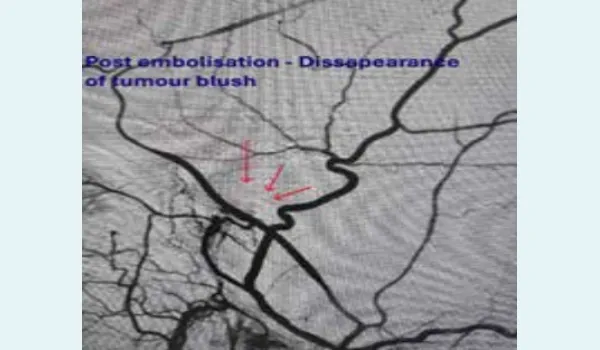
Figure 3C: Postembolisation status.
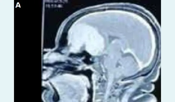
Figure 4A: Postembolisation magnetic resonance imaging (MRI) axial view revealing hypointense areas with decreased contrast uptake, indicating reduced tumour vascularity.
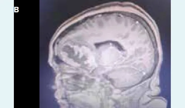
Figure 4B: Postembolisation magnetic resonance imaging (MRI) coronal view showing further reduction in contrast enhancement, consistent with successful embolisation.
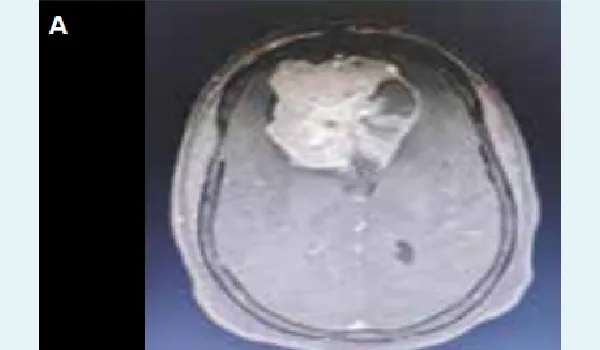
Figure 5A: Postoperative computed tomography (CT) axial view demonstrating satisfactory removal of the anterior skull base meningioma following bifrontal craniotomy.
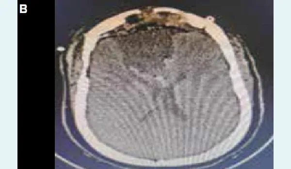
Figure 5B: Postoperative computed tomography (CT) coronal view confirming near-complete tumour resection with no significant residual lesion.
Postembolisation of feeders revealed the disappearance of tumour blush on both sides, indicating satisfactory tumour embolisation (Figure 3). Postembolisation MRI showed hypointense areas of decreased contrast uptake suggestive of decreased tumour vascularity (Figure 4).
Post endovascular embolisation, the patient underwent definitive surgery (bifrontal craniotomy) and tumour decompression via subfrontal approach. Intraoperatively, there was a significant reduction in tumour vascularity, which contributed to a near complete tumour resection (Figure 5) along with decreased blood loss. Postoperatively, the patient was stable with no new neurological deficits and was discharged subsequently
Discussion
Anterior skull base meningiomas can have a varied and complex vascular supply. The anterior and/or posterior ethmoidal arteries are the major arterial feeders in more than 13% of skull base meningiomas. The vascular supply of meningiomas in the olfactory groove and planum sphenoidale can arise from dural, transosseous, and even pial feeders.1 These tumours may also be fed by art these feeders is done through special microcatheters. However, aggressive embolisation can be fraught with permanent postprocedural neurological deficits in view of external and internal carotid artery anastomoses, which may sometimes not be visible. eries that supply the anterior branch of the MMA, the recurrent meningeal branches of the ophthalmic artery (OphA) and lacrimal artery, the meningeal branches of the internal carotid artery (ICA), the tentorial branch of the meningohypophyseal trunk (MHT), and the terminal branches of the internal maxillary artery (IMA), accessory meningeal artery.1,5 Super selective embolisation of these feeders is done through special microcatheters. However, aggressive embolisation can be fraught with permanent postprocedural neurological deficits in view of external and internal carotid artery anastomoses, which may sometimes not be visible.
Tumour embolisation decreases the tumour vascularity and initiates tumour necrosis, making the tumour consistency soft and more amenable to surgical resection. The necrotic areas can be identified on the postembolisation MRI as hypointense areas of decreased contrast uptake, which were noted in this case.
Conclusion:
Preoperative embolisation of skull base meningiomas is increasingly used to improve surgical time, reduce bleeding, and enhance the neurosurgeon's confidence while minimising the manipulation of healthy brain tissue during the surgical procedure. Endovascular approaches using super selective embolisation of the tumour feeders form the baseline treatment in these cases.
Daljit Singh, Abhishek Katyal, Simranjeet Singh. Preoperative Embolisation and Surgical Resection of a
Giant Anterior Skull Base Meningioma. MMJ. 2025, September. Vol 2 (3).
References
- Yoon N, Shah A, Couldwell WT, et al. Preoperative embolization of skull base meningiomas: current indications, techniques, and pearls for complication avoidance. Neurosurg Focus. 2018;44(4):E5.
- Sacdalan DRL, Ignacio OR, Ignacio GMG. Pre- and Postoperative Parameters among Patients Undergoing Preoperative Embolization of Meningioma: a Tertiary Referral Center Experience. Acta Med Philipp. 2025;59(5):52–7.
- de Jesús Gutiérrez-Baños J, Castillo-Rangel C, RodriguezPereira MI, et al. Presurgical Embolization of Meningiomas: Analysis of Safety and Efficacy in a Mexican National Center and Proposal for a Classification of Preoperative Embolization of Meningiomas Based on Embolic Agent Penetration. World Neurosurg. 2025;195:123663.
- Noor MM, Rahman MM. Angiographic Features of Meningiomas Predicting Extent of Preoperative Embolization. Neurosurgery. 2025;96(2):e35–6.
- Cecchini G. Anterior and posterior ethmoidal artery ligation in anterior skull base meningiomas: a review on microsurgical approaches. World Neurosurg. 2015;84(4):1161−5.