 WhatsApp Us (24/7)
 +91 926 888 0303 (24/7)


Abstract: Pneumocystis jiroveci pneumonia (PJP) remains a critical opportunistic infection in haematopoietic stem cell transplantation (HSCT) recipients, traditionally occurring late post-transplant. This report presents two rare cases of early-onset PJP during the peri-engraftment phase in high-risk patients undergoing haploidentical HSCT — one a paediatric case with inborn error of immunity due to dedicator of cytokinesis 8 ( DOCK8) deficiency and the other, an adult with Janus kinase 2 (JAK2) positive primary myelofibrosis. Both patients developed acute respiratory symptoms and characteristic radiographic findings within 10–22 days post-transplant. PJP was confirmed through bronchoalveolar lavage (BAL) and direct immunofluorescence assay. Prompt initiation of trimethoprim-sulfamethoxazole (TMP-SMX) in therapeutic doses led to clinical improvement and successful haematological recovery. These cases underscore the potential for PJP to emerge earlier than expected, challenging existing guidelines that recommend prophylaxis primarily after engraftment. Earlier initiation of TMP-SMX may be associated with a risk of delayed engraftment and graft failure. These cases highlight that the timing of prophylactic interventions, potentially initiating before or during the engraftment period, may enhance patient outcomes. Radiological imaging and early bronchoscopy remain essential for timely diagnosis. Further multicentric studies and registry analyses are warranted to inform evidence-based prophylaxis protocols and improve early detection strategies in this vulnerable patient population.
Key words: Pneumocystis jiroveci Pneumonia, Haematopoietic Stem Cell Transplantation, Early Onset, Haploidentical Transplant, Trimethoprim-Sulfamethoxazole (TMP-SMX) Prophylaxis.
Introduction
Pneumocystis jiroveci pneumonia (PJP), formerly known as Pneumocystis carinii pneumonia (PCP), is a potentially fatal opportunistic fungal infection predominantly affecting immunocompromised individuals, particularly those undergoing haematopoietic stem cell transplantation (HSCT). Despite significant advances in supportive care and antimicrobial prophylaxis, PJP remains a substantial cause of morbidity and mortality in this vulnerable population. The profound immunosuppression resulting from myeloablative conditioning regimens, prolonged neutropenia, graft-versus-host disease (GVHD), and the use of corticosteroids or other immunosuppressive therapies contributes to a high susceptibility to PJP in HSCT recipients.1
The clinical presentation of PJP in HSCT patients often diverges from that seen in human immunodeficiency virus (HIV) infected individuals, typically manifesting as an acute, rapidly progressive pneumonia characterised by nonproductive cough, dyspnoea, hypoxaemia, and bilateral interstitial infiltrates on imaging. These atypical and rapidly evolving features often delay diagnosis, underscoring the need for high clinical suspicion and timely diagnostic interventions, such as bronchoalveolar lavage (BAL) and polymerase chain reaction assays.2
Prophylactic strategies, particularly trimethoprimsulfamethoxazole (TMP-SMX) or cotrimoxazole, have proven highly effective in reducing the incidence of PJP, yet challenges remain in determining optimal duration and managing drug-related toxicities.3 Recent reports also indicate emerging resistance and breakthrough infections, even in the context of prophylaxis, highlighting the importance of vigilance and reassessment of existing protocols.4
We present unusual cases of early-onset of this potentially life-threatening opportunistic infection that can occur during the peri-engraftment period following HSCT.
Case 1
A 7-year-old girl, a case of hyper-immunoglobulin E (IgE) syndrome with chromosome 9 deletion and dedicator of cytokinesis 8 (DOCK8) deficiency, presented with a history of recurrent episodes of pneumonia and ear infections since the age of 1 year. Subsequently, she developed bronchiectasis with pulmonary hypertension at the age of 6 years and was on sildenafil and torsemide. The patient also developed multiple crusted skin lesions and erythema of the skin since the age of 1 year. Serum IgE was markedly elevated at 2230.37 IU/mL (normal < 100). The immunodeficiency panel was normal, showing natural killer (NK) and T-cell populations with a preserved CD4:CD8 ratio, normal neutrophil and monocyte populations, and a reduced B-cell population. Baseline complete blood count (CBC) prior to HSCT showed haemoglobin 11.2 g/dL, total white blood cell (WBC) count 25.3 x 10³ /L, and a differential cell count comprising neutrophils 58%, lymphocytes 7%, monocytes 6%, eosinophils 28%, and a platelet count 263 x 103 /L.
The patient underwent haploidentical HSCT from the mother as the donor, who did not have any pathogenic variant on genetic evaluation. The patient received fludarabine 150 mg/m2, treosulphan 42 g/m2, and total body irradiation (TBI) 2 Gy as myeloablative conditioning and post-transplant cyclophosphamide 100 mg/kg and tacrolimus/mycophenolate mofetil based graft-versus-host disease (GVHD) prophylaxis. The patient was transfused with 10.9 x 10⁶/kg peripheral blood stem cells from the mother as donor.
On Day +10 of HSCT, she developed high-grade fever, sudden onset of breathlessness, tachypnoea, tachycardia, and was placed on non-invasive ventilation. HRCT of the chest (Figure 1A and 1B) revealed diffuse, bilateral groundglass opacities with mosaic attenuation, predominantly in perihilar, middle, and lower lung zones, along with thin-walled cysts. It also showed atelectatic changes with traction bronchiectasis. The patient was evaluated for infective aetiology and bronchoscopy with BAL performed on Day +12, in which direct immunofluorescence assay was positive for Pneumocystis jiroveci cysts and trophozoites.
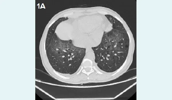
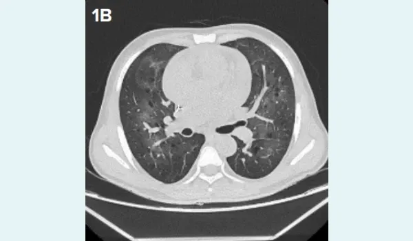
Figure 1A and 1B High-resolution computed tomography: A. Axial HRCT chest showing diffuse, bilateral ground-glass opacities with mosaic attenuation, B. Coronal HRCT chest showing diffuse, bilateral ground-glass opacities with thin-walled cysts and traction bronchiectasis.
The patient was treated with a therapeutic dose of injectable TMP-SMX from Day +14 for PJP for 1 week and later changed to oral when the patient became haemodynamically stable and given for a total duration of 3 weeks. Neutrophil engrafted on Day +14 of HSCT. The patient showed symptomatic improvement after 6 days of non-invasive ventilation. The patient’s postengraftment chimerism on Day +20 and Day +60 was 99% with normal haematological recovery.
Case 2
A 53-year-old male with a case of primary myelofibrosis, Janus kinase 2 (JAK2) positive, presented with a history of left hypochondrium pain, early satiety, significant weight loss and progressive generalised weakness for a year. The patient received thalidomide and ruxolitinib and did not show any significant improvement and was planned for HSCT.
The patient underwent haploidentical HSCT with fludarabine 150 mg/m2 , busulphan 6.4 mg/kg, TBI 2 Gy reduced-intensity conditioning and post-transplant cyclophosphamide 100 mg/kg and tacrolimus/ mycophenolate mofetil-based GVHD prophylaxis. Increased bronchovascular markings were seen in both lungs on the chest X-ray (Figure 2A), indicating that the bronchial tubes in the lungs were more visible than usual. The patient was transfused with 10.2 x 10⁶/kg peripheral blood stem cells from his brother as a donor. The patient developed Grade III gut and oral mucositis and neutropenic fever.
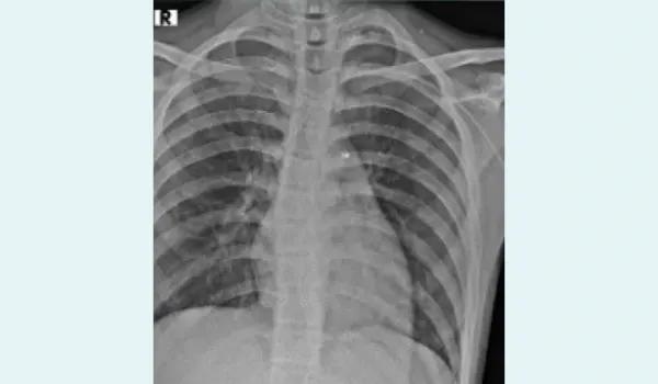
Figure 2A: Normal chest X-ray with increased bronchovascular marking.
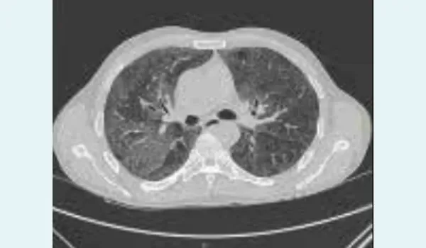
Figure 2B: Patchy and confluent areas of ground-glass opacities in bilateral lungs with relative peripheral sparing with multiple thin-walled air-filled cysts.
Neutrophil engrafted on Day +19. On Day +20, the patient developed a sudden onset of breathlessness, cough with haemoptysis, hypoxemia and bilateral crackles. HRCT chest (Figure 2B) was suggestive of patchy and confluent areas of ground-glass opacities in bilateral lungs with relative peripheral sparing, with multiple thin-walled air-filled cysts scattered in bilateral lung parenchyma. Sputum direct immunofluorescence assay positive for Pneumocystis jiroveci cysts and trophozoites. The patient was started on injectable TMP-SMX followed by oral treatment at therapeutic doses total of 3 weeks. His post-engraftment chimerism on Day +30 was 94% and Day +60 was 95%. The patient had mild cytopenia, which improved following completion of PJP treatment.
Discussion
PJP remains a significant opportunistic infection in HSCT recipients, typically manifesting late in the posttransplant course — often beyond 100 days — particularly in patients receiving prolonged immunosuppression, corticosteroids, or with chronic GVHD. The American Society for Transplantation and Cellular Therapy (ASTCT) and European Society for Blood and Marrow Transplantation (EBMT) guidelines recommend PJP prophylaxis — preferably with TMP-SMX for at least 6–12 months post-transplant and until the cessation of immunosuppressive therapy.5 However, the occurrence of PJP during the peri-engraftment phase, as seen in our cases, is exceptionally rare and underreported.
Both patients in this report developed PJP within the first 3 weeks post-transplant, during the neutropenic and engraftment phases. The early onset raises concern about a vulnerable period before prophylaxis is typically initiated or becomes effective. Nazir et al. reported similar early-onset cases in haploidentical transplant recipients with familial haemophagocytic lymphohistiocytosis (HLH), suggesting that profound immune dysregulation and mucosal barrier damage during conditioning may permit early fungal invasion.6 Additionally, the EBMT Paediatric Diseases Working Party has highlighted that atypical pathogens, including PJP, may present even within the first 30 days, particularly in high-risk patients or those with prior pulmonary compromise.7 Studies estimate PJP incidence in HSCT recipients at 3%–5% without prophylaxis, but lower with routine TMP-SMX use.8 However, breakthrough infections have been noted, possibly due to suboptimal absorption, emerging resistance, or atypical immune responses.9
A retrospective cohort analysis from the Multidisciplinary Digital Publishing Institute (MDPI) Journal of Clinical Medicine described ground-glass opacities and cystic changes in PJP similar to those seen in our HRCT findings, underlining the importance of radiographic suspicion even early post-transplant.10 These cases underscore the need to revisit current prophylactic protocols and consider pre-engraftment initiation of TMP-SMX, particularly in high-risk scenarios. Both of our cases showed timely haematological recovery following haploidentical HSCT, with no persistent cytopenia observed after therapeutic administration of TMP-SMX. Moreover, heightened clinical suspicion, early BAL, and polymerase chain reaction (PCR)-based diagnostics are crucial for timely diagnosis. Multicentric surveillance and data from HSCT registries are imperative to better understand the incidence and optimal preventive timing for early PJP.
Conclusion
PJP can occur unusually early in the post-transplant course of HSCT, even during the peri-engraftment phase, especially in high-risk HSCT settings like haploidentical HSCT. The two cases presented highlight the critical need for heightened clinical vigilance and early diagnostic measures in high-risk patients. These findings challenge current prophylactic practices, emphasising the potential value of initiating TMP-SMX before engraftment in select populations. Imaging and timely bronchoalveolar diagnostics played a pivotal role in rapid identification and successful treatment. Multicentre studies and registry-based surveillance are essential to optimise prevention strategies and improve outcomes in this vulnerable cohort.
Neelkumar Patel, Sanjeev Sharma, Anamika Bakliwal, Viral Jain, Kumar Animesh. Early-Onset
Pneumocystis jiroveci Pneumonia During the Periengraftment Phase in Haploidentical Haematopoietic
Stem Cell Transplantation. MMJ. 2025, September. Vol 2 (3).
References
- Thomas CF, Limper AH. Pneumocystis pneumonia. N Engl J Med. 2004;350(24):2487–98.
- Roux A, Gonzalez F, Roux M, et al. Update on pulmonary Pneumocystis jirovecii infection in non-HIV patients. Med Mal Infect. 2014;44(5):185–98.
- Green H, Paul M, Vidal L, et al. Prophylaxis for pneumocystis pneumonia (PCP) in non-HIV immunocompromised patients. Cochrane Database Syst Rev. 2007;(3):CD005590.
- Fillatre P, Decaux O, Jouneau S, et al. Incidence of Pneumocystis jirovecii pneumonia among groups at risk in a single-center study. Eur J Clin Microbiol Infect Dis. 2014;33(1):131–6.
- Christopeit M, Schmidt-Hieber M, Sprute R, et al. 2020 EBMT update: Infections after autologous hematopoietic stem cell transplantation. Ann Hematol. 2021;100(2):345–60.
- Nazir HF, Alawi FSB, Al Hosni S, et al. T cell-depleted haploidentical HSCT in HLH: Infectious complications. Biol Blood Marrow Transplant. 2020;26(10):1871–9.
- Ifversen M, Meisel R, Sedlacek P, et al. Pediatric supportive care after HSCT: EBMT recommendations. Front Pediatr. 2021;9:705179.
- Fillatre P, Decaux O, Jouneau S, et al. Incidence of Pneumocystis jirovecii pneumonia among risk groups. Eur J Clin Microbiol Infect Dis. 2014;33(1):131–6.
- Sweiss K, Anderson J, Wirth S, et al. Pentamidine prophylaxis and breakthrough PJP. Biol Blood Marrow Transplant. 2017;23(9):1465–70.
- Astashchanka A, Ryan J, Lin E, et al. Pulmonary complications in HSCT: Radiologic clues and diagnosis. J Clin Med. 2021;10(15):3227.