 WhatsApp Us (24/7)
 +91 926 888 0303 (24/7)


Abstract: Cholesterol granulomas are benign granulomatous lesions caused by a tissue reaction to a foreign body. Lesion formation is caused by cholesterol crystals and occurs secondary to insufficient ventilation, drainage disorder, haemorrhage, and/or chronic inflammation. Cholesterol granulomas may involve the middle ear, the mastoid bone, and the petrous apex. However, cholesterol granulomas presenting as a mass obstructing the external ear canal are relatively rare and can be a diagnostic challenge, especially in a post-mastoidectomy case.
We report a case of cholesterol granuloma occupying the roof of the mastoid cavity and presenting as a mass in the external auditory canal (EAC). Temporal bone computed tomography showed a soft tissue mass along the roof of the mastoid cavity with rarefaction of the tegmen mastoideum/roof of the mastoid air cells. The cholesterol granuloma was removed surgically, and characteristic pathological findings confirmed the diagnosis.
Key words: Cholesterol Granuloma, External Auditory Canal (EAC), Mastoid Cavity, Post-Mastoidectomy.
Introduction
Cholesterol granuloma can occur anywhere in the body, with the most common site being the temporal bone.1 Cholesterol granuloma, or cholesterol cyst, is a clinical entity that was first reported by Manasse in 1894.2,3 Cholesterol granulomas are benign granulomatous lesions caused by a tissue reaction to a foreign body.3 These lesions develop secondary to insufficient ventilation, drainage disorders, haemorrhage or chronic inflammation. Cholesterol granulomas have been reported in various regions of the body where cholesterol crystals may form, such as the lungs, breast, peritoneum, mediastinum, liver, spleen, thyroid, kidneys, lymph nodes, testis, facial skeleton, skull and temporal bone. Within the temporal bone, cholesterol granuloma is known to occur most commonly in the petrous apex.4 Very few cases of cholesterol granuloma presenting as a mass in the external auditory canal (EAC) have been reported, and occlusion of the entire EAC by a cholesterol granuloma
Case Report
A 50-year-old male presented to the Ear, Nose and Throat (ENT) outpatient department with complaints of bilateral ear discharge and decreased hearing for the past few months. He had no history of ear pain, tinnitus, vertigo, headache or any neurological complaints. He had a history of hypertension, left mastoidectomy 20 years earlier, and right mastoidectomy 18 years ago. On otomicroscopic examination, the right tympanic membrane could not be visualised due to a mass completely obstructing the EAC (Figure 1). The left ear showed a mastoid cavity with discharge, with an intact and normal tympanic membrane. The remaining ENT examination was within normal limits, including the facial nerve. The audiogram showed bilateral moderate conductive hearing loss. Highresolution computed tomography (HRCT) of the temporal bone showed a well-defined abnormal soft tissue along the roof of the mastoid cavity, measuring approximately 1.5 cm in cephalo-caudal extent (Figure 2) and 1.8 cm x 1.6 cm in maximal axial dimension (Figure 3).
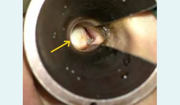
Figure 1: Otomicroscopic Examination — mass obstructing the external auditory canal (yellow arrow).
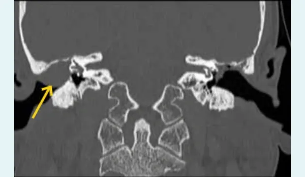
Figure 2: High-resolution computed tomography of the temporal bone — coronal section, showing cephalo-caudal extension of the mass in the external auditory canal (yellow arrow).
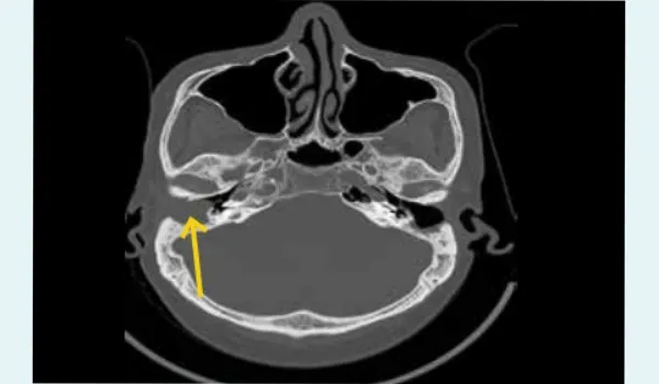
Figure 3: High-resolution computed tomography of the temporal bone — axial section, showing the horizontal extent of mass in the external auditory canal (yellow arrow).
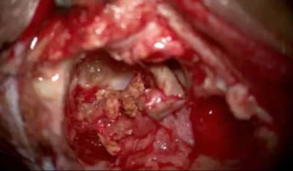
Figure 4: Cyst wall of cholesterol granuloma.
Rarefaction of the tegmen mastoideum (roof of the mastoid air cells) was seen. Post-mastoidectomy status was noted in both ears, with non-visualisation of the ossicular chain except for the footplate of the stapes and handle of the malleus. Mucosal thickening was noted in both mastoid cavities. The preoperative blood work-up was normal.
Based on the diagnosis of cholesterol granuloma from medical imaging, revision mastoidectomy under general anaesthesia was performed via a postauricular approach. A cystic lesion containing dark, chocolate-coloured fluid was found in the operated mastoid cavity, extending to the EAC. The cyst wall of cholesterol granuloma was removed along the EAC, and a large cholesteatoma was cleared from the mastoid air cells and middle ear cavity (Figure 4). The mastoid cavity smoothened. Ossiculoplasty was performed, and the attic was repaired using conchal cartilage. After the mass was completely excised, the meatal flap was repositioned.
The postoperative period was uneventful. The excised tissue was sent for histopathological examination. The histopathology report confirmed the diagnosis of cholesterol granuloma. The cyst had a fibrous lining and contained cholesterol crystals, haemosiderin and fibrin, which were surrounded by foreign body giant cells. Follow-up of the patient during the postoperative course was uneventful, with no neurological complications.
Discussion
The first theory about the pathogenesis of cholesterol cyst was proposed by Friedman in 1974, according to which haemorrhage and the catabolism of haemoglobin produce cholesterol crystals and iron.5 Sade, in 1979, emphasised the role of low ventilation and hypoxia. Many more theories have since been formulated.6 Summarising, there are three major factors regarded as responsible for the formation of cholesterol granuloma — local haemorrhage occurring during an inflammatory process, obstruction of ventilation and poor drainage of cavities.7
Cholesterol granuloma represents the most common cystic lesion of the temporal bone. It is often found in young to middle-aged patients with a history of chronic otitis media, temporal bone surgery, or temporal bone trauma.8 When a ventilation problem occurs, oedema of the mucosal membrane and rupture of blood vessels may result, secondary to the negative pressure created in the closed space. The degradation products of haemoglobin may then be exposed to the surrounding tissue, causing a foreign body-type reaction.9 With repeated inflammatory reactions and additional haemorrhage, a cholesterol granuloma can form. In our patient, a cholesterol granuloma formed a large mass in the EAC, which appears to support the local haemorrhage theory proposed by Friedman. The lesion may have originated inside the tympanic bone of EAC and expanded into the mastoid and EAC.4
Microscopically, the cyst presents with cholesterol clefts (the cholesterol dissolving during tissue processing) that are surrounded by histiocytes and foreign body giant cells.7 In fact, cholesterol crystals stimulate the accumulation of giant cells, which are responsible for the tissue reaction.10
Patients with cholesterol granuloma may present with hearing loss (conductive when concerning the middle ear or sensorineural when located in the petrous apex), tinnitus, vertigo, cranial nerve deficits, headache, facial pain/numbness, diplopia, and cerebrospinal fluid (CSF) leak.7 Clinically, the differential diagnosis in a post-operated ear with a bulge in the EAC includes cholesteatoma and brain herniation. Computed tomography (CT) scan and magnetic resonance imaging (MRI) play a significant role in the diagnosis of cholesterol granuloma. The cholesterol granuloma appears as a well-marginated lesion on CT, isodense with brain tissue, with no significant contrast enhancement, while bone erosion may be present.
Cholesterol granulomas do not routinely require surgery. However, surgical intervention may be considered based on the size and location of the lesion, the presence of symptoms and the need for diagnostic confirmation.4,11,12,13 In this case, the mass was excised through mastoidectomy. The patency of the EAC was maintained without postoperative complications such as necrosis and stenosis.
This case highlights the importance of long-term followup after cholesteatoma surgery.
Conclusion
Cholesterol granulomas are benign granulomatous lesions caused by cholesterol crystals, which occur secondary to haemorrhage, chronic inflammation, and/or insufficient ventilation. They may involve the middle ear, mastoid bone, and the petrous apex. Cholesterol granulomas of the mastoid cavity presenting as a mass obstructing the EAC are a rare entity, particularly in post-mastoidectomy patients. This case underscores the importance of considering cholesterol granuloma in the differential diagnosis of EAC masses, especially when associated with a history of chronic otitis media or prior ear surgery. Early imaging with HRCT and, when necessary, MRI can aid in prompt diagnosis, while surgical excision remains a safe and effective treatment for symptomatic or diagnostically uncertain lesions. Long-term postoperative follow-up is essential to monitor for recurrence and to preserve auditory function.
Vaishali Agarwal, Manohar Shaan. A Case Report of Cholesterol Granuloma in Mastoid Cavity. MMJ.
2025, September. Vol 2 (3).
References
- Ko MC, Jeong JY, Hwang SK, et al. A case of temporal bone cholesterol granuloma extending to the posterior cranial fossa without middle ear involvement. Korean J Otorhinolaryngol-Head Neck Surg. 2020;63(7):324–9.
- Manasse P. Ueber Granulationsgeschwülste mit Fremdkörperriesenzellen. Archiv f. pathol. Anat. 1894;136:245–63
- Nager GT, Vanderveen TS. Cholesterol granuloma involving the temporal bone. Ann Otol Rhinol Laryngol. 1976;85(2):204–9.
- Kong JS, Kim MS, Lee KY. A case of a cholesterol granuloma occluding the external auditory canal in a 12-year-old girl. Korean J Audiol. 2014;18(2):89–92.
- Friedmann I. Pathology of the ear. Oxford: Blackwell Scientific; 1974:103–9.
- Sadé J, Halevy A, Klyman A, et al. Cholesterol granuloma. Acta Otolaryngol 1980;89(3-4):233–9.
- Nikolaidis V, Malliari H, Psifidis D, et al. Cholesterol granuloma presenting as a mass obstructing the external ear canal. BMC Ear Nose Throat Disord. 2010;10:4.
- Ashman PE, Jyung RW. Cholesterol granuloma of the external auditory canal and skull base. Ear Nose Throat J. 2022;101(2):NP36–NP38.
- Royer MC, Pensak ML. Cholesterol granulomas. Curr Opin Otolaryngol Head Neck Surg. 2007;15(5):319–22.
- Altes AJK. Cholesterol granuloma in the tympanic cavity. J Laryngol and Oto. 1966;80(7):691–98.
- Shih TY, Prendes BL, Parsa AT, et al. Erosive cholesterol granuloma. Otol Neurotol. 2013;34(4):e26–7.
- Shihada R, Brand R, Segev Y, et al. Spontaneous regression of petrous apex cholesterol granuloma. Otol Neurotol. 2012;33:e9–10.
- Martin N, Sterkers O, Mompoint D, et al. Cholesterol granulomas of the middle ear cavities: MR imaging. Radiology. 1989:172(2):521–5.