 WhatsApp Us (24/7)
 +91 926 888 0303 (24/7)


Background: Anaesthesia management begins with the pre-anaesthetic checkup and continues throughout the perioperative treatment. It requires not only determining the appropriate type of anaesthesia for the surgery but also taking into account the patient’s various abnormalities and comorbidities, their severity, progression and ongoing treatment. This presents a significant challenge and varies from case to case.
Challenges may include haemodynamic stress due to cardiac changes, respiratory changes, renal impairment, or other systemic abnormalities.
The surgeon’s requirement must also be considered. For instance, the surgeon may require relaxation of the operating field, or conversely, may request no relaxant to monitor nerve integrity. Surgical demands related to operative exposure, patient positioning, or intraoperative monitoring can themselves present additional challenges — for example, fibreoptic intubation, one lung anaesthesia, prevention of ocular injury in the prone position, avoidance of optic nerve damage or cerebral oedema in extreme Trendelenburg positioning, as well as preventing hypothermia or inducing it when required.
Drug interactions and the implications of concurrent treatments must also be carefully evaluated, with the end goal of delivering safe anaesthesia. All factors must be anticipated, planned, and executed systematically to achieve an optimal outcome. In this paper, we present seven challenging cases, each requiring individualised anaesthetic management.
Case Report – 1
Intraoperative manifestation of superior vena cava syndrome (SVCS) in a renal transplant recipient with an indwelling haemodialysis catheter.
Introduction
SVCS arises from obstruction of venous return from the upper body to the heart, typically caused by extrinsic compression or intraluminal thrombosis. It is most commonly associated with malignancies like bronchogenic carcinoma, lymphoma, or with prolonged chemotherapy.1,2 However, with the increasing use of central venous catheters (CVC) for long-term dialysis, non-malignant causes like catheter-induced thrombosis have become more prevalent.3-5 SVCS presents with facial and upper-limb oedema, dyspnoea, and engorged neck veins.6,7 The anaesthetic implications are profound, especially when SVCS is first diagnosed intraoperatively, as it may precipitate airway obstruction, cardiovascular collapse, or cerebral hypoperfusion.7 This case report describes the intraoperative diagnosis and management of SVCS in a 45-year-old woman undergoing a livingdonor renal transplant.
Case discussion
A 45-year-old woman with end-stage renal disease (ESRD) on maintenance haemodialysis for one year via a right subclavian Permacath was scheduled for a livingdonor renal transplant. Her medical history included hypertension, hypothyroidism, anaemia of chronic kidney disease, and a prior episode of infective endocarditis caused by Providencia rettgeri.
Because of a positive crossmatch, the patient received rituximab therapy and underwent plasmapheresis one week prior to surgery. Post-plasmapheresis, she developed hypocalcaemia and hypokalaemia, both of which were corrected. She underwent a routine dialysis one day before surgery. Preoperative evaluation showed stable vitals (heart rate [HR]: 78 beats per minute [bpm], blood pressure [BP]: 100/60 mmHg, SpO2: 98%), normal airway assessment, and clear lung fields on auscultation. Investigations, including complete blood count (CBC), kidney function test (KFT), liver function test (LFT), prothrombin time (PT), activated partial thromboplastin time (APTT), international normalised ratio (INR), thyroid function tests (TFT), electrocardiogram (ECG), and chest X-ray (CXR) were within normal limits.
Patient was premedicated preoperatively with intravenous (IV) ranitidine, metoclopramide, and methylprednisolone. Anaesthesia was induced with IV fentanyl (100 µg), propofol (100 mg), and cisatracurium (10 mg). A 7.0-mm cuffed endotracheal tube (ETT) was inserted, with correct placement confirmed by bilateral equal air entry. Intraoperatively, invasive haemodynamic monitoring was instituted using left radial arterial canulation for blood pressure and central venous pressure (CVP) measurement by the existing right subclavian Permacath. Simultaneously, an abdominal ultrasound was performed to evaluate fluid status; the inferior vena cava (IVC) measured 1.5 cm in diameter with more than 50% collapsibility during intermittent positive pressure ventilation (IPPV), findings consistent with functional hypovolaemia.
The CVP was initially maintained between 10–14 cm H₂O. However, over the next two hours, after administration of only 200 mL of IV fluid, the CVP rose progressively to 35–45 cm H₂O. This was accompanied by profound hypotension that was unresponsive to further fluid resuscitation, along with the sudden onset of facial and upper limb swelling. Intraoperative transoesophageal echocardiography (TEE) revealed impaired right atrial filling, a dilated SVC with a thrombus, and an empty left ventricle, confirming the diagnosis of SVCS.
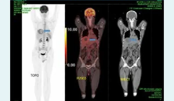
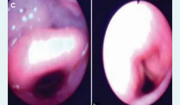
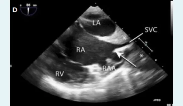
Figure 1A-D: Clinical and intraoperative findings of superior vena cava syndrome (SVCS) in a renal transplant recipient: A. Preoperative appearance with no significant swelling, B. Perioperative onset of facial puffiness suggestive of venous congestion, C. Bronchoscopic image showing oedematous airway secondary to upper body venous obstruction, D. Intraoperative transoesophageal echocardiography (TEE) demonstrating a dilated superior vena cava (SVC) with intraluminal thrombus.
Major anaesthetic challenges included airway oedema with risk of obstruction, haemodynamic instability, central venous access difficulties, preservation of renal perfusion, and the potential for cerebral and pulmonary complications.
Management
The patient was placed in a head-up tilt position to reduce venous congestion. Invasive arterial blood pressure (IBP) and CVP monitoring were used to guide therapy. Femoral vein cannulation was performed to avoid unpredictable circulation times through upper-extremity lines. Haemodynamic instability was managed with cautious fluid administration and a norepinephrine infusion. Corticosteroids were given to reduce airway oedema. Ventilation was managed using low tidal volumes and high fraction of inspired oxygen (FiO2) to minimise intrathoracic pressure and optimise oxygenation. Postoperative imaging and vascular team review confirmed SVCS secondary to thrombotic occlusion associated with the indwelling catheter. The right subclavian Permacath was removed, and the patient was started on anticoagulation therapy. Gradual improvement in urine output and renal function was observed.
Conclusion
This case illustrates the critical importance of early recognition and prompt intraoperative management of catheter-associated SVCS in a renal transplant recipient. Although the patient was asymptomatic preoperatively, general anaesthesia and positive pressure ventilation unmasked a latent venous obstruction, leading to sudden haemodynamic collapse and airway compromise. This highlights how central venous thrombosis can remain clinically silent until physiological changes during surgery precipitate acute decompensation.
Case Report – 2
Anaesthetic challenges in a patient with a large ventricular septal defect (VSD) and pulmonary hypertension undergoing emergency lower segment caesarean section (LSCS).
Introduction
Anaesthesia management in parturients with a large VSD and pulmonary hypertension (World Health Organisation [WHO] risk class IV cardiac disease) involves many challenges.1,2 These patients are at risk of haemodynamic stress due to cardiocirculatory changes, contributing to maternal morbidity and mortality.1-3 Effective management involves an understanding of the type, severity, and progression of the underlying disease. We report the case of a term primigravida with a previously undiagnosed large VSD who underwent emergency LSCS under general anaesthesia (GA) with a successful maternal and neonatal outcome.
Case discussion
A 31-year-old primigravida presented to the emergency department at 36 weeks of gestation with dyspnoea at rest and active labour. She had a two-month history of progressive exertional breathlessness (New York Heart Association [NYHA] III) that had not been investigated.
The preoperative pre-anaesthesia check-up (PAC) revealed a pansystolic murmur best heard at the cardiac apex. Her vital signs recorded were as follows: HR 107 bpm, BP 138/92 mmHg, SpO 97% on room air, and respiratory rate 54 per min. Routine laboratory investigations were within normal limits. The ECG showed a right bundle branch block (RBBB) with right ventricular hypertrophy. Bedside 2D echocardiography (Figure 2) demonstrated a large perimembranous VSD measuring 2.2 cm with a left-to-right shunt, left ventricular ejection fraction (LVEF) 50%–55%, pulmonary valve thickening with a peak gradient of 140 mmHg, moderate tricuspid regurgitation (TR), and biventricular hypertrophy. High-risk consent was obtained (American Society of Anaesthesiologists [ASA] class IV E) and the patient was immediately shifted to the operation theatre for emergency LSCS.
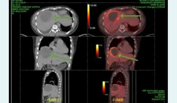
Figure 2: Parasternal long-axis view on 2D echocardiography showing a perimembranous ventricular septal defect (VSD) with mosaic flow jet from the subaortic region of the left ventricle to the right ventricle.
Abbreviations: Ao: aorta; LA: Left Atrium; LV: Left Ventricle; RV: Right Ventricle; VSD: Ventricular Septal Defect.
Ranitidine and metoclopramide were administered IV stat for aspiration prophylaxis. A 15° left lateral tilt was maintained to prevent supine hypotension. The patient was preoxygenated with 100% oxygen for three minutes followed by rapid sequence induction (RSI) using etomidate 16 mg IV and succinylcholine 150 mg IV. The patient was intubated with a 6.5 mm cuffed oral ETT and put on volume-controlled ventilation (VCV). Milrinone (phosphodiesterase-3 inhibitor), norepinephrine, and epoprostenol were kept ready to manage pulmonary hypertension.
Anaesthesia was maintained with sevoflurane in oxygen and air at minimum alveolar concentration (MAC) 1. Cisatracurium IV (8 mg, 50% of the intubation dose) was administered for neuromuscular blockade. Oxytocin 3 international units (IU) bolus followed by 10 IU in 500 ml Ringer’s lactate (RL) was given after delivery. Fentanyl 120 µg IV and paracetamol (PCM) 1 g IV were also administered after delivery. RL was infused at 60 mL/hour intraoperatively using a dial flow. Urine output at the end of surgery was 300 mL.
A live male infant weighing 2.4 kg was delivered within 8 minutes of induction of GA, with an Apgar score of 8 and 10 at 1 and 5 min, respectively. An arterial blood gas (ABG) analysis performed prior to extubation revealed no significant abnormalities. Esmolol 10 mg IV was given to attenuate tachycardia. The patient was extubated successfully, awake, responding to verbal commands, and breathing spontaneously.
Conclusion
The incidence of cardiac disease in pregnant patients in developing countries ranges from 3% to 5%, with around 44% of affected women developing pulmonary oedema in the third trimester.1 In this case, the patient presented at term with a large perimembranous VSD (2.2 cm2) left-to-right shunt and moderate pulmonary hypertension that had previously gone undiagnosed. Such patients are at greatest risk of maternal mortality during labour, delivery, and the first month postpartum, when venous return is diminished and right ventricular filling decreases.3 GA was selected for better control of ventricular and airway pressures.
Case Report – 3
Combined spinal-epidural (CSE) anaesthesia for bipolar hemiarthroplasty in a patient with hypertrophic obstructive cardiomyopathy (HOCM) and recent lower respiratory tract infection (LRTI)
Introduction
HOCM is a genetic disorder of the myocardium that carries significant perioperative risk due to dynamic left ventricular outflow tract (LVOT) obstruction, particularly when compounded by acute respiratory infections.1-3 We report the successful use of CSE anaesthesia for bipolar hemiarthroplasty in a high-risk patient with HOCM (resting LVOT gradient 55 mmHg) and recent LRTI. The anaesthetic challenges include a high incidence of intraoperative LVOT obstruction, diastolic dysfunction, myocardial ischaemia, and atrial arrhythmias.4
Case discussion
An 80-year-old woman with a history of type 2 diabetes mellitus (DM), hypertension (HTN), and coronary artery disease (CAD) presented with a fractured neck of the femur following a fall. She was planned for bipolar hemiarthroplasty after orthopaedic assessment. The patient had a recent LRTI that was clinically resolving at admission. She had received antibiotics for 5 days and had remained afebrile for 48 hours, with no clinical signs of active infection.
On examination, her HR was 88 bpm, BP 130/80 mmHg, and SpO2 was 95% on supplemental oxygen at 2 L/min via nasal prongs. Preoperative workup included CBC, LFT, KFT, cardiac assessment, and respiratory evaluation.
A CSE technique was selected as the preferred anaesthetic approach to optimise perioperative haemodynamic stability. A femoral nerve block was performed to attenuate the nociceptive response to positioning for the CSE. An epidural catheter was placed to allow for titrated postoperative analgesia and controlled sympathectomy, further enhancing haemodynamic stability. Phenylephrine was selectively administered to maintain systemic vascular resistance (SVR) without increasing myocardial contractility, thereby reducing the risk of dynamic LVOT obstruction (Figure 3).
The patient received comprehensive postoperative management focused on analgesia and respiratory support. Continuous epidural analgesia was maintained using 0.125% bupivacaine, supplemented by a transdermal fentanyl patch delivering 25 µg/hr to ensure adequate pain control. This multimodal analgesic approach effectively managed surgical pain while minimising systemic opioid requirements.
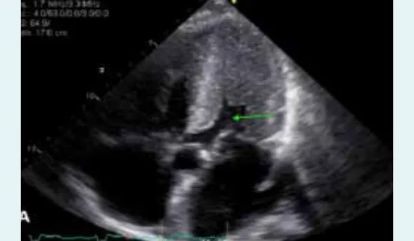
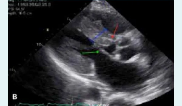
Figure 3 A-B: Echocardiographic view. A. Marked left ventricular hypertrophy (LVH) with a narrow chamber (green arrow), B. Irregular septal hypertrophy with partial left ventricular outflow tract (LVOT) obstruction (blue line), systolic anterior motion (SAM) causing LVOT (green arrow), and a speckled or granular myocardial appearance (red arrow).
Conclusion
HOCM presents significant anaesthetic challenges due to its complex pathophysiology, dynamic LVOT obstruction, diastolic dysfunction, myocardial ischaemia, and arrhythmogenic potential. This case highlights the critical importance of comprehensive anaesthetic planning for patients with HOCM and systolic anterior motion (SAM) undergoing non-cardiac surgery. CSE anaesthesia represents an optimal approach in such complex cases, offering dual benefits of maintaining haemodynamic stability while minimising respiratory risks associated with GA in a patient with LRTI.
Case Report – 4
Anaesthesia management of a child with Rett syndrome undergoing open reduction and internal fixation (ORIF) of the femur.
Introduction
Rett syndrome is a rare genetic neurodevelopmental disorder that can present with unique challenges in the perioperative period such as difficult airway management, seizure disorder, DM, heightened sensitivity to anaesthetic agents, prolonged QT interval, and risk of sudden death.1,2 This case report describes the anaesthetic management of a 15-year-old girl with Rett syndrome scheduled for ORIF of the femur. It also outlines the associated organ system involvement and discusses anaesthetic strategies tailored to this patient population.
Case discussion
A 15-year-old female weighing 30 kg with RETT syndrome was scheduled for ORIF of the left femur. PAC was done the day before surgery and included a detailed history obtained from her mother. Birth history revealed she had full-term normal delivery with an uncomplicated antenatal period. She had normal developmental motor milestones (walked without support) till 24 months of age, followed by regression of motor and language milestones. By 34 months, genetic testing confirmed Rett syndrome. Six months prior, lumbar scoliosis was diagnosed, and she was started on physiotherapy during which she sustained a left femoral fracture.
On examination, the child was pale, spoke only occasional monosyllabic words, and had lumbar scoliosis with multiple limb contractures (both upper and lower limbs). Laboratory investigations revealed anaemia (haemoglobin 7.9 g/dL); other investigations were normal. One unit of packed red blood cell was transfused the day before surgery.
Total IV anaesthesia (TIVA) using target-controlled infusion (TCI) was planned, along with an ultrasound guided suprainguinal fascia iliaca block for postoperative pain relief. Preoperatively, IV glycopyrrolate 0.2 mg and IV midazolam 1mg were administered via an existing peripheral line. GA was induced with IV fentanyl 60 μg (2 μg/kg) followed by propofol TCI. Bag-mask ventilation was provided without difficulty, followed by neuromuscular blockade with IV atracurium 15 mg (0.5 mg/kg). Intubation was done with C-MAC and a 5.5 mm cuffed ETT was placed.
Tranexamic acid was administered for the prevention of fibrinolysis (50 mg/kg bolus, followed by 5 mg/kg/hr infusion). Crystalloids were given as maintenance fluid. Positioning of the patient was done with utmost care due to the presence of multiple contractures. Surgical duration was around 90 minutes with minimal blood loss and uneventful. She was then transferred to the post-anaesthesia care unit and monitored closely. She remained hospitalised for three days without any further complications and was subsequently discharged in stable condition.
Conclusion
Rett syndrome is a neurodevelopmental disorder that affects the grey matter of the brain. It is caused by mutations in the methyl-CpG-binding protein 2 (MECP2) gene, which is located on the X chromosome.3 Because MECP2 is essential for survival, the condition is usually fatal in males before birth, making Rett syndrome primarily a disorder seen in females.3
Special consideration should be given during anaesthesia administration for these patients due to airway challenges, autonomic instability, seizure risk, and abnormal sensitivity to anaesthetic drugs. In this case, preoperatively midazolam and intraoperatively propofol were used due to their anticonvulsant properties.1-3
Case Report – 5
Locked jaw, high stakes: Navigating anaesthesia in paediatric temporomandibular joint (TMJ) ankylosis.
Introduction
Anaesthetic management of patients with TMJ ankylosis poses significant challenges due to limited or absent mouth opening, which restricts access for conventional intubation techniques. Children with TMJ ankylosis often exhibit retrognathia, mandibular hypoplasia, and altered oropharyngeal anatomy, making direct laryngoscopy, supraglottic airway insertion, and even bag-mask ventilation challenging. These factors increase the risk of airway obstruction, hypoxia, and failed intubation, especially under GA with muscle relaxation. In such cases, awake fibreoptic intubation is the preferred technique, as it enables controlled airway access while preserving spontaneous respiration and airway reflexes. In our case, it was further compounded by our patient being an international patient with a language barrier. Sedation with propofol, opioids, or benzodiazepines carries a risk of respiratory depression and airway obstruction.1 Dexmedetomidine, an α2-adrenergic agonist, provides anxiolysis, sedation, and modest analgesia without significant respiratory depression, allowing the patient to remain calm and arousable with preserved airway reflexes.
Case discussion
A 10-year-old international patient, weighing 22 kg, presented with congenital restricted mouth opening and feeding difficulties. The child was referred for further evaluation and management. On examination, child had misaligned dentition along with severely hypoplastic left mandible. The mouth opening was 8 mm and the Mallampatti grading assessment was not possible due to limited oral aperture (Figure 4). All preoperative lab investigations were within normal limits. After multidisciplinary discussion, right TMJ release with right interpositional arthroplasty was planned.
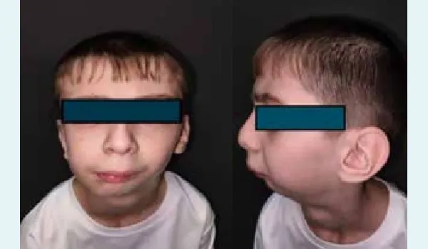
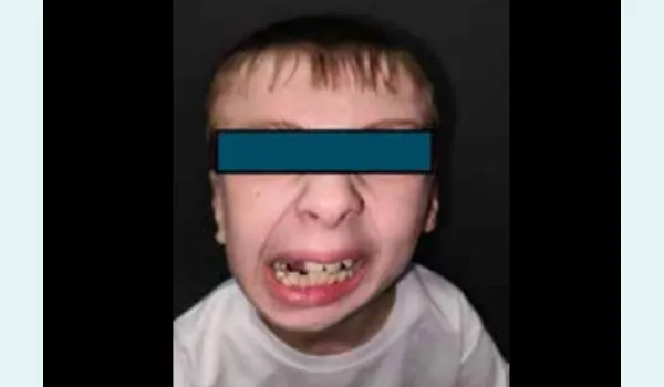
Figure 4: Preoperative photographs depicting restricted mouth opening and retrognathia.
Preoperatively, IV access was secured, and IV glycopyrrolate 0.2 mg, ranitidine 25 mg, dexamethasone 2 mg, and ondansetron 2 mg were administered. Nebulisation of the airway was done with 2 mL of 2% lignocaine in the preoperative room. A loading dose of dexmedetomidine (3 µg/kg) was infused over 10 minutes. Oxygen enrichment was done with a face mask held just above the child face. The fibreoptic bronchoscope was introduced without difficulty and clear visualisation of the glottic opening was achieved. Lidocaine 1% was sprayed onto the vocal cords, and a lubricated 5.0 flexometallic ETT was advanced over the bronchoscope into the trachea. Correct tube placement was confirmed using capnography and bilateral chest auscultation.
Anaesthesia was maintained with an admixture of oxygen and nitrous oxide (50:50) with sevoflurane, while muscle relaxation was achieved with cisatracurium. Multimodal analgesia included IV paracetamol and diclofenac. At the end of the procedure, check laryngoscopy was performed to confirm adequate mouth opening, and to ensure preparedness for potential reintubation if required. Thorough oral suctioning was carried out, and after confirming adequate muscle tone, limb power, and protective airway reflexes, the patient was extubated with a satisfactory air blast (Figure 5).
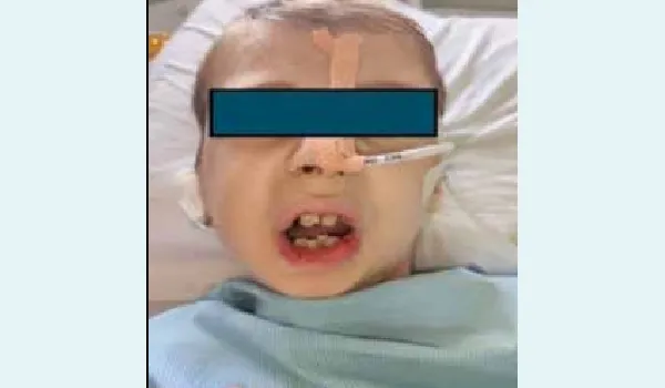
Figure 5: Postoperative Day 1 after the corrective surgery.
Conclusion
Airway management in paediatric patients with TMJ ankylosis poses significant challenges due to restricted mouth opening and the potential for difficult mask ventilation and intubation.2 The combination of dexmedetomidine and ketamine has proven effective for fibreoptic intubations in such scenarios, due to their ability to maintain spontaneous ventilation and airway reflexes.3 This approach maintained spontaneous ventilation, provided adequate sedation and analgesia, and avoided the need for more invasive techniques. The combination of these agents may be considered in similar challenging paediatric airway scenarios.
Case Report – 6
Anaesthetic management of a 65-year-old female posted for total conservative parotidectomy with facial nerve monitoring under TIVA.
Introduction
Parotidectomy procedures present unique anaesthetic challenges, particularly when intraoperative facial nerve monitoring is required.1,2 Management of parotid masses typically involves imaging, diagnostic needle aspiration, and surgical excision.1,2 Depending on the aetiology, surgical approaches range from extracapsular excision to radical parotidectomy with facial nerve sacrifice, with facial nerve injury being a feared complication. The use of TIVA ensures minimal interference with neuromonitoring.2,3 We present the perioperative management of a 65-year-old obese, diabetic female with anaemia and a predicted difficult airway who underwent total conservative parotidectomy under TIVA, with intraoperative facial nerve monitoring.
Case discussion
A 65-year-old female with a history of type 2 DM, chronic hepatitis C infection, and anaemia (haemoglobin: 10.5 g/dL) was scheduled for right total parotidectomy for a suspected neoplasm. The patient was obese (weight: 75 kg, height: 156 cm, BMI: 30.8 kg/m2), and a difficult airway was anticipated (Mallampati Grade-III). Preoperative evaluation was unremarkable apart from her comorbidities. After standard monitoring was established, anaesthesia was induced using fentanyl 2 µg/kg, propofol 2 mg/kg, and 10% lignocaine spray was applied orally to blunt the intubation reflex. Anaesthesia was maintained with TIVA using TCI of propofol (plasma concentration: 2–3 µg/mL), supplemented with fentanyl 50 µg/hr for analgesia. A rightsided total parotidectomy was performed. Intraoperative facial nerve stimulation (IFNM) confirmed intact responses in all five branches (Figure 6) of the facial nerve. A rotational flap was placed for contour restoration at the surgical site. Postoperatively, facial nerve function was evaluated and found to be intact in all branches.
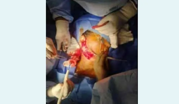
Figure 6: Intraoperative facial nerve monitoring (IFNM).
Parotidectomy carries an inherent risk of facial nerve injury due to the nerve’s anatomical course through the gland. Complications can range from transient paresis to permanent paralysis, depending on factors such as surgical technique, anatomical variations, or accidental trauma. The use of IFNM (Figure 6) has significantly improved nerve preservation, reducing permanent deficits to under 2% in experienced centres.2,3
Case Report – 7
Neuraxial anaesthesia in Lutembacher syndrome — a rare case.
Introduction
Lutembacher syndrome is a rare cardiac condition defined by the coexistence of a congenital atrial septal defect (ASD) and mitral stenosis (MS).1,2 The ASD may be either an ostium primum defect, or more commonly, an ostium secundum defect. It affects females more frequently than males.1,2 It is reported to be more prevalent in developing countries where incidence of rheumatic fever is high.1-3
We also encountered a 65-year-old female with Lutembacher syndrome, left ventricular dysfunction, and a low ejection fraction of 30%, who was posted for modified radical mastectomy (MRM). The main challenge was the patient and attendant’s refusal for GA. This report highlights the perioperative anaesthetic management and its outcome in this case.
Case discussion
A 65-year-old lady posted for right MRM presented to the PAC room. She had history of rheumatic heart disease, 40 years back, and was diagnosed with Lutembacher syndrome with an ostium secundum ASD and MS. She also had heart failure with a reduced ejection fraction of 30%. Additional comorbidities included chronic liver disease (CLD) and Hepatitis C virus (HCV) positivity, though there was no evidence of portal hypertension, oesophageal varices, ascites, or drug allergy.
On physical examination, she appeared afebrile and clinically stable. Her vital signs were within normal range, with BP 110/70 mmHg, and HR 90 bpm. Respiratory examination was unremarkable. Cardiovascular examination revealed irregular pulse on radial artery palpation. Further evaluation with TTE showed: mitral valve area of 0.9 cm2, LVEF 30%, dilated left atrium, and thickened mitral valve (Figure 7). A CXR revealed cardiomegaly. On head and neck examination, she had Mallampati Grade I, with mouth opening of > 3 finger breadths, thyromental distance of > 6 finger breadths and adequate neck movement.
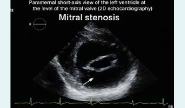
Figure 7: 2D echocardiography showing mitral stenosis and reduced valve area.
After painting and draping, the surgery was commenced under GA. A bolus of 5 mL of 2% lignocaine was administered prior to incision. Intraoperatively, BP remained stable around 110/70 mmHg with a HR of 75–80 bpm. At 30 minutes, a bolus of 5 mL of 0.5% bupivacaine was given to maintain the block. The surgery lasted 1.5 hrs and was completed uneventfully. Only 200 mL of IV Kabilyte was required intraoperatively. The postoperative period was uneventful.
Conclusion
Administration of GA in patients with Lutembacher syndrome can be unpredictable due to complex intracardiac shunting and compromised ventricular function.2,3 Epidural anaesthesia offers several advantages:
- Graded sympathetic block, minimising sudden drop in SVR, which can worsen shunting or compromise coronary perfusion
- Slow onset permits better BP control compared to spinal or GA induction agents3
- Good analgesia, reducing stress induced tachycardia. It provides continuous pain relief postoperatively, it reduces the catecholamine surges, which increases peripheral vascular resistance (PVR) and HR
- Beneficial for patients with underlying lung conditions, like chronic obstructive pulmonary disease (COPD) and restrictive lung diseases, where GA carries high risk
The successful use of epidural anaesthesia in a patient with Lutembacher syndrome highlights the importance of individualised anaesthetic planning based on the patient’s unique cardiac physiology
Arun Puri, Vikky Jaiswal, Smriti Sahu, Manoj Sinha, Naveen Gupta, Devender Dua, Mallika Kulkarni,
Anurag Chaturvedi, Sony Mathew, Anoop VP, B Kanmani, Deepak, Nishant Sood, Prachi Sharma, Satish
Gajula, Sanjeev Sharma, Pragya Singh. A Series of Challenging and Interesting Cases for Anaesthesia
Management. MMJ. 2025, September. Vol 2 (3)
References
Case 1
- Wilson LD, Detterbeck FC, Yahalom J. Clinical practice. Superior vena cava syndrome with malignant causes. N Engl J Med. 2007;356(18):1862–9.
- Kishi K, Sonomura T, Mitsuzane K, et al. Self-expandable metallic stent therapy for superior vena cava syndrome: clinical observations. Radiology. 1993;189(2):531–5.
- Sheth RA, Niekamp A, Quencer KB, et al. Superior vena cava syndrome—a contemporary review of a historic disease. Cardiovasc Diagn Ther. 2016;6(6):580–8.
- Verso M, Agnelli G. Venous thromboembolism associated with long-term use of central venous catheters in cancer patients. J Clin Oncol. 2003;21(19):3665–75.
- Dabbagh O, Oza A, Prakash S, et al. Cardiac tamponade and superior vena cava syndrome from central venous catheter thrombosis. Am J Med Sci. 2008;335(5):390–4.
- Bagan P, Dujon A, Berna P, et al. Superior vena cava syndrome: modern diagnostic and therapeutic aspects. Ann Thorac Surg. 2002;73(1):328–32.
- Seelig MH, Oldenburg WA, Klingler PJ, et al. Superior vena cava syndrome in patients with benign disease. Ann Thorac Surg. 1997;64(2):559–62.
Case 2
- Krishna R, Goneppanavar U. Anaesthetic management of caesarean section in a patient with double outlet right ventricle. J Obstet Anaesth Crit Care. 2012;2:50–2
- ESC Guidelines on the management of cardiovascular diseases during pregnancy. Eur Heart J. 2011;32:3147–97.
- Cunningham FG, Leveno KJ, Bloom SL. Cardiovascular Disorders. In: Cunningham, F Gary, editor. William Obstetrics. 24th Edition. New York: McGraw-Hill Education/Medical; 2014. p. 973–99.
Case 3
- Gersh BJ, Maron BJ, Bonow RO, et al. 2011 ACCF/AHA guideline for the diagnosis and treatment of hypertrophic cardiomyopathy. J Am Coll Cardiol. 2011;58(25):e212–e260.
- Elliott PM, Anastasakis A, Borger MA, et al. 2014 ESC guidelines on diagnosis and management of hypertrophic cardiomyopathy. Eur Heart J. 2014;35(39):2733–79.
- Maron BJ, Ommen SR, Semsarian C, et al. Hypertrophic cardiomyopathy: present and future, with translation into contemporary cardiovascular medicine. J Am Coll Cardiol. 2014;64(1):83–99.
- Maron BJ, Rowin EJ, Casey SA, et al. Risk stratification and outcome of patients with hypertrophic cardiomyopathy ≥60 years of age. Circulation. 2013;127(5):585–93.
Case 4
- Kako H, Martin DP, Cartabuke R, et al. Perioperative management of a patient with Rett syndrome. Int J Clin Exp Med. 2013;22;6(5):393–403.
- Lee AR, Lee HK., Kim YU, et al. Anesthetic management of a pediatric patient with Rett syndrome – A case report. Anesthesia and Pain Medicine. 2018;13(2):173–5.
- Tofil NM, Buckmaster MA, Winkler MK, et al. Deep sedation with propofol in patients with Rett syndrome. J Child Neurol. 2006;21:857–60.
Case 5
- Bhalotra AR, Balyan R, Manchanda G, et al. Opioid-free anaesthesia in children with severe mandibular hypoplasia and TMJ ankylosis with sleep apnoea for mandibular distraction osteogenesis. Indian J Anaesth. 2019;63(5):412– 14.
- Kaban LB, Perrott DH, Fisher K. A protocol for management of temporomandibular joint ankylosis. J Oral Maxillofac Surg. 1990;48(11):1145–51.
- Jagannathan N, Sohn LE, Ramsey MA, et al. A randomized trial comparing the Ambu® Aura-i™ with the Air-Q® intubating laryngeal airway as conduits for fibreoptic intubation in children. Br J Anaesth. 2012;109(5):699–705.
Case 6
- Lynam J, Anderson B, Thomson P. Total intravenous anaesthesia and facial nerve monitoring in parotid surgery. Anaesthesia. 2008;63(10):1141–2.
- Guntinas-Lichius O, Klussmann JP, Wittekindt C, et al. Parotidectomy for benign parotid disease at a university teaching hospital: outcome of 963 operations. Laryngoscope. 2006;116(4):534–40.
- Albergotti WG, Nguyen SA, Zenk J, et al. Extracapsular dissection for benign parotid tumors: a meta-analysis. Laryngoscope. 2012;122(9):1954–60.
Case 6
- Shivanna S, Tejesh C, Manjunath A, et al. Sciaticofemoral nerve block for varicose vein surgery in the patient with eventration of diaphragm, dextroposition of the heart and bicuspid aortic valve. J Anaesthesiol Clin Pharmacol. 2012;28(2):277–9.
- Belinkoff S, York N. Complications following anesthesia in a case of Lutembacher’s syndrome. Anesth Analg. 1946;25:172–5.
- Gordon C, Collard CD, Pan W. Intraoperative management of pulmonary hypertension and associated right heart failure. Curr Opin Anaesthesiol. 2010;23:49–56.