 WhatsApp Us (24/7)
 +91 926 888 0303 (24/7)


Abstract: Intractable lower back pain is increasingly adding to the morbidity of the general population. Conservative therapy failure and refusal for operative procedures has led to the use of regional pain relief options in the form of caudal epidural blocks. Caudal epidural steroid injections, once popular among anaesthetists for lower abdominal surgeries in paediatric patients, have transitioned from the operating theatres to the outpatient departments (OPD) and become an important part of orthopaedic practice for pain relief. Previously, these procedures were performed blindly, which was accepted by both the medical community and patients, despite a failure rate of 5%-25% and varying degrees of success. However, with the advent of musculoskeletal (MSK) dedicated ultrasound (US) machines, the accuracy and success rates of these procedures have significantly improved primarily because of reduction in complication rates, more accurate needle placement in the sacral hiatus, and consequently, a lower percentage of failed blocks.
Key words: Ultrasonography, Caudal Epidural, Pain Relief, Sacral Hiatus, Needle Placement
Source of support: Waldman SD
Conflict of interest: Nil
Introduction
There are many pathways described for effective pain relief, and we are moving towards more and more sophisticated techniques, ultrasound (US)-guided procedures being one of them.
The term interventional US refers to a wide and heterogeneous range of invasive procedures performed percutaneously using ultrasound guidance.
Unfortunately, blind procedures are still being performed for various reasons. Despite the free availability of a newer modality which is non-radiation based, cost effective and one as easily available as US, it’s a pity that the clinicians should not be adopting it for effective pain management.
This article describes the basic technique of performing a caudal epidural block with tips and tricks for accurate needle placement under US guidance for effective pain relief for patients with intractable low back pain which has not responded to conservative treatment in an OPD setting.
Indications for Caudal Epidural Block
Out -patient procedure: for relief of low back pain (acute, chronic, or following failed back surgeries).
The technique can be used for accurate needle placement for spinal anaesthesia in:
- Skin grafting on the lower limbs
- Procedures involving the anus and rectum
- Orthopaedic surgery on the pelvic girdle
- Hernia repair
- Lower limb surgery
- Obstetric analgesia
- 2nd stage or instrumental deliveries
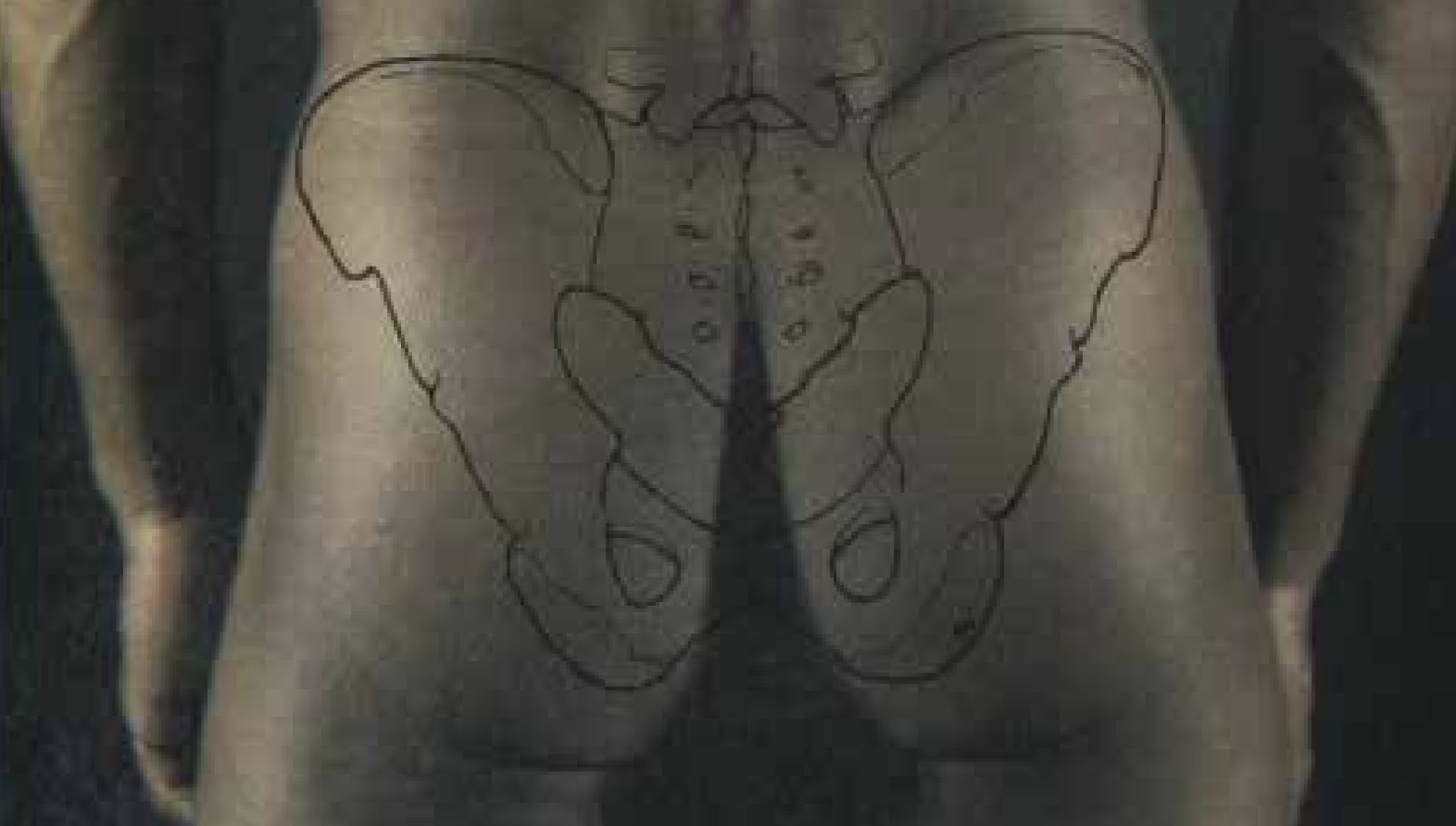
Figure 1: Surface markings of caudal (sacrococcygeal) region (posterior view)
Anatomic Landmarks
The sacrum is an inverted triangular bone composed of five fused sacral vertebrae, with an anterior concave and a posterior convex surface. The sacrum articulates obliquely with the two iliac bones on either side, forming the sacroiliac joints and connects superiorly with the fifth lumbar vertebra and caudally with the coccyx. (Figure 1). The sacral hiatus is formed by the incomplete midline fusion of the posterior elements of the lower portion of the S4 and the entire S5 vertebrae. This inverted U-shaped space is covered posteriorly by the sacrococcygeal ligament, which is also an important soft tissue landmark when performing a caudal epidural block. Penetration of sacrococcygeal ligament provides direct access to the epidural space of the sacral canal.
The vestigial osseous remnants of the inferior articular processes at S4 project downward on each side of the sacral hiatus forming coarse button like projections. These bony projections are called the sacral cornua and represent important osseous landmarks for localizing the sacral hiatus when performing a caudal epidural block in the short axis scanning.
The triangular coccyx is made up of three to five rudimental vertebrae. The tip of the coccyx is / was considered an important clinical landmark when performing a blind caudal epidural block. However, when performing the procedure under US guidance it’s the sacral cornu and sacral hiatus that are the important bony anatomical landmarks.
A continuation of the lumbar spinal canal, the sacral canal continues inferiorly to terminate at the sacral hiatus. The sacral canal contains the inferior termination of the dural sac, which ends between S1 and S3 and should not be punctured whilst doing the procedure. The five sacral nerve roots and the coccygeal nerve all traverse the canal as does the, filum terminale. The anterior and posterior rami of the S1-4 nerve roots exit through the respective sacral foramina and provide sensory and motor innervation to their respective dermatomes and myotomes. The pelvic organs, including the uterus, fallopian tubes, bladder and prostate also receive partial innervation from these nerve roots.
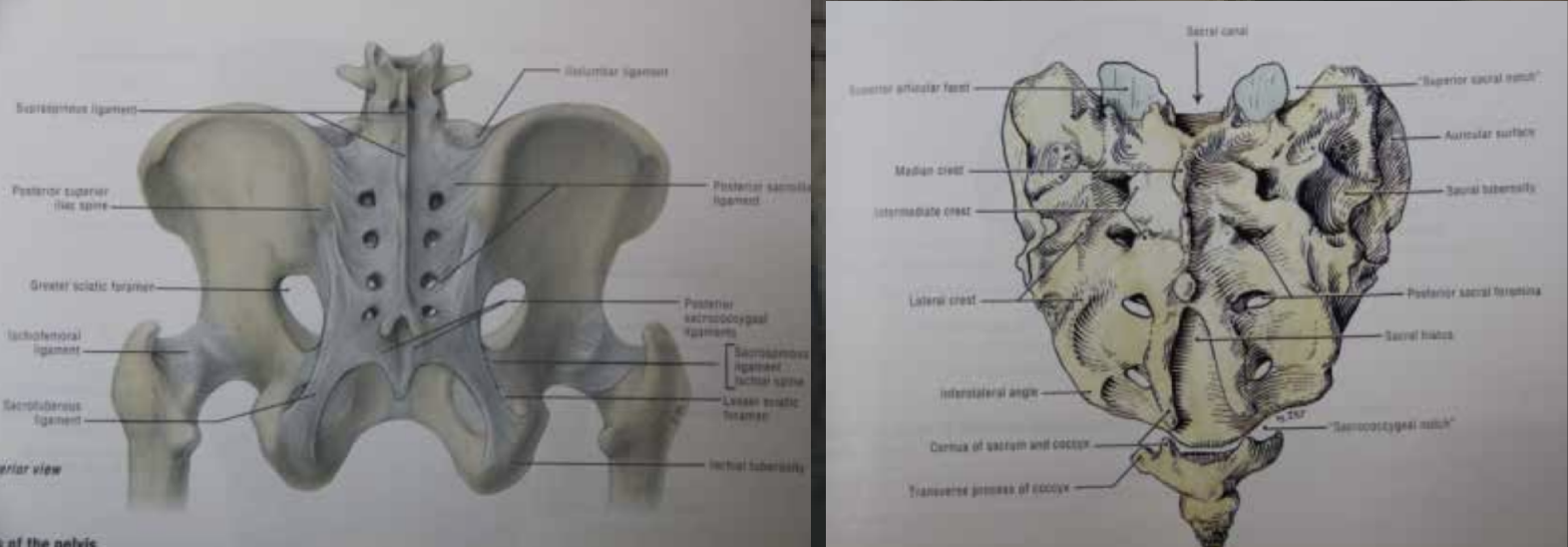
Figure 2: (a) Anatomical bony landmarks of sacrum (b) Detailed bony lands marks of pelvis (posterior view)
Sonoanatomy
Scanning is initiated at the midline with the probe placed in a transverse orientation at the proximal end of the median crest, which is seen as an echogenic bony prominence. As we proceed caudad from this level, two semi-circular convex echogenic shadows are visible on each side of the midline, corresponding anatomically to the sacral cornua. (Figure 2a) A transverse hypoechoic band, 2-3mm in thickness, is noted in between the sacral cornu, known as the sacral hiatus. Both sacral cornua are covered by a hypoechoic band called the sacrococcygeal ligament, and posteriorly, the sacral hiatus is bordered by a dense echogenic line corresponding to the posterior surface of the sacral bone. (Figure 2b)
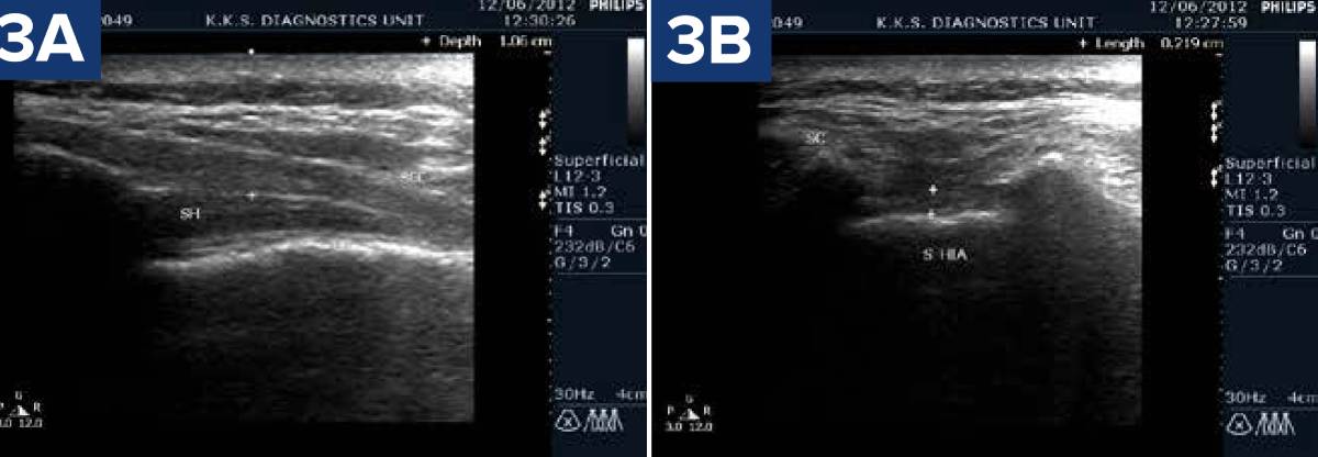
In the long-axis view, with the probe oriented longitudinally, the sacral hiatus appears as a dark, hypoechoic beak-like structure, enclosed between the sacrococcygeal ligament superiorly and the sacral bone inferiorly. (Figure 3 a,b)
Contraindications
NOTE: There are no absolute contraindications except for local skin infection, a history of coagulopathies, and lignocaine allergy.
Coagulation disorders: Bleeding abnormalities are an absolute contraindication to caudal epidural injections. Other contraindications include, a history of haemophilia, idiopathic thrombocytopenic purpura (ITP), tumours, disseminated intravascular coagulation (DIC) from sepsis, or the administration of anticoagulants such as heparin or warfarin.
Infection: Caudal epidural injections should not be performed if there is an active infection at the injection site, either on the skin surface or surrounding region. • Unstable cardio-vascular status: Conditions such as labile blood pressure and/or heart rate are contraindications. • Congenital anatomic anomalies: In cases of congenital anomalies of the spinal cord or vertebral bodies, such as spina bifida, caudal epidural block should not be attempted, as the spinal cord may be tethered within the spinal canal.
Scoliosis: Although scoliosis is not an absolute contraindication to a caudal epidural block, it may make the injection technically more difficult.
Decreased cardiac function: The dose of local anaesthetic must be carefully controlled in patients with decreased cardiac function, as is often the case of patients with muscular dystrophy.
Relative contraindications
Diabetes mellitus, hypertension and glaucoma.
Pearls
- Temporary worsening of local symptoms can occur because of the use of steroids, needle trauma, or microcrystalline synovitis.
- Patients should be counselled about the potential side effects of steroid injections, which may include flushing, patches of de-pigmentation or local fat resorption. The volume of the sacral canal, with all of its contents removed, averages approximately 34 mL in dried bone specimens. It should be emphasized that only a small volume of local anaesthetic should be used in combination with steroids in day-to-day practice. The use of large volumes of local anaesthetic may result in unacceptable anaesthetic-induced side effects, such as incontinence and urinary retention.
- The sacral canal contains the epidural venous plexus, which generally ends at S4 but may extend inferiorly. Most of these vessels are concentrated in the anterior portion of the canal. Both the dural sac and epidural vessels are susceptible to trauma if the needles are advanced cephalad into the sacral canal, particularly if the procedure is performed blindly. The remainder of the sacral canal is filled with fat, which is subject to an age-related increase in volume and density. Some investigators believe this change is responsible for the increased incidence of “spotty” caudal epidural nerve blocks in adults.
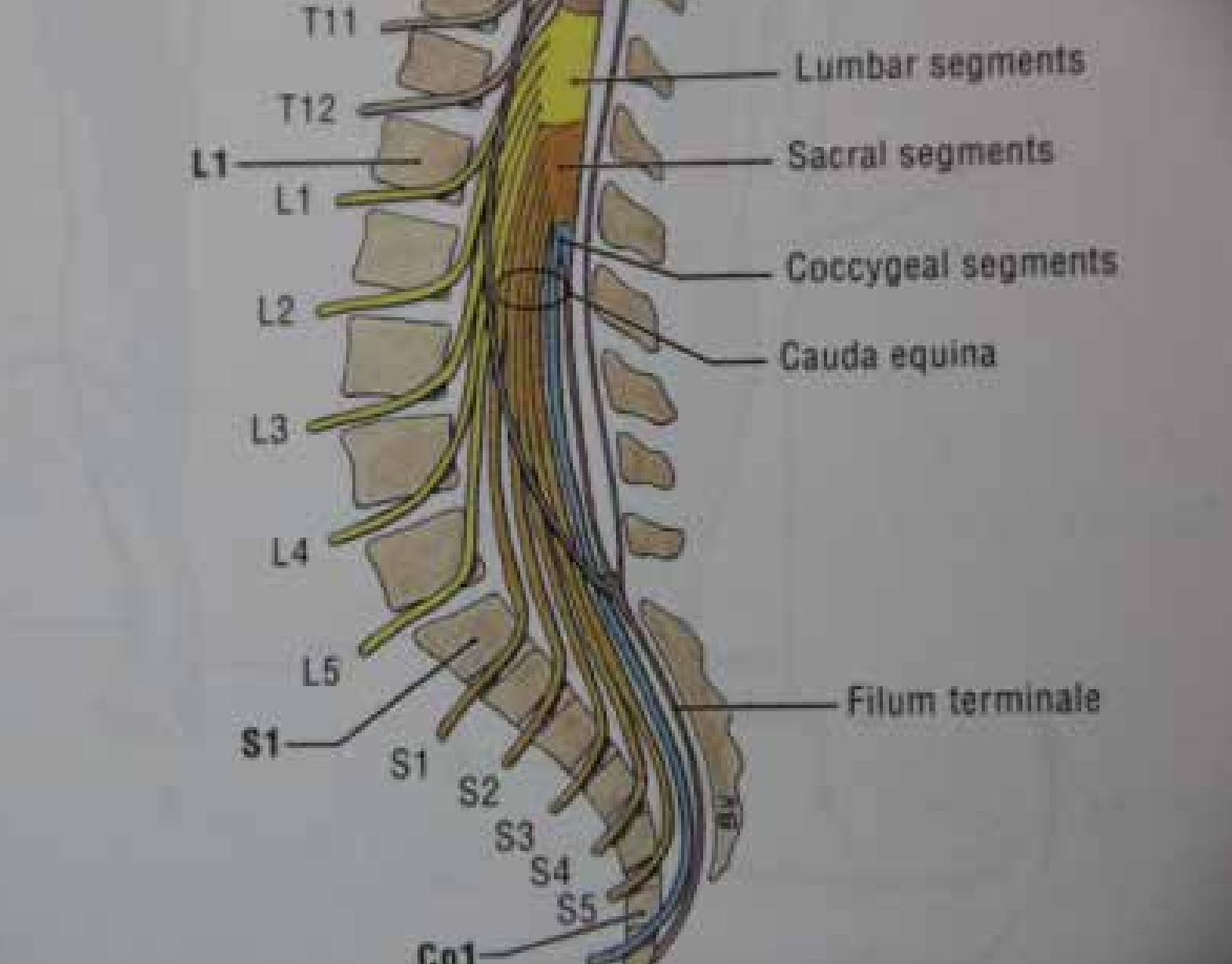
Figure 4: Diagrammatic representation of caudal epidural anatomy (Lateral View): Grant Atlas of Anatomy
Positioning
A caudal epidural block can be performed in the prone or decubitus position in an OPD setting with proper patient consent taken by the consultant performing the procedure. The possibility of complications which under routine practice are negligible, should be discussed gently without alarming the patient.2 (Figure 4). In an US-guided caudal epidural block, the prone position is preferred to optimize comfort for both the patient and the consultant, ensuring better manoeuvrability for probe and needle placement. The patient’s head should be placed on a pillow and turned away from the side of the consultant performing the procedure. The legs and heels should be abducted to prevent tightening of gluteal muscles, which can make identification of the sacral hiatus difficult.
The preparation begins with a wide area of skin being cleansed with antiseptic solution. A fenestrated sterile drape is then placed to an avoid contamination by the operating hand and to create a sterile field.
As with any US-guided interventional procedure, a screening US exam of the affected region should be performed prior to creating a sterile field to identify an accurate path of needle approach. It is essential to find an ergonomically comfortable position for both the patient as well as the ultrasonologist. A rolled-up sheet or pillow is placed under the patient in the prone position to eliminate the lumbar lordotic curve. This helps in optimal probe positioning and also to visualize the bony as well as soft tissue landmarks accurately. In obese and older patients, the prone position should be maintained for the shortest interval of time for the risk of precipitating hypotension or compromising the respiratory system.
Separating the gluteal masses, either with the help of doublesided tape or an assistant increases the accuracy of needle placement particularly in obese patients. The patient should be informed verbally about the procedure, including why and how it is being conducted, before starting.
Procedure
An ultrasound probe with a frequency range between 7.5-12 MHz (linear probe) is placed transversely at the level of the sacral dip in the midline to locate the sacral hiatus. The sacral cornua are important sonographic osseous landmarks. The sacral hiatus appears as a hypoechoic band, a horizontal space of a few millimeters depth between the two cornua. In obese patients, a convex probe (3-5MHz) may be used for better visualization.
At this level, the transducer is then rotated by 90 degrees to bring in a longitudinal orientation. The sacrococcygeal ligament, a relatively echogenic band is visualized superior to the hiatus. A dense echogenic shadow which is the posterior cortical surface of the sacral bone is seen deep to it. Between these two linear shadows, is the beak shaped sacral hiatus in the long-axis orientation. (Figure 5)
After locating the sacral hiatus, the sacrococcygeal ligament is pierced with a 25-gauge, 1 ½- inch long needle through the anesthetized area at a 45-degree angle to the skin surface. Theuse of a longer needle increases the probability of complications, like intravascular injection and inadvertent dural puncture, without adding any advantage to the overall success of this technique.
A “pop” sensation, which indicates the give-way of the sacrococcygeal ligament as it is pierced, is a classical sign relied upon in blindly performed procedures, and remains a useful adjunct when the procedure is being performed under US guidance. As soon as the needle enters the hiatus, the needle shaft within the hiatus cannot be visualized as it is obscured by the overlying bony sacral posterior wall. On short axis of the hiatus, needle can be seen as an echogenic pinhead, on gentle jiggling of the needle. The soft tissue movement under dynamic US guidance can further confirm the accurate needle placement.
n air acceptance test, traditionally used in blind procedures, still holds some value under US guidance, especially for the novices. This can be performed by injecting of 1 mL of air. There should be no bulging or crepitus of the tissues overlying the sacrum. The force required for injection should not exceed that necessary to overcome the resistance of the needle. If initial resistance to injection is encountered, the needle should be rotated 180 degrees, as the needle bevel may be occluded by the internal surface of the sacral canal. Any significant pain or sudden increase in resistance during injection suggests incorrect needle placement, and the consultant should stop injecting immediately to reassess the position of the needle.
When the needle is satisfactorily positioned, a syringe containing 5ml of 1.0% preservative-free lignocaine with 40mg Tricort (Triamcinolone) and 80mg of Depo-Medrol reconstituted to 20mL in normal saline is attached to the needle hub. For patients with post-spinal surgery low back pain, 5ml of Hyalase (Hyaluronidase) is added to the injectate to helps with fibrotic changes that may interfere with the even spread of the injectate.
If the aspiration test is positive for either spinal fluid or blood, the needle is repositioned, and the aspiration test repeated. If the test is negative, injection of 0.5-mL increments of local anaesthetic- steroid is pushed gently with colour Doppler used to look for free flow of injectate in the hiatus. In obese patients, there is a small probability of the needle finding its way in the subcutaneous fat. A colour Doppler application helps to localize the flow of injectate more accurately. A careful observation for signs of local anaesthetic-steroid toxicity or subarachnoid spread of injectate during and after the procedure is advised. The use of smaller volumes of local anaesthetic has markedly decreased the incidence of local anaesthetic related side effects.
Technical Considerations
The most important sign of correct needle placement is the ease with which the injectate flows. If the local anaesthetic-steroid solution can be injected with no resistance, it is mostly likely in the correct space. If there is initial resistance or resistance develops over the course of the injection, the injection should be stopped and the needle location reassessed. There will be some increase in resistance as the potential space of the caudal epidural space expands, but this should be negligible.
A word of caution, preparation of the injectate should be done without making the patient conscious of it.
There are two ways of directing the needle under US guidance:
- Indirect technique – where US is utilized to localize the puncture site and the depth of the target, but not to guide advancement of the needle under real time scanning.
- Real time technique – probe is placed close to the puncture site and the needle is advanced under US guidance. This approach can be lateral or co-axial to the probe. In the lateral approach, we can see the entire length of the needle shaft. The optimal visualization of needle is dependent upon the gauge of the needle and the angle to the skin surface at which the needle is inserted. Thicker the needle and lesser the angle, better is the needle visualization in the soft tissues.
With co-axial approach, only the needle tip is visualised, and confirmation can be achieved by a little lignocaine or air instillation showing tissue dissection or movement.
A biopsy guide attachment can be utilized by those who are not ambidextrous. However, use of such equipment makes the probe manipulation less flexible as compared to free hand technique.
Regardless of the technique used, it is important to closely assess the soft tissues structures along the needle path to avoid incidental damage to the nerves, tendons or vessels.
Anaesthetic agents provide instantaneous pain relief, lasting approximately 24-36 hours. Following this, the steroid’s antiinflammatory action takes over offering prolonged pain relief.
It’s important to note that the accuracy of injection in the caudal epidural space is directly proportional to the efficacy of treatment.
Ultrasound allows precise needle placement within the small space thus reducing injury to the adjacent structures and the side effects related to the extra-hiatal instillation of the drugs.
Although fluoroscopy, computed tomography (CT) or magnetic resonance imaging (MRI) can be used as alternative imaging modalities for accurate needle placement, these are time consuming, cumbersome, and involve radiation exposure or require special loss of resistance needles in order to achieve the same purpose. The cost benefit ratio cannot be over-stated.
Complications
The complications of caudal epidural block can be classified as5:
- Failed or incomplete block. Between 5%-25 % of caudal epidural blocks can be considered "failed or incomplete". There is a considerable variation in the anatomy of the sacral hiatus, which may account for the small percentage of caudal epidural block failures. A sound knowledge of anatomic landmarks with variations and sono-anatomy is mandatory for the success of accurate needle placement
- Unilateral block: is less common than with lumbar epidural blocks because the sacral/ caudal epidural space is larger and requires more volume to fill. Patchy or one-sided blocks are rare with caudal epidural blocks but can result from too rapid an injection. Injection of drugs should be done slowly over 2 minutes after test dose.
- Local anaesthetic toxicity: intravascular injections can occur as the extra-dural veins have no valves, allowing the injectate to enter the circulation, potentially producing convulsions even at doses lower than the recommended maximum safe doses. The consultant should remain vigilant for signs of intravascular injection throughout the procedure.
- Dural puncture (intra-thecal injection). The dural sac can extend up to the level of the third or fourth sacral vertebrae, which may lead to an intrathecal injection. This can result in a 'total spinal block', characterized by sudden apnoea, unconsciousness, and dilated pupils
- Intra-osseous injection. An intra-osseous injection is equivalent to an intravenous injection and can be fatal. A very strong resistance to the injection should raise a redflag for a potential intraosseous injection. In the elderly with osteopenia, the resistance may not be as strong as expected.
- Bleeding and infection: While haematoma and abscess formation are very uncommon after a caudal epidural block, patients must be counselled of the probability
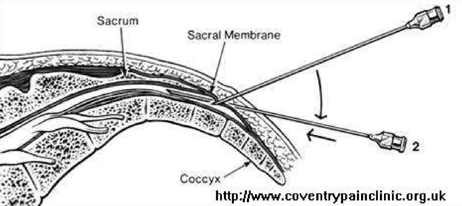
Figure 5: Bony anatomical landmarks (sagittal view) with needle trajectory for caudal epidural injection with patient in supine position.
Although uncommon, infections are an ever-present possibility, especially in the immune-compromised or cancer patient. Early detection of infection is crucial to avoid potentially lifethreatening sequelae.
Few tips to reduce complications:
- Performing needle placement by direct technique under real time US guidance with the use of colour Doppler for tracking the needle tip and injectate path.
- Careful observation of the patient during and after the procedure is mandatory.
- Use of smaller volumes of local anaesthetic with steroid.
- Use of shorter, smaller-gauge needles
Why an Ultrasound Guidance
There is a high probability of inaccurate needle placement when performing caudal epidural block blindly.
- The needle may be placed outside the sacral canal, resulting in an injection into the subcutaneous tissues, especially in an obese patient. The subcutaneous fat is very low in this region; however, needle placement is difficult due to large gluteal masses. An increased resistance to injection accompanied by pain is noted. Similar findings are noted when the needle goes a little deeper but stays within the sacrococcygeal ligament.
- The needle tip may be placed into the periosteum of the sacral wall. A mal-positioned needle at this site causes considerable pain on injection, a very high resistance, and an inability to inject more than a few millilitres of the drug.
- Possible needle mal-positioning can occur if the needle tip is forced into the marrow cavity of the sacral vertebra, resulting in very high blood levels of local anaesthetic. This needle mal-position is detected by the initial ease in injecting a few millilitres of local anaesthetic, followed by a rapid increase in resistance to injection as the noncompliant bony cavity fills with the local anaesthetic.
- Last but not the least, the most serious needle mal-position is when the needle is inserted through the sacrum or lateral to the coccyx into the pelvic cavity and beyond. This can result in the needle entering either the rectum or cervix anteriorly, resulting in contamination of the needle. The repositioning of the contaminated needle into the sacral canal carries a great danger of serious infection.
References
- Alder RS, Sofka CM. Percutaneous ultrasound guided injections in musculoskeletal system. Ultrasound Quarterly. 2003;19(1):3-12
- O’Dwyer HM, Lyon SM, Fotheringham T, et al. Informed consent for interventional radiology procedures: a survey detailing current Eurpoean practice Cardiovascular and Interventional Radiology. 2003;26(5):428-433.
- Saker MB, Kane RA, MatalonTA. Factors affecting and techniques to improve needle visualization . InSeminars in interventional radiology 1997;14:471-475.
- Sofka CM, Collins AJ, Alder RS. Use of ultrasonographic guidance in interventional musculoskeletal procedures :a review from a single institution Journal of ultrasound in medicine. 2001;20(1):21-6.
- Edler A, Wellis VG. Caudal epidural anesthesia for pediatric patients: a safe, reliable and effective method in developing countries. 2003:15-19.
- Vadodaria B, Conn D. Caudal epidural anaesthesia. Update in Anaesthesia. 1998;8:14-7.
- Waldman SD: Caudal epidural nerve block In: Atlas of interventional pain Management, ed. 2 .Philadelphia, Saunders, 2004.