 WhatsApp Us (24/7)
 +91 926 888 0303 (24/7)


Abstract: Sarcomatoid carcinoma or spindle cell carcinoma is an uncommon and aggressive form of breast cancer. It is characterized by an atypical combination of epithelial and mesenchymal components exhibiting sarcomatoid features. However, due to its rarity, there is limited documentation. Herein, we present the case of a 52-year-old lady who had previously undergone simple mastectomy with axillary nodal clearance for spindle cell neoplasm, which had recurred within a month after primary surgery. The patient subsequently underwent a left-sided wide excision of the recurrent lesion followed by a parascapular flap reconstruction. Adjuvant chemotherapy consisting of ifosfamide and adriamycin was administered, followed by locoregional radiation therapy. This case report is valuable in providing insights into the management of such uncommon and challenging cases, given the high frequency of recurrence and the difficulties associated with understanding the clinical and morphological features of this disease.
Key words: Breast, Mass, Spindle Cell Neoplasm, Phyllodes, Malignant, Sarcomatoid, Recurrent Breast Lump
Introduction
Metaplastic carcinoma (MC) is a rare breast cancer subtype, comprising approximately 1% of breast neoplasms.1 It includes sarcomatoid carcinoma, characterized by a spindle cell component and weak keratin positivity and variable association with ductal carcinoma, impacting metastatic potential.2,3
Case report
A 52-year-old perimenopausal woman presented with a recurring lump on her mastectomy scar. She was previously diagnosed at another facility with a 12×12cm malignant spindle cell neoplasm in the upper quadrant of her breast, that tested positive for cytokeratin (CK) and tumour protein 63 (p63). A simple mastectomy with axillary node clearance was subsequently performed, revealing 9×7.5×7cm malignant phyllodes with a 1cm close posterior margin. The tumour exhibited mitotic activity of 10-12 per 10 high-power fields (hpf) and moderate to marked nuclear pleomorphism. All 28 dissected lymph nodes tested negative for metastasis. Following this, margin revision surgery, including pectoral muscle excision and lateral dog ear correction was performed. One month later, a lump of 7x7 cm was detected on the lateral aspect of the scar. She visited our centre for evaluation. Positron emission tomography (PET) scan showed no metastasis. Biopsy revealed malignant spindle cell tumour with a mitosis of 10- 14/10 hpf. The patient underwent a left-sided wide excision of the tumour with a parascapular flap reconstruction following a multidisciplinary team consensus.
Histopathology confirmed high-grade malignant spindle cell tumour that had an immune profile of a sarcomatoid carcinoma. Grossly, a pearly white tumour measuring 6x5.5x5 cm with clear margins of more than 2cm was noted. Microscopy showed high-grade pleomorphic spindled and epithelioid cells with myxoid stroma and areas of necrosis with abundant bizarre and tumour giant cells. A mitotic count of 28-30/10 hpf with atypical mitosis and no glandular or squamous component was noted. Immunohistochemistry (IHC) revealed positivity for vimentin, cluster of differentiation (CD)10, CK and focally for p63 (Figure 1) and were negative for oestrogen receptor (ER), progesterone receptor (PR), Human Epidermal Growth Factor Receptor 2 (Her2neu), smooth muscle actin (SMA), desmin, S100 and CD34. Kiel 67 (Ki67) labelling index indicated a proliferation rate of 60%-70%.
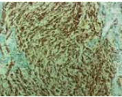
A: CD10
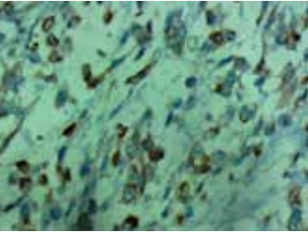
B: Cytokeratin
Figure 1: Histopathological analysis images. A: CD10; B: Cytokeratin
The patient received adjuvant chemotherapy (ifosfamide and adriamycin), followed by locoregional radiotherapy. However, a month later, she developed breathlessness and imaging revealed right nodular lung lesions that were biopsy-proven to be metastasis. Palliative chemotherapy was initiated, but her condition worsened due to the underlying heart failure. Despite intensive care, she ultimately succumbed within one week.
Discussion
Metaplastic carcinomas are rare, characterized by the presence of various components, including squamous, spindle, or mesenchymal components in addition to adenocarcinoma and they range from low to high grade depending on cellular morphology.4 Although triple negative (TNBC), MC with a dominant spindle cell component have a worse prognosis than typical TNBC.5
Sarcomatoid carcinoma is a rare variant of MC, clinically described as firm, nodular and well-circumscribed, accounting for less than 0.5% of all breast cancers (World Health Organization Histological Classification 8032/3).6–9 A study involving 29 sarcomatoid cases reported tumours ranging from 1.5 to 15 cm, with a median size of 4 cm. Axillary nodal metastasis occurred in 1 in 20 cases, with lung and bones being common sites of distant metastasis.3 Our patient presented with a 12×12cm mass clinically, with a pathological tumour size measuring 9×7.5×7 cm showing a significant spindle cell component, indicating sarcomatoid (spindle cell) neoplasm.
Due to the rarity and heterogeneity of MC, there are no established guidelines for their management.10,11 However, the current recommendation remains wide surgical excision with clear margins, followed by radiation with or without chemotherapy.3 The absence of hormone receptors and Her2/ Neu limits the efficacy of targeted as well as conventional chemotherapy.12 In contrast, Leibl and Moinfar et al, revealed in their audit on MC that most express Her1 but not Her2 in contrast to the typical breast carcinoma and suggested that protein kinase inhibitors (such as gefitinib) might be beneficial.13 A recent study on the management of MC revealed that postlumpectomy radiotherapy was associated with improved overall survival rates in all stages, whereas survival benefit with adjuvant radiotherapy was only seen in the advanced stage.14 The rapidity of recurrences in our patient exemplifies the aggressive nature of sarcomatoid carcinoma and the management challenges.
Metaplastic carcinoma, including sarcomatoid carcinoma, have lower disease-free and overall survival rates compared to adenocarcinoma,15,16 with a reported 5-year survival rate of 64%.7 Prognostic factors influencing survival outcomes in MC include symptom duration, tumour size and axillary nodal involvement.17
CONCLUSION:
Sarcomatoid carcinomas have a poor prognosis and there is no standardized management approach due to limited understanding. However, surgical-wide excision with clear margins followed by adjuvant therapy is recommended based on existing literature.
References
- Smitt MC. Metaplastic Breast Cancer. Clin Breast Cancer. 2003;4(3):210-11.
- Kumar M, Nigam JS, Khanna P, et al. Metaplastic carcinoma of the breast – Case report and review of literature. Int J Surg Open. 2020;24:43-46.
- Carter MR, Hornick JL, Lester S, et al. Spindle Cell (Sarcomatoid) Carcinoma of the Breast: A Clinicopathologic and Immunohistochemical Analysis of 29 Cases. Am J Surg Pathol. 2006;30(3):300-309.
- Sinn HP, Kreipe H. A Brief Overview of the WHO Classification of Breast Tumors, 4th Edition, Focusing on Issues and Updates from the 3rd Edition. Breast Care. 2013;8(2):149-54.
- Lee H, Jung SY, Ro JY, et al. Metaplastic breast cancer: clinicopathological features and its prognosis. J Clin Pathol. 2012;65(5):441-46.
- Lakhani S, Ellis I, Schnitt S, et al. World Health Organization Classification of Tumours of the Breast. 4th ed. IARC Press; 2012.
- Wargotz ES, Does PH, Norris HJ. Metaplastic carcinomas of the breast. II. Spindle cell carcinoma. Hum Pathol. 1989;20(8):732- 740.
- Ellis IO, Bell J, Ronan JE, et al. Immunocytochemical investigation of intermediate filament proteins and epithelial membrane antigen in spindle cell tumours of the breast. J Pathol. 1988;154(2):157-65.
- Khan HN, Wyld L, Dunne B, et al. Spindle cell carcinoma of the breast: a case series of a rare histological subtype. Eur J Surg Oncol EJSO. 2003;29(7):600-03.
- Maemura M, Iino Y, Oyama T, et al. Spindle cell carcinoma of the breast. Jpn J Clin Oncol. 1997;27(1):46-50.
- Tzanninis IG, Kotteas EA, Ntanasis-Stathopoulos I, et al. Management and Outcomes in Metaplastic Breast Cancer. Clin Breast Cancer. 2016;16(6):437-43.
- Lee AHS. Recent developments in the histological diagnosis of spindle cell carcinoma, fibromatosis and phyllodes tumour of the breast: Spindle cell and fibroepithelial lesions. Histopathology. 2007;52(1):45-57.
- Leibl S, Moinfar F. Metaplastic breast carcinomas are negative for Her-2 but frequently express EGFR (Her-1): potential relevance to adjuvant treatment with EGFR tyrosine kinase inhibitors? J Clin Pathol. 2005;58(7):700-04.
- Haque W, Verma V, Naik N, et al. Metaplastic Breast Cancer: Practice Patterns, Outcomes, and the Role of Radiotherapy. Ann Surg Oncol. 2018;25(4):928-36.
- Drekolias D, Mamounas EP. Metaplastic breast carcinoma: Current therapeutic approaches and novel targeted therapies. Breast J. 2019;25(6):1192-97.
- Gortman A, Aherne NJ, Westhuyzen J, et al. Metaplastic carcinoma of the breast: Clinicopathological features and treatment outcomes with long-term follow up. Mol Clin Oncol. 2021;15(3):178.
- Chao TC, Wang CS, Chen SC, et al. Metaplastic carcinomas of the breast. J Surg Oncol. 1999;71(4):220-25.