 WhatsApp Us (24/7)
 +91 926 888 0303 (24/7)


Abstract:
Cytotoxic lesions of the corpus callosum (CLOCCs) represent a distinct radiological entity that can mimic ischaemic stroke on neuroimaging. We present a case of a 36-year-old male with persistent headaches and altered sensorium whose magnetic resonance imaging (MRI) revealed hyperintense lesions on diffusion-weighted imaging with corresponding hypointensity on apparent diffusion coefficient maps in the splenium of the corpus callosum. The patient had no identifiable vascular risk factors, suggesting an alternative aetiology to stroke. CLOCCs can be associated with various conditions including infections, metabolic disorders, and drug toxicity. Recognition of this entity can prevent misdiagnosis as ischaemic stroke and guide appropriate management. Follow-up imaging confirmed the typically transient nature of these lesions, with complete resolution observed at four weeks
Key words: Cytotoxic Lesions, Corpus Callosum, MRI, Headaches, Altered Sensorium, Stroke Mimic.
Introduction
The corpus callosum, the largest white matter tract in the brain connecting the cerebral hemispheres, can be affected by various pathological processes. Cytotoxic lesions of the corpus callosum (CLOCCs) represent a distinct radiological entity characterised by transient lesions with restricted diffusion on magnetic resonance imaging (MRI).1 These lesions predominantly affect the splenium of the corpus callosum and have been associated with numerous aetiologies, including infectious diseases, metabolic disturbances, drug toxicity, and other systemic conditions.4
CLOCCs are often misdiagnosed as acute ischaemic stroke due to their appearance on diffusion-weighted imaging (DWI), potentially leading to inappropriate management strategies. Despite their alarming radiological appearance, these lesions typically resolve completely within weeks to months without specific treatment.2 Understanding the characteristic imaging features and clinical contexts in which CLOCCs occur is crucial for accurate diagnosis and management.
This case report describes a patient presenting with persistent headaches and altered sensorium who was found to have CLOCCs on neuroimaging. We discuss the clinical presentation, diagnostic approach, pathophysiological mechanisms, and management considerations for this radiological entity.
Case Report
A 36-year-old male presented to our emergency department with complaints of persistent, moderate to severe headache for three days and progressive altered sensorium over the preceding 24 hours. The headache was described as diffuse, non-pulsatile, and unresponsive to over-the-counter analgesics. There was no history of fever, recent infections, trauma, or substance abuse. The patient had no known comorbidities and was not on any regular medications.
On examination, the patient was afebrile with stable vital signs. Neurological examination revealed a Glasgow Coma Scale score of 13/15 (E3V4M6), with the patient being disoriented to time and place. There were no focal neurological deficits, meningeal signs, or papilloedema. Routine laboratory investigations, including complete blood count, renal function tests, liver function tests, and serum electrolytes, were within normal limits. Cerebrospinal fluid analysis was unremarkable.
Brain MRI was performed, which revealed oval-shaped lesions in the splenium of the corpus callosum characterised by hyperintensity on DWI and hypointensity on T1-weighted images. The lesions showed restricted diffusion with corresponding hypointensity on apparent diffusion coefficient (ADC) maps and hyperintensity on T2- FLAIR sequences. There was no gadolinium enhancement or mass effect (Figure 1). Magnetic resonance angiography was normal with no evidence of vascular occlusion.
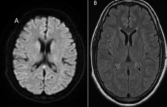
Figure 1: Magnetic resonance imaging (MRI) findings showing cytotoxic lesions in the splenium of the corpus callosum, characterised by restricted diffusion and hyperintensity on T2-FLAIR images.
Based on the clinical presentation and characteristic MRI findings, a diagnosis of CLOCCS was made. The patient was managed conservatively with intravenous fluids and symptomatic treatment for headache. His sensorium gradually improved over the next 48 hours, and he was discharged after five days of hospitalisation. Follow-up MRI performed four weeks later showed complete resolution of the lesions, confirming the transient nature of CLOCCs.
Discussion
CLOCCs represent a distinct radiological entity characterised by transient lesions with restricted diffusion on MRI. These lesions predominantly affect the splenium of the corpus callosum, as observed in our patient. The term "cytotoxic" refers to the pathophysiological mechanism involving intracellular oedema due to dysfunction of cellular fluid homeostasis.5
Pathophysiology
The pathophysiology of CLOCCs is not fully understood but is thought to involve excitotoxicity due to elevated glutamate levels, leading to cytotoxic oedema. This process results in water influx into neurons and glial cells, causing cell swelling and restricted diffusion seen on MRI1 . The splenium of the corpus callosum may be particularly vulnerable due to its high density of glutamate receptors and specific cellular architecture that makes it susceptible to excitotoxic injury.6
Alternative mechanisms proposed include intramyelinic oedema due to separation of myelin layers, inflammatory infiltrates, and oxidative stress. The transient nature of these lesions suggests that the underlying pathology is reversible, unlike the permanent damage typically seen in ischaemic stroke.3
Aetiology
CLOCCs have been associated with numerous conditions, broadly categorised as:
-
Infections: Viral encephalitis (particularly influenza, rotavirus, adenovirus), coronavirus disease 2019 (COVID-19), Epstein-Barr virus, and other infectious processes.
-
Metabolic disorders: Hypoglycaemia, hypernatraemia, hyponatraemia, and hepatic encephalopathy.
-
Drug-related: Antiepileptic medications (particularly carbamazepine, oxcarbazepine), metronidazole, 5-fluorouracil, and immunosuppressants.
-
Others: High-altitude cerebral oedema, malignancy, eclampsia, and systemic inflammatory conditions.4
In our patient, the absence of identifiable risk factors suggests either an undiagnosed viral infection or an idiopathic aetiology, which has been reported in approximately 15%-20% of cases.2
Clinical Presentation
The clinical presentation of patients with CLOCCs varies widely, ranging from asymptomatic cases discovered incidentally to severe encephalopathy. Common symptoms include altered mental status, seizures, headache, and ataxia. The severity of symptoms does not necessarily correlate with the extent of lesions on imaging.1
Our patient presented with persistent headache and altered sensorium, which are consistent with previous reports. The absence of focal neurological deficits, despite the presence of restricted diffusion on MRI, is a key clinical feature that helps differentiate CLOCCs from acute ischaemic stroke.
Imaging Characteristics
The radiological hallmark of CLOCCs is the presence of oval or round lesions in the splenium of the corpus callosum with restricted diffusion on MRI. These lesions are hyperintense on DWI and hypointense on ADC maps, similar to acute ischaemic stroke. However, unlike stroke, CLOCCs typically do not conform to a vascular territory and often resolve completely on follow-up imaging.5
Based on morphology, CLOCCs can be classified into two patterns:
-
Type 1: Small, oval lesion in the centre of the splenium
-
Type 2: Larger, more extensive lesion involving most of the corpus callosum
Our patient exhibited the more common Type 1 pattern. The absence of gadolinium enhancement and mass effect further supports the diagnosis of CLOCCs rather than a neoplastic or inflammatory process.
Management and Prognosis
Management of CLOCCs is primarily supportive and directed at the underlying aetiology when identified. In most cases, the lesions resolve spontaneously within weeks to months without specific treatment. Follow-up imaging is recommended to confirm resolution and exclude alternative diagnoses if symptoms persist.3
The prognosis for patients with CLOCCs is generally favourable, with complete clinical and radiological recovery in most cases. However, the underlying aetiology may influence the outcome, with poorer outcomes reported in cases associated with severe metabolic derangements or encephalitis.2
Conclusion
CLOCCs represent an important differential diagnosis for patients presenting with neurological symptoms and restricted diffusion on MRI. Recognition of this entity can prevent misdiagnosis as ischaemic stroke and avoid unnecessary interventions such as thrombolysis. The case presented highlights the characteristic imaging features and clinical course of CLOCCs, emphasising their typically benign and reversible nature.
Clinicians should maintain a high index of suspicion for CLOCCs when encountering lesions with restricted diffusion in the corpus callosum, particularly in patients with a clinical presentation inconsistent with ischaemic stroke. A thorough evaluation for potential underlying aetiologies and follow-up imaging are warranted to confirm resolution of the lesions.
Further research is needed to elucidate the exact pathophysiological mechanisms underlying CLOCCs and to develop targeted therapeutic strategies for cases with persistent symptoms or incomplete resolution.
Abhishek Dixit, Man Mohan Mehndiratta, Tanzeel Wani, Deepinder Kaur Maini. Cytotoxic Lesions of the
Corpus Callosum: A Neuroradiological Stroke Mimic. MMJ. 2025, June. Vol 2 (2).
References
- Starkey J, Kobayashi N, Numaguchi Y, et al. Cytotoxic lesions of the corpus callosum that show restricted diffusion: mechanisms, causes, and manifestations. Radiographics. 2017;37(2):562-576.
- Moors S, Nakhostin D, Ilchenko D, et al. Cytotoxic lesions of the corpus callosum: a systematic review. Eur Radiol. 2024;34(8):4628-4637.
- Kontzialis M, Soares BP, Huisman T. Lesions in the splenium of the corpus callosum on MRI in children: a review. J Neuroimaging. 2017;27(6):549-561.
- Garcia-Monco JC, Martínez A, Brochado AP, et al. Isolated and reversible lesions of the corpus callosum: a distinct entity. J Neuroimaging 2011;21(3):259-266.
- Takanashi J. Two newly proposed infectious encephalitis/ encephalopathy syndromes. Brain Dev. 2009;31(7):521-528.
- Doherty MJ, Jayadev S, Watson NF, et al. Clinical implications of splenium magnetic resonance imaging signal changes. Arch Neurol. 2005;62(3):433-437.