 WhatsApp Us (24/7)
 +91 926 888 0303 (24/7)


Abstract:
A 27-year-old male patient, a case of Peutz-Jeghers syndrome (PJS) with hyperpigmented lesions and the presence of intestinal polyps, was diagnosed on colonoscopy. The patient had a history of intussusception, which was operated on in the past. He presented with severe iron deficiency anaemia and breathlessness at rest. Despite correction of haemoglobin, the breathlessness persisted, and he subsequently developed left-sided effusion, which was diagnosed as tubercular effusion. As pulmonary tuberculosis is common in India, any patient who develops a unilateral pleural effusion should be suspected of having tuberculosis and managed accordingly.
Key words: Intestinal Polyposis, Pleural Effusion, Colonoscopy, USG-guided Pleural Tapping, Anti-tuberculosis Regimen.
Introduction
Peutz-Jeghers syndrome (PJS) is an inherited condition with an autosomal dominant pattern of inheritance.1 Mutations in the STK11 gene, located on chromosome 19p13, are among the germline variants found in patients with PJS.2 About 17% -50% of cases are solitary and have no associated family history.3
The syndrome is characteristically marked by flat, hyperpigmented, cutaneous lesions which are usually present on the eyelids, sclera, periorbital regions, lower lip, perioral regions and in the buccal mucosa. These skin lesions are mostly benign and do not pose a risk for malignant transformation. Multiple small and large polyps are found, which are hyperplastic, hamartomatous in nature. These polyps are located within the mucosal epithelium and consist of dendritic cells and contain smooth muscle fibre bundles from the muscularis mucosa— known as Peutz-Jeghers polyps.4
These polyps are most commonly found in the mucosa of the stomach, small intestine (particularly the jejunum), colon and rectum. Other common areas are the nasal passages, urinary tract and lungs.3 After recent research, it has been recognised that the hamartomas may possess malignant potential, and in some of them, adenomatous changes and foci of adenocarcinoma may be seen.4 The first presentation of
polyps occurs around the age of 11-13 years. During the first three decades, features of obstruction, intussusception, anaemia, and bleeding per rectum are commonly seen.5 Almost half of the patients with PJS suffer from small bowel intussusception throughout their lives.6 In the younger age group in patients without a family history, PJS is often diagnosed when they suffer from intestinal obstruction and need emergency care.7
Case Report
A 27-year-old male patient presented with complaints of low-grade fever, cough, breathlessness at rest, malaise, anorexia, and swelling of the limbs. On examination, he had a pulse rate of 124 beats per minute and a blood pressure of 90/60 mmHg. His temperature was 98°F, respiratory rate 28 /min and an oxygen saturation of 98% on room air
He had multiple hyperpigmented patches over the sclera and in the buccal mucosa (Figure 1), Grade II clubbing (Figure 2) pedal oedema, and scrotal swelling were noted (Figure 3). Overall, he appeared cachectic, with muscle wasting and pallor. On respiratory system examination, air entry was absent on the left side of the chest, and the trachea was deviated to the right. Vocal resonance was diminished on the left side, with no added sounds. Other systemic examinations were found to be normal. He had a history of intussusception, for which he had undergone surgery in the past.
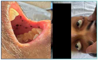
Figure 1 Hyperpigmented macules over buccal mucosa and sclera.
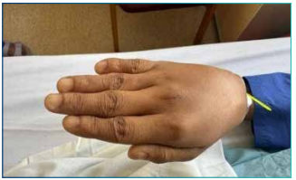
Figure 2 Digital clubbing.
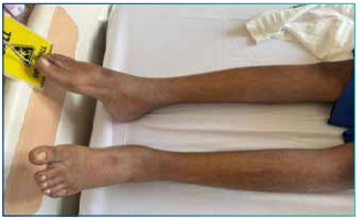
Figure 3: Bilateral pedal pitting oedema. Laboratory investigations revealed the following (Table 1):
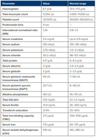
Table 1: Laboratory analysis of the patient.
Urine microscopy was normal, while stool occult blood tested positive. Chest X-ray showed left-sided pleural effusion (Figure 5). Lung ultrasonography (USG) confirmed a massive left-sided pleural effusion. Diagnostic and therapeutic pleural tapping was performed under ultrasound guidance. Pleural fluid analysis revealed—hazy appearance with clot formation. The fluid protein was 1.79 g/dL, and glucose was <0.24 g/dL. The total nucleated cell count was 6476 cells/μL, 68% polymorphs, 32% lymphocytes. The adenosine deaminase (ADA) level was 70.4 U/L (Normal: < 40U/L). GeneXpert testing showed the presence of Mycobacterium tuberculosis. Lactate dehydrogenase (LDH) in the pleural fluid was 2610 U/L. Malignant cells were absent. Culture showed the presence of Escherichia coli, which was pan-sensitive.
An ultrasound of the abdomen was performed, revealing multiple jejunal polyps, following which a colonoscopy was ordered. On colonoscopy, numerous medium and large-sized polyps were observed (Figure 5), which were surgically managed with resection of affected areas.
Figure 4: Left-sided pleural effusion.
Figure 5: Colonoscopy suggestive of multiple intestinal polyps.
The patient was administered two units of packed red blood cells, an albumin injection infusion for 3 days and high-protein diet was advised, after which his oedema started reducing gradually. He was given an anti-tuberculosis regimen with antibiotics, and other supportive management was provided. Gradually, his symptoms started improving—absence of pedal oedema, absence of breathlessness, increased appetite, and absence of fever. His vital parameters and laboratory values came back to normal, and he was then discharged after receiving adequate treatment.
Discussion
As per the World Health Organisation (WHO), PJS is diagnosed if any of the two following features are present:4
- Two or more histologically confirmed PJS-type hamartomatous polyps
- Family history of diagnosed PJS or similar clinical features
- Presence of hyperpigmented mucosal lentigos.
In this case, we see that two criteria are fulfilled. The patient had a positive history of multiple hamartomatous polyps with mucocutaneous pigmentation on the buccal mucosa and sclera.
Most frequently observed complications of PJS are obstruction or intussusception of the intestinal lumen due to polyps. These polyps can lead to chronic blood loss, leading to anaemia.8 Up to 69% of patients with PJS experience intussusception. The risk of intussusception reaches 50% by age 20 in these patients.9
Our patient was breathless despite correcting haemoglobin by blood transfusion. Breathlessness was because of left-sided pleural effusion, which developed during admission. Bilateral pleural effusion would have been transudative. Mycobacterium tuberculosis is a well-known major public health issue and a leading cause of death globally.10
Patients with PJS are more prone to developing other systemic illnesses and should be promptly investigated in such cases so that the appropriate treatment may be started.
The deranged iron profile was suggestive of iron deficiency anaemia. The patient was then immediately started on treatment with the anti-tuberculosis regimen, which was not multidrugresistant. The patient started showing signs of improvement and was then discharged.
Conclusion
As patients of PJS are susceptible to different malignancies of the gastrointestinal tract, oesophagus, uterus, ovaries, breast, pancreas, lung, testis, surveillance should be done at regular intervals so that early diagnosis can be achieved.4 Nowadays, surveillance of the large bowel by colonoscopy or flexible sigmoidoscopy with the use of barium enema is usually recommended every 2-3 years starting from adulthood. Upper gastrointestinal endoscopy should be done every 2-3 years.3
Patients and relatives should be informed about the possible complications and risks of developing other systemic comorbid illnesses like tuberculosis, especially in areas which have high prevalence. Early surveillance would help in the prompt diagnosis and treatment of the patient.
Nitin Rathod, Andrea Janice Fonseca, Mohammed Amin Hamidani, Snehal Sadanandan.
An Unusual Presentation of Pulmonary Tuberculosis and Iron Deficiency Anaemia in a Case of Peutz-Jeghers Syndrome. MMJ. 2025, June. Vol 2 (2).
References
- Beggs AD, Latchford AR, Vasen HF, et al. Peutz-Jeghers syndrome: a systematic review and recommendations for management. Gut. 2010;59(7):975–86.
- Klimkowski S, Ibrahim M, Ibarra Rovira JJ, et al. Peutz-Jeghers syndrome and the role of imaging: Pathophysiology, diagnosis, and associated cancers. Cancers (Basel). 2021;13(20):5121.
- Sreemantula HS, Joseph CA, Jamal F, et al. Intussusception caused by Peutz-Jeghers syndrome. Cureus. 2022;14(4).
- Yamamoto H, Sakamoto H, Kumagai H, et al. Clinical guidelines for diagnosis and management of Peutz-Jeghers syndrome in children and adults. Digestion. 2023;104(5):335–47.
- Nasri S, Kellil T, Chaouech MA, et al. Intestinal intussusception in Peutz-Jeghers syndrome: a case report. Annals of Medicine and Surgery. 2020;54:106–8.
- Shukla RM, Tiwari P, Dariya S, et al. Peutz–Jeghers Syndrome: Lessons to be Learned in the Clinical Diagnosis. Journal of Indian Association of Pediatric Surgeons. 2023;28(3):218–22.
- Jiang YL, Zhao ZY, Li BR, et al. Early screening of the small bowel is key to protect Peutz-Jeghers syndrome patients from surgery: a novel mutation c.243delG in STK11 gene. BMC Gastroenterology. 2019;19:1–5.
- Shakil S, Aldaher Z, DiValentin L. Peutz-Jeghers syndrome presenting with anemia: a case report. Cureus. 2022;14(7).
- Shenoy V, Park C, Nguyen A, et al. 2624 A classic case of Peutz-Jeghers syndrome presenting as recurrent intussusception. Official journal of the American College of Gastroenterology | ACG. 2019;114:S1443–4.
- Chauhan A, Parmar M, Dash GC, et al. The prevalence of tuberculosis infection in India: a systematic review and meta-analysis. Indian Journal of Medical Research. 2023;157(2&3):135–51.