 WhatsApp Us (24/7)
 +91 926 888 0303 (24/7)


Abstract:
Neuromyelitis optica (NMO) is a devastating demyelinating syndrome characterised by optic neuritis (ON) and myelitis, occurring either simultaneously or in sequential relapses. Classical symptoms of NMO include ON, myelitis or area postrema syndrome. The key diagnostic test is the NMO antibody (anti aquaporin-4 antibody [AQP4]) with a sensitivity of 70%-80% and specificity close to 100%. There may be overlap syndromes with Sjögren’s syndrome, systemic lupus erythematosus (SLE), and positive vasculitis antibodies. Cerebrospinal fluid (CSF) analysis shows oligoclonal bands in only 10% of patients, with a mild elevation in white blood cell (WBC) count and protein being commonly observed. There is no cure for NMO, but it is treatable. Some patients recover, but many are left with impaired vision and limb function, which can be severe in some cases. A high dose of intravenous (IV) methylprednisolone pulse is given for 5 days, followed by a prednisolone taper. Depending on treatment response, plasmapheresis or IV immunoglobulin (IVIG) may be used for relapses with incomplete recovery after steroids. Azathioprine, mycophenolate mofetil, rituximab, cyclophosphamide, and mitoxantrone have all been used as preventive treatments. Some improvement may be seen within a few weeks, but disability may persist. Unlike multiple sclerosis (MS), NMO rarely progresses to a secondary progressive phase. Approximately 20% of patients with monophasic NMO experience permanent visual loss, and 30% develop permanent paralysis in one or both legs.
Key words: Neuromyelitis Optica (NMO), Demyelinating Syndrome, Autoimmune Disorders.
Introduction
Neuromyelitis optica (NMO) is a devastating demyelinating syndrome characterised by optic neuritis (ON) and myelitis, occurring either simultaneously or in sequential relapses. It was coined by Eugène Devic in the late 1800s. In 2004, NMO was considered a severe variant of multiple sclerosis (MS). However, in 2004, the anti-aquaporin-4 (AQP4) antibody was identified in most patients with the clinical syndrome of NMO with high specificity.1 Thus, it was reclassified as a distinct entity from MS.1
Pathophysiology
NMO antibody (AQP4)
AQP4 is a transmembrane protein that facilitates water transport in the central nervous system (CNS). It is expressed at the astrocytic foot processes at the bloodbrain-barrier (BBB). There is complement activation, leading to a “rim and rosette” pattern of immune complexes in NMO lesions, causing astrocyte death and tissue necrosis in both grey and white matter. There is accumulation of T cells, B cells, and macrophages, similar to MS, but with additional infiltration by neutrophils and eosinophils, which also contribute to tissue damage.2,3
Epidemiology
NMO is a rare condition, with a prevalence of 2-4/100,000 in most populations. The female: male ratio is 9:1. The age of onset is older, with a median age of 40 years. It is more common in non-Caucasian populations, especially those of Hispanic and African descent. The course of NMO is more devastating, with greater disability accumulated through relapses, including total blindness and paralysis, which are uncommon in MS. The majority of NMO patients have no affected relatives, and it is regarded as a non-familial condition.4
Clinical features
Classical symptoms of NMO are ON, myelitis or area postrema syndrome.5
Optic neuritis
Is typically, severe and more often bilateral; recovery may be incomplete, and total blindness can also occur.
Longitudinally extensive transverse myelitis (LETM)
Spinal cord relapses are a hallmark of NMO. LETM is defined as a spinal cord lesion that is contiguous and spans more than 3 vertebral segments. It often extends from the cervicomedullary junction to the cervical and thoracic spinal cord, with motor, sensory, and/or bladder and bowel function involvement.
Area postrema syndrome
Although NMO typically spares the brain, involvement is seen in 10% of patients. Brainstem symptoms include intractable nausea, vomiting, hiccups (area postrema syndrome), double vision, dysphagia and respiratory compromise. Cerebral symptoms include motor or sensory deficits and encephalopathy.
Diagnosis
The key diagnostic test is the NMO antibody (AQP4) with a sensitivity 70%-80% and specificity close to 100%. There may be overlap syndromes with Sjögren’s syndrome and systemic lupus erythematosus (SLE), with vasculitis antibodies being positive. Cerebrospinal fluid (CSF) analysis shows oligoclonal bands in only 10% patients, with a mild elevation in white blood cell (WBC) count and protein commonly observed.
Wingerchuk diagnostic criteria (2015)
A diagnosis of NMO can be made in a patient who is AQP4-IgG positive and has a history of one or more core clinical syndromes (ON, myelitis, area postrema syndrome, brainstem syndrome, narcolepsy, acute diencephalic syndrome, or symptomatic cerebral syndrome), with exclusion of alternative diagnoses (Table 1).
Diagnostic criteria for NMOSD with AQP4 IgG 5
- At least one core clinical characteristic
- Positive AQP4 IgG test
- Exclusion of other alternative diagnosis
Diagnostic criteria for NMO without AQP4 IgG OR NMOSD with unknown AQP4 IgG status
- At least two core clinical characteristics occurring because of one or more clinical attacks and meets all requirements:
- At least one core clinical characteristic; acute LETM or area postrema syndrome.
- Dissemination in space (2 or more different clinical characteristics)
- Fulfilment of additional MRI requirements
- Negative test for AQP4 IgG or unavailable
- Exclusion of alternate diagnosis
Core clinical characteristics
- Optic neuritis (ON)
- Acute myelitis (LETM)
- Acute postrema syndrome: episodes of unexplained hiccups or nausea and vomiting
- Acute brainstem syndrome
- Symptomatic narcolepsy or acute diencephalic syndrome with NMOSD typical diencephalic MRI lesions
- Symptomatic cerebral syndrome with NMOSD typical brain lesions39
Additional MRI requirements for NMOSD without AQP4 IgG and NMOSD with unknown AQP4-IgG status
- Acute ON: Brain MRI showing a) normal findings or only non-specific white matter lesions or b) optic nerve MRI with T2 hyperintense lesions or T1-weighted gadolinium-enhancing lesions extending over more than half the length of the optic nerve, or involving the optic chiasma.
- Acute myelitis: associated intramedullary MRI lesions over >3 contiguous segments of focal spinal cord atrophy in patients with history of acute myelitis
- Acute postrema syndrome: Associated lesions in the dorsal medulla/area postrema
- Acute brainstem syndrome: Associated periependymal brainstem lesions
Table 1: Diagnostic criteria for NMO spectrum disorder (NMOSD) with AQP4 IgG5
Abbreviations: AQP4 IgG: Aquaporin-4 Immunoglobulin G; LETM: Longitudinally Extensive Transverse Myelitis; MRI: Magnetic Resonance Imaging; NMOSD: Neuromyelitis Optica Spectrum Disorder; ON: Optic Neuritis.
Myelin oligodendrocyte glycoprotein (MOG)-associated autoimmune disorders vs NMOSD (Table 2)6-9
- MOG antibody is seen in a subset of AQP4- seronegative NMOSD patients6,7
- Transient seropositivity of MOG antibody is associated with a monophasic disease course
- Persistently high titres are associated with a higher risk for relapsing disease
- MOG-associated disorders also include ON, myelitis, and brain stem syndrome, with subtle differences compared to NMO disorders 8,9
- More common in males
- Acute disseminated encephalomyelitis (ADEM) is a common presenting feature
- ON: presents with disc oedema; involves a long section of the optic nerve and is often bilateral
- Myelitis: Typically affects the lower spinal cord (can be LETM or involve only a short segment)
- Area postrema syndrome: Less common
- Prognosis: Better than AQP4-IgG-associated disorders.
- Relapses: Less likely to result in severe neurological deficits. Most patients retain motor function, though bladder, bowel, or erectile dysfunction may be permanent6,9
| Feature | NMOSD | MOG disease |
|---|---|---|
| Mean age at onset | 40-46 years | 27-37 years |
| Female to male ratio | 7.2:1-10:1 | 1:1.6-1.3:1 |
| Median EDSS at last follow up | 4.0-5.8 | 0-1.5 |
| Coexisting autoimmune disease | 16%-45% | 6%-11% |
| Localisation of optic nerve lesions | Orbital, chiasma | Orbital, canalicular, intracranial |
| Features of optic neuritis | OCT: prominent RNFL thinning | Severe optic nerve swelling at onset; frequently simultaneous or rapidly sequential optic neuritis and LETM |
| Localisation of spinal cord lesions | Cervical, thoracic | Thoracic, lumbar, conus |
| MRI brain lesions | Frequently lesions in medulla oblongata, area postrema (Figure 1) | ADEM-like brain lesions; deep grey matter lesions; lesions in pons, thalamus |
Table 2 Differences between NMOSD and MOG DISEASE
Abbreviations:
ADEM: Acute Disseminated Encephalomyelitis; EDSS: Expanded Disability Status Scale; LETM: Longitudinally Extensive Transverse Myelitis; MOG: Myelin Oligodendrocyte Glycoprotein; MRI: Magnetic Resonance Imaging; NMOSD: Neuromyelitis Optica Spectrum Disorder; OCT: Optical Coherence Tomography; RNFL: Retinal Nerve Fibre Layer
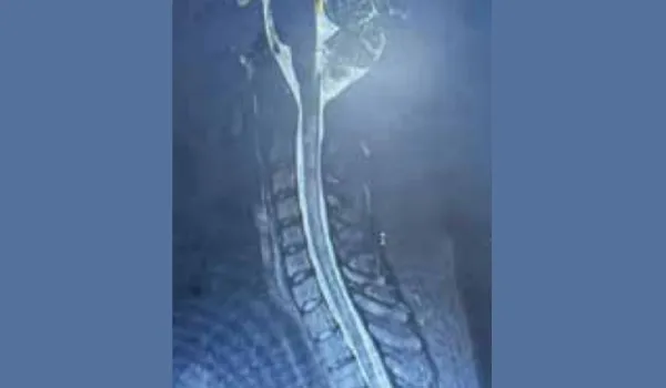
Figure 1: Preventive treatment for NMOSD
Management
The approach to treatment in both disorders is identical and consists of three parts:
a)Treatment of relapse
b)Prevention of relapse
c)Treatment of chronic symptoms
Treatment of relapse10
- Treatment should be prompt and aggressive.
- There is no cure for NMO, but it is treatable. Some patients recover, but many are left with impaired vision and limb function, which can be severe in some cases.
- High-dose IV methylprednisolone pulse therapy is given for 5 days, followed by a prednisolone taper
- Depending on the treatment response; plasmapheresis or IVIG may be used for relapses with incomplete recovery after steroids.
- Early plasmapheresis is encouraged for patients with serious deficits or those who do not respond to steroids. Treatment of subsequent attacks with plasmapheresis has shown better outcomes. This treatment involves the patient’s own blood being pumped out, blood cells being removed from the plasma and mixed with a solution, and then the new blood mixture being pumped back in.
Prevention of relapse10,11
Low dose prednisolone or prednisone
- Low-dose oral corticosteroids are used in many neurological diseases. Tapering doses of oral steroids are given for a few months
- Data on long-term use of steroids are limited. A few studies show a decrease in annual relapse rate by low-dose steroid therapy.
- It is known from treatment with azathioprine that additional oral prednisone is effective to reduce disease activity during the first 3-6 months until azathioprine reaches full efficacy.
- In MOG disease, low treatment failure rates were achieved with oral prednisone. There can be occurrence of relapses post tapering and stopping of IV methyprednisolone. But a long-term therapy should be carefully used in view of known side effects.9
- Azathioprine (is a purine analog), mycophenolate mofetil, rituximab, cyclophosphamide, and mitoxantrone all have used for preventive treatments (Table 3)
- Eculizumab, a complement inhibitor is now US FDA-approved for the treatment of NMOSD. It showed a 94% risk reduction for new relapses when added on to patients who were on placebo and continued their previous disease-modifying treatment (DMT).11
- Satralizumab and inebilizumab both showed marked reduction in new NMOSD relapses versus the comparison group.12
- Multiple DMTs used in MS, like interferons and natalizumab can worsen the disease course of NMO. Therefore, early and accurate diagnosis is critical in the management of NMO.
| Drug | Mechanism of action | Note |
|---|---|---|
| Azathioprine | Inhibits purine metabolism | Reported effective in 1998; a mainstay of treatment combined with steroids due to delayed onset of action (takes months) |
| Mycophenolate mofetil | Inhibits purine metabolism | Better efficacy and tolerability. Combined with steroids due to delayed onset of action |
| Methotrexate | Inhibits folate metabolism | - |
| Cyclophosphamide | DNA cross-linker | |
| Rituximab | Anti-CD20 antibody; depletes B cells | Most commonly used treatment for NMOSD |
| Intravenous immunoglobulins | - | - |
| Mitoxantrone | Inhibits DNA synthesis/repair | - |
| Haematopoietic stem cell transplantation | - | Used in severe case of NMO. Provides short term benefit by inflammation reduction. But most patients relapse within 5 years |
Table 3: Preventive treatment for NMOSD
It is important to note that interferon beta; fingolimod; natalizumab and alemtuzumab worsen NMO disease progression and should not be used to treat NMO.12
Prognosis
Some improvement typically occurs in a few weeks, but disability may persist. Although it has a monophasic phase, approximately 85% have a relapsing course with repeated episodes of transverse myelitis and/or optic neuritis. In the monophasic form, transverse myelitis and optic neuritis occur simultaneously, but in the relapsing form, there are typically weeks or months between initial attacks, and patients have better motor recovery after the initial transverse myelitis event. Around 55% have a relapse in the first year and 90% in the first five years. Unlike MS, NMO rarely enters a secondary progressive phase. Approximately 20% with monophasic NMO have permanent visual loss, and 30% experience permanent paralysis in one or both legs.5
Conclusion
It is very important to diagnose NMO and MOG-associated disorders as early as possible to ensure proper management and avoid complications and poor long-term prognosis. IVIG, plasma exchange and IV rituximab have revolutionised the management and early recovery of most cases, resulting in reduced disability and an intermediate prognosis. MOG disorders carry a more favourable prognosis compared to NMO, with better recovery with supportive care. IVIG and plasma exchange, when administered in good neurology intensive care units, worldwide, are associated with low mortality.
Manoj Khanal. Neuromyelitis Optica (NMO) and Myelin Oligodendrocyte Glycoprotein (MOG) Associated
Autoimmune Neurological Disorders and Recent Management. MMJ. 2025, June. Vol 2 (2).
References
- Lennon VA, Wingerchuk DM, Kryzer TJ, et al. A serum autoantibody marker of neuromyelitis optica: distinction from multiple sclerosis. Lancet. 2004;364(9451):2106-2112.
- Lucchinetti CF, Mandler RN, McGavern D, et al. A role for humoral mechanisms in the pathogenesis of Devic’s neuromyelitis optica. Brain. 2002;25(Pt 7):1450-61.
- Jacob A, McKeon A, Nakashima I, et al. Current concepts of neuromyelitis optica (NMO) and NMO spectrum disorders. J Neurol Neurosurg Psychiatry. 2013;84(8):922-30.
- Flanagan EP, Cabre P, Weinshenker BG, et al. Epidemiology of aquaporin – 4 autoimmunity and neuromyelitis optica spectrum. Ann Neurol. 2016;79(5):775-783.
- Wingerchuk DM, Banwell B, Bennett JL, et al. International consensus diagnostic criteria for neuromyelitis optica spectrum disorders. Neurology. 2015;85(2),177-189.
- Hennes EM, Baumann M, Lechner C, et al. MOG Spectrum Disorders and Role of MOG- Antibodies in Clinical practice. Neuropediatrics. 2018;49(1):3-11.
- Chen JJ, Tobin WO, Majed M, et al. Prevalence of Myelin Oligodendrocyte Glycoprotein and Aquaporin – 4 Ig G in patients in the Optic Neuritis treatment Trial. JAMA Ophthalmol. 2018;136(4):419-422.
- Weber MS, Derfuss T, Metz I, et al. Defining distinct features of anti-MOG antibody associated central nervous system demyelination. Ther Adv Neurol Disord. 2018:11:1756286418762083.
- Jurynczyk M, Messina S, Woodhall MR, et al. Clinical presentation and prognosis in MOG-antibody disease: a UK study. Brain. 2017;140(12):3128-3138.
- Kleiter I, Gahlen A, Borisow N, et al. Neuromyelitis optica: Evaluation of 871 attacks and 1,153 treatment courses. Ann Neurol. 2016;79(2):206-16.
- Kimbrough DJ, Fujihara K, Jacob A, et al. Treatment of Neuromyelitis optica: Review and Recommendations. Mult Scler Relat Disord. 2012;1(4):180-187.
- Collongues N, Ayme-Dietrich E, Monassier L, et al. Pharmacotherapy for Neuromyelitis optica spectrum disorders: Current management and future options. Drugs. 2019;79(2):125-142.