

Abstract:
The objective of the balloon Eustachian tuboplasty (BET) is to widen the cartilaginous part of the Eustachian tube (ET). It improves the physiological functions with minimal or no complications. This minimally invasive technique has proved to be feasible and safe in the treatment of ET dysfunction. The postsurgical pain is minimal, and patients can return to normal activities the next day.
Key words: Eustachian Dysfunction, Endoscopic Balloon Eustachian Tuboplasty, Minimally Invasive Technique.
Introduction
Endoscopic Eustachian tube balloon dilatation is a novel technique aimed to address Eustachian tube (ET) dysfunction. ET dysfunction is a common clinical entity which is usually seen in about 1% of the adult population. Its treatment has ever been challenging to otorhinolaryngologists.1,2 The natural history of ET dysfunction is poorly understood, and evidence for current treatments are limited.1
Dysfunction of the ET is defined as failure of the functional valve of the ET to open and/or close properly. The aetiopathogenesis of ET dysfunction is allergic rhinitis, laryngopharyngeal reflux, mechanical obstruction by nasopharyngeal growth, primary ciliary dyskinesia, and neuromuscular dysfunction.1-3 A new technique of rigid nasal endoscopy for dilating ET was developed by Yamashita. Long-term ventilation tubes carry the risk of permanent tympanic membrane perforation.3 As the cartilaginous portion of the ET is the most likely site of pathology, an intervention in this area should bring about better outcome. The evolution of rigid endoscopes for endoscopic sinus surgery and inflatable balloons has opened new possibilities for treatment of ET dysfunction.
Symptomatology
Obstructive ET dysfunction (OETD) may include:4,5
- Otitis media with effusion (OME)
- Tympanic membrane retraction, or perforation
- Middle ear atelectasis
- Chronic otitis media (COM)
- Cholesteatoma
In milder cases, OETD may only be apparent in situations of baro-challenge (inability to equalise with rapid barometric pressure changes), with otherwise normal function in stable ambient conditions. Symptoms of OETD may include aural fullness, otalgia, tinnitus, and hearing loss. These symptoms may or may not be reversible after Valsalva manoeuvre.4,5
Methodology
Seven patients were treated by endoscopic ET balloon dilatation. All patients pre-operatively underwent detailed clinical examinations, pure tone audiometry and tympanometry. Patients underwent standard endoscopic examinations of the nose and nasopharynx to rule out any pathology in the nose and nasopharynx. Pre-operative and post-operative assessment of clinical symptoms, Valsalva manoeuvre, tympanometry, and post-operative complications were analysed.
Surgical procedure
The procedure was done under general anaesthesia with topical application of nasal decongestants. The nasopharynx was examined endoscopically (Figure 1). A specially designed balloon was introduced into the ET for a length of about 20 mm via a special insertion instrument aided by rigid nasal endoscope (Figures 2 and 3). After insertion, the balloon was inflated using sterile water (Figure 4). Pressure of about 10 Bars was maintained for 2 minutes (Figure 5). The balloon was withdrawn after deflation (Figure 6). The tympanic membrane was examined after surgery (Figure 7). Success was defined by an improvement in tympanogram type: Type B or C to Type A, or Type B to Type C. Pre- and post-operative tympanograms were further analysed using middle ear pressure values.
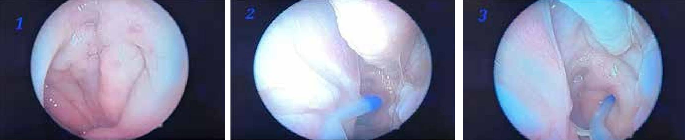
Figure 1: The nasopharynx was examined endoscopically; Figure 2: A specially designed balloon was introduced into the Eustachian tube (ET) for a length of about 20 mm via a special insertion instrument aided by rigid nasal endoscope; Figure 3: A specially designed balloon was introduced into the ET for a length of about 20 mm via a special insertion instrument aided by rigid nasal endoscope.
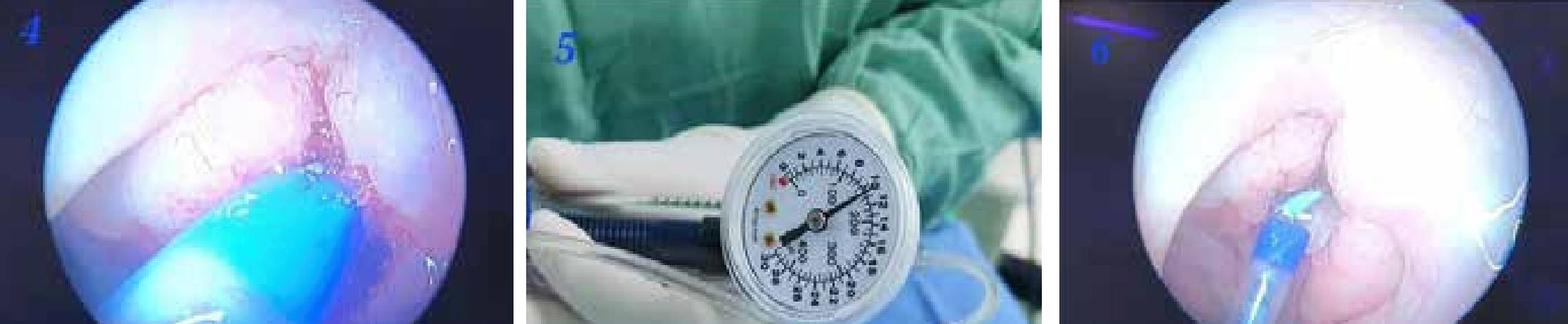
Figure 4: The balloon was inflated using sterile water; Figure 5: Pressure of about 10 Bars was maintained for 2 minutes; Figure 6: The balloon was withdrawn after deflation.
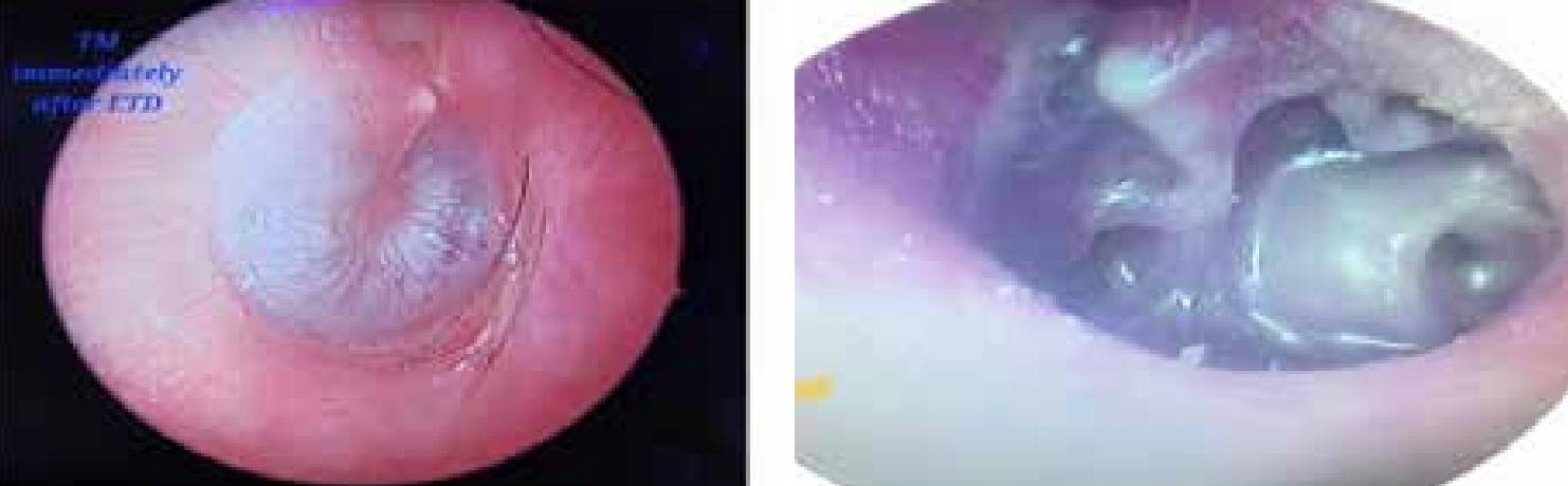
Figure 7: The tympanic Membrane was examined after surgery; Figure 8: Severely atelectatic tympanic membrane.
Discussion
The ET is an uncommon location for intervention because of its anatomical site, uncertain function and anecdotal reports for catastrophic situations like rupture of internal carotid artery. The average length of ET in adults is 37.5 mm long with bony and cartilaginous parts, extending from nasopharynx to the middle ear.1,5-7 The physiological functions of the ET are ventilation, drainage of middle ear secretions, pressure regulation of middle ear, and protection of the middle ear from sound pressure and nasopharyngeal secretions.6,7 The current research utilising video endoscopy has helped to improve the understanding of ET function and its role in middle ear pathology, identifying the common site for dysfunction at cartilaginous part.
ET dysfunction is the failure of the functional valve of the ET to open or close properly. The aetiopathogenesis of the ET dysfunction are mechanical obstruction by nasopharyngeal growth, laryngopharyngeal reflux, allergic rhinitis, primary ciliary dyskinesia, and neuromuscular dysfunction.1,8 Clinical presentation of ET dysfunction includes fullness of ear, otalgia, decreased hearing, tinnitus and vertigo. Medical management of ET dysfunction includes antihistamines, nasal decongestants and oral or nasal steroids. When conservative management of ET dysfunction does result in the desired changes, surgical intervention like ET dilatation is done.
Conclusion
Endoscopy guided balloon dilatation of ET is a novel technique to perform minimal invasive ET dilation for improvement of ET dysfunction. The objective of the balloon Eustachian tuboplasty is to widen the cartilaginous part of ET. It improves the physiological functions with minimal or no complications. This minimally invasive technique can prove to be feasible and safe in the treatment of the ET dysfunction. The postsurgical pain is minimal, and patients can return to normal activities the next day. ET balloon dilation is a safe procedure and offers great improvement in tympanogram values. Further refinement of patient selection and standardisation of technique is required to optimise the effect of this therapy. Long-term follow-up data will clarify the persistence of the effect.
Rajesh Mishra. Endoscopic Eustachian Tube Balloon Dilatation. MMJ. 2025, March. Vol 1 (5).
References
- McCoul ED, Anand VK. Eustachian tube balloon dilation surgery. Int Forum Allergy Rhinol. 2012;2(3):191-8.
- Ockermann T, Reineke U, Upile T, et al. Balloon dilatation eustachian tuboplasty: a clinical study. Laryngoscope. 2010;120(7):1411-6.
- Poe DS, Silvola J, Pyykkö I. Balloon dilation of the cartilaginous eustachian tube. Otolaryngol Head Neck Surg. 2011;144(4):563- 9.
- Tisch M, Störrle P, Danz B, et al. Zum Stellenwert der Bildgebung vor Tubendilation mit dem Bielefelder Tubenkatheter [Role of imaging before Eustachian tube dilation using the Bielefeld balloon catheter]. HNO. 2013;61(6):488-91. German.
- Catalano PJ, Jonnalagadda S, Yu VM. Balloon catheter dilatation of Eustachian tube: a preliminary study. Otol Neurotol. 2012;33(9):1549-52.
- Schröder S, Reineke U, Lehmann M, et al. Chronisch obstruktive Tubenfunktionsstörung des Erwachsenen: Langzeitergebnisse der Ballondilatation der Tuba Eustachii [Chronic obstructive eustachian tube dysfunction in adults: long-term results of balloon eustachian tuboplasty]. HNO. 2013;61(2):142-51. German.
- McCoul ED, Anand VK, Christos PJ. Validating theclinical assessment of eustachian tube dysfunction: the EustachianTube Dysfunction Questionnaire (ETDQ-7). Laryngoscpe. 2012;122,1137–1141.
- McCoul ED, Singh A, Anand VK, et al. Balloon dilation of the eustachian tube in a cadaver model: technical considerations, learning curve, and potential barriers. Laryngoscope. 2012;122(4):718-23.