 WhatsApp Us (24/7)
 +91 926 888 0303 (24/7)


Abstract:
Ectopic superior mediastinal parathyroid adenoma is an uncommon cause of primary hyperparathyroidism (PHPT). Accurate localisation of ectopic mediastinal parathyroid adenomas can be achieved through imaging modalities such as Technetium-99m (99mTc) Sestamibi parathyroid scan, combined with single-photon emission computed tomography (SPECT), and contrast-enhanced computed tomography (CECT) of the neck and thorax. This report presents a case of a 40-year-old female who experienced right knee pain and swelling, and generalised weakness over a period of four months. Biochemical tests revealed elevated serum calcium (10.6 mg/dL) and intact parathyroid hormone (iPTH) levels (370 pg/mL), indicative of PHPT. Imaging studies identified an ectopic parathyroid adenoma in the superior mediastinum, which could not be accessed via the neck. The patient underwent video-assisted thoracoscopic surgery (VATS) for the excision of the adenoma. Post-surgical follow-up showed normalisation of serum calcium and iPTH levels, with an uncomplicated recovery. The VATS approach proved to be an effective minimally invasive technique for the resection of mediastinal parathyroid adenomas.
Key words: Parathyroid Adenoma, Video-Assisted Thoracoscopic Surgery (VATS), Sestamibi Parathyroid Scan.
Introduction
The parathyroid glands were first identified in humans by Swedish medical student Ivar Sandström in 1879.1 Typically, there are two superior and two inferior parathyroid glands. These glands are bean-shaped, pale yellow, approximately the size of a grain of rice (3-5 mm), and weigh about 30-50 mg.2 They consist of chief cells, oxyphil cells, adipose tissue, and fibrovascular stroma. Chief and oxyphil cells produce parathyroid hormone (PTH), which regulates blood calcium and phosphate levels.
The superior parathyroid glands develop from the fourth branchial pouch and are located near the lateral aspects of the thyroid lobes. The inferior parathyroid glands originate from the third branchial pouch, are closely associated with the thymus, and have a longer embryologic descent. The anatomical positions of the inferior parathyroid glands are more variable; they can be found as high as the upper carotid sheath or as low as the pericardium in the mediastinum.2
However, they are most commonly located near the inferior pole of the corresponding thyroid lobe. The superior parathyroid glands undergo minimal migration and maintain a relatively constant location. Supernumerary parathyroid glands occur in approximately 13% of patients, while fewer than four glands are found in less than 3% of cases.2,3,4 The majority of parathyroid adenomas are found within the parathyroid glands themselves. However, up to 22% of cases involve ectopic parathyroid adenomas.3 These ectopic adenomas are most commonly located in the thymus (38%), followed by the retroesophageal region (31%), the intrathyroidal region (18%), the mediastinum (6%), and the carotid sheath (3%).5
Primary hyperparathyroidism (PHPT) arises from an intrinsic abnormality of the parathyroid glands. The condition is typically caused by a parathyroid adenoma, an abnormal enlargement of the gland that secretes PTH inappropriately. This excess PTH leads to elevated serum calcium levels. About 85% of patients with PHPT have a single parathyroid adenoma,2 while 3% to 5% have a double gland adenoma, and 10% to 15% exhibit hyperplasia of all four glands.6 Risk factors for parathyroid adenoma include childhood exposure to radiation in the head and neck region, and certain familial syndromes such as multiple endocrine neoplasia type 1 (MEN1) and multiple endocrine neoplasia type2A (MEN2A).6 Parathyroid carcinoma is a rare cause of PHPT, occurring in 0.7% to 2.1% of cases.2
Secondary hyperparathyroidism occurs due to the physiological secretion of PTH by the parathyroid glands in response to hypocalcaemia, which often occurs due to vitamin D deficiency or chronic renal failure. This condition is typically managed with medical treatment, and surgery (subtotal parathyroidectomy) is rarely necessary, only in cases where medical therapy is ineffective.
Tertiary hyperparathyroidism is an advanced stage of secondary hyperparathyroidism and is observed in patients with long-term chronic renal failure or those who have undergone kidney transplantation but continue to have excessive, autonomous secretion of PTH. This condition leads to elevated serum calcium and PTH levels and often requires surgical intervention.
Case Report
A 40-year-old female presented with complaints of pain and swelling in her right knee, along with generalised weakness for the past four months. Upon evaluation, she was found to have an elevated serum calcium level of 10.6 mg/dL (normal range: 8.6-10.3 mg/dL) and osteoarthritic changes in the right knee joint, as seen on X-ray, with a deficient vitamin D level of 9.17 ng/mL (considered deficient at <10 ng/mL).
Her biochemical investigations revealed persistently elevated serum calcium, a raised intact parathyroid hormone (iPTH) level of 370 pg/mL (normal range: 12-88 pg/mL), and a low serum phosphorus level of 2.0 mg% (normal range: 2.5-4.6 mg%), indicating PHPT. Although an ultrasound of the neck did not detect any parathyroid enlargement, a Technetium-99m (99mTc) Sestamibi parathyroid scan revealed an ectopic parathyroid adenoma in the mediastinum (Figure 1). For accurate localisation, a contrast-enhanced computed tomography (CT) of the neck and thorax was performed which revealed it to be in the superior mediastinum in the prevascular space (Figure 2). The patient subsequently underwent video-assisted thoracoscopic surgery (VATS) for the excision of the ectopic adenoma.
The patient was positioned supine under general anaesthesia with the right shoulder abducted and extended. An 11 mm optical port was inserted into the right 6th intercostal space along the mid-axillary line. Using this as a guide, a 5 mm port was placed in the right 4th intercostal space along the anterior axillary line, and a third 5 mm port was inserted into the right 8th intercostal space along the anterior axillary line to provide access to the mediastinum with CO2 insufflation. The pneumothorax pressure was maintained at 5-6 mmHg throughout the procedure.
The prevascular space was identified, and the parathyroid adenoma was localised in front of the arch of the aorta. The pleura was incised to gain entry into the mediastinum. The tumour was further localised intraoperatively using indocyanine green (ICG) dye, which highlighted the solitary parathyroid adenoma with a greenish coloration (Figure 3).
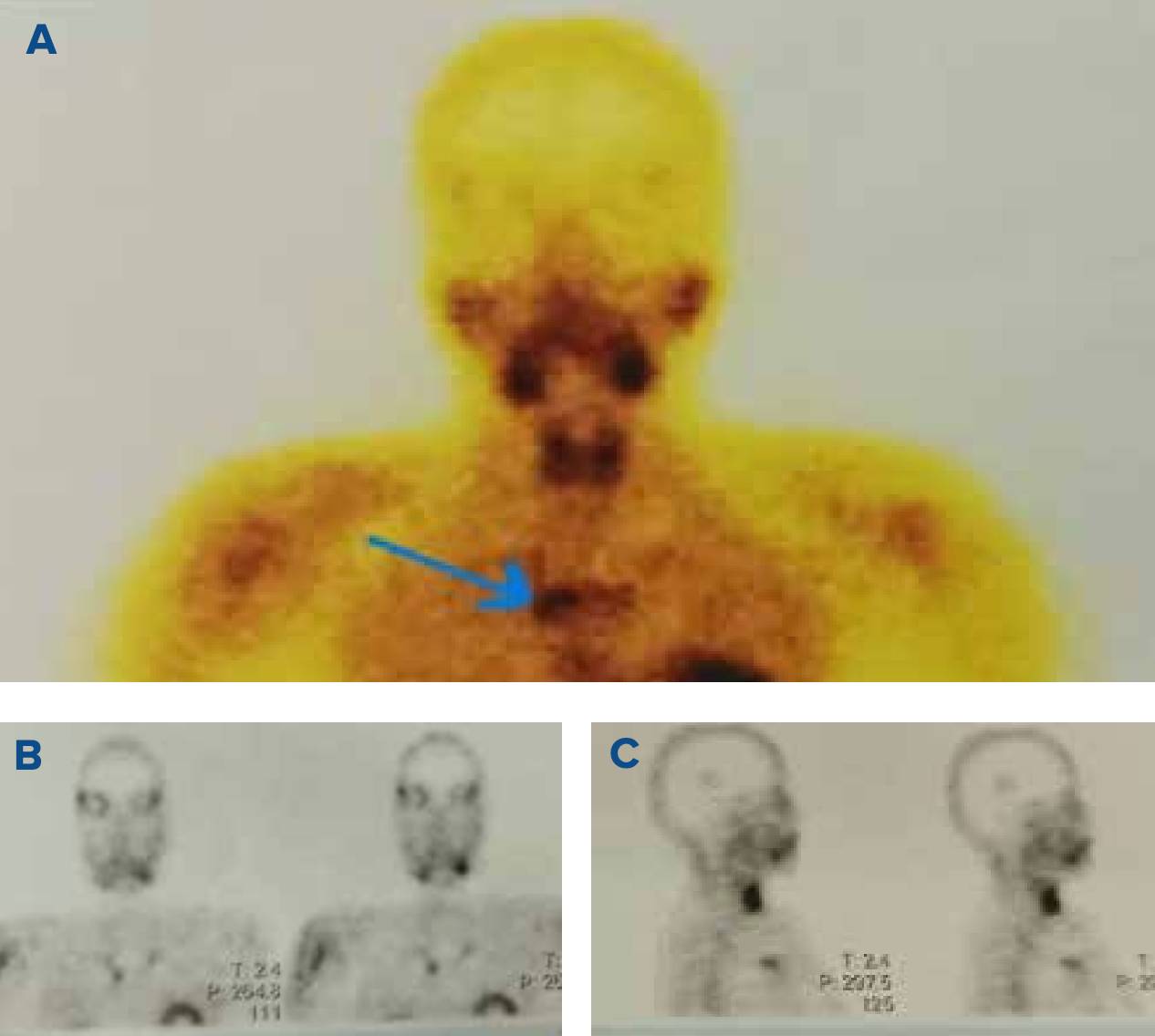
Figure 1 (A, B and C): Showing an ectopic mediastinal parathyroid adenoma in 99mTc Sestamibi parathyroid scintigraphy along with a single-photon emission computed tomography (SPECT) (Indicated with arrow).
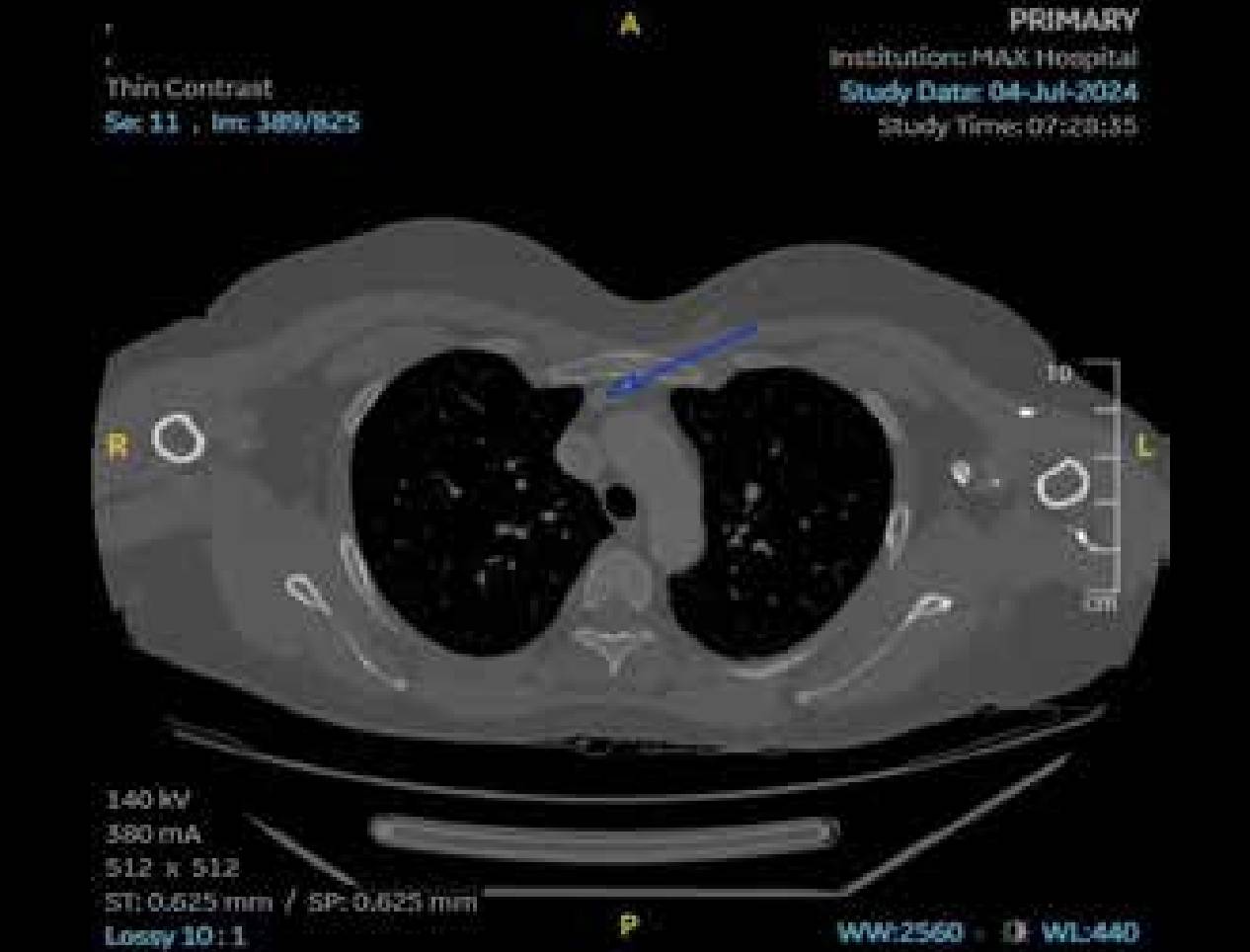
Figure 2: Showing an ectopic mediastinal parathyroid adenoma in contrast-enhanced computed tomography (CT) of the neck and thorax (shown with an arrow).
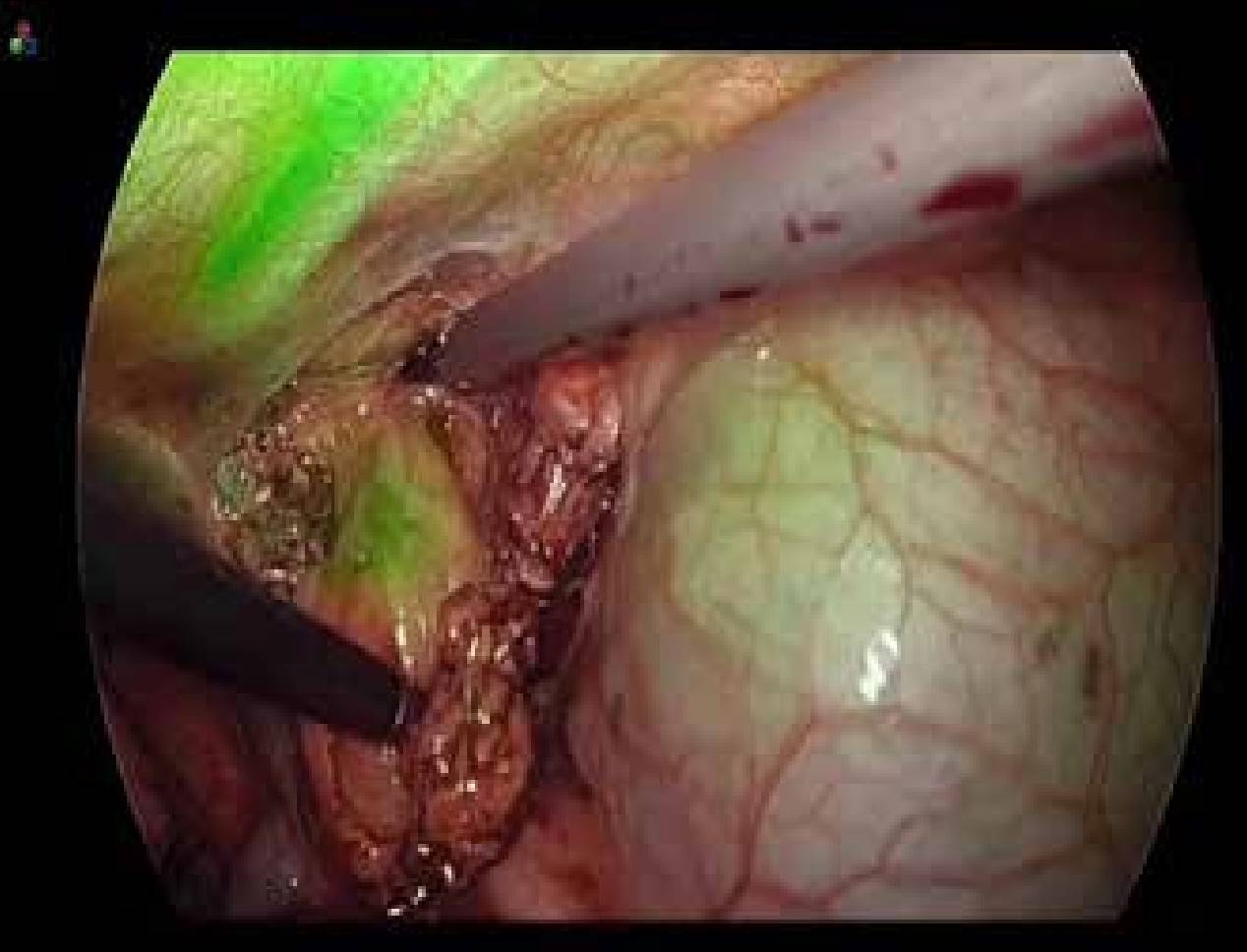
Figure 3: Intra-operative indocyanine green (ICG) image.
The tumour was carefully dissected from the surrounding tissues, with its vascular supply interrupted while avoiding injury to the superior vena cava, arch of the aorta, phrenic nerve, and pericardium. The tumour was then retrieved intact using an endo-bag. The intraoperative rapid iPTH level dropped to 16.2 pg/mL. After ensuring haemostasis, a 28F chest tube was placed, and the wound was sutured. The patient was extubated at the end of the procedure, and her recovery was uneventful. Histopathological examination of the specimen confirmed the diagnosis of a parathyroid adenoma. The drainage tube was removed 48 hours later, and the patient was discharged. At the time of discharge, her corrected serum calcium level was 8.6 mg/dL, and her intact PTH level was 64.9 pg/mL (normal range: 12-88 pg/mL), indicating a successful cure of the disease.
Discussion
The incidence of mediastinal ectopic parathyroid adenoma is approximately 1%-2% of cases. Preoperative localisation of these adenomas is crucial as it helps identify patients who may be candidates for minimally invasive procedures, such as video-assisted thoracoscopic surgery. Technetium-99m (99mTc) Sestamibi scintigraphy is a non-invasive imaging technique that selectively targets abnormal parathyroid glands. When combined with single photon-emission computed tomography (SPECT), Sestamibi scintigraphy has become the most used preoperative imaging study for parathyroid disease.7,8,9 Additionally, combining Sestamibi with CT enhances the detection of smaller parathyroid lesions that might not be visible with Sestamibi alone.7
Injecting ICG intraoperatively helps localise ectopic adenomas. This method is a simple and safe way to visually identify mediastinal ectopic parathyroid adenomas during surgery.5 However, while ICG is a useful visual marker, it does not replace intraoperative PTH monitoring.
VATS is a minimally invasive surgery that provides adequate access to mediastinally located glands, optimal visualisation of mediastinal structures and has a high resection success rate with less morbidity than open approaches.11 The combination of precise preoperative localisation, intraoperative PTH monitoring, and the use of VATS has improved cure rates to 95%-98%. VATS offers several significant advantages, including better visualisation of the tumour, less postoperative pain, shorter hospital stays, and superior cosmetic outcomes.9,10 We found the ectopic adenomas in the prevascular space within the superior mediastinum (Figure 1 and 2).
Intraoperative PTH monitoring involves measuring PTH levels at three key points: preoperatively, before excision, and 30 minutes after removing the suspected adenoma. Blood samples are taken from a peripheral vein or the internal jugular vein. A reduction in intraoperative PTH levels by more than 50% of the pre-excision value is a positive predictor of cure.
Conclusion
Ectopic mediastinal parathyroid adenoma is a rare condition that can be accurately localised using modern cross-sectional imaging techniques. CO2 insufflation and the intraoperative use of ICG are also safe and effective for precisely localising the ectopic parathyroid tissues. VATS is a safe and effective minimally invasive procedure for removing ectopic mediastinal parathyroid adenomas.
Kaushal Yadav, Sagir Ahamed. Video-Assisted Thoracoscopic Surgery for Excision of Ectopic Mediastinal
Parathyroid Adenoma. MMJ. 2025, March. Vol 1 (5).
References
- Organ CH Jr. The history of parathyroid surgery, 1850-1996: the Excelsior Surgical Society 1998 Edward D Churchill Lecture. J Am Coll Surg. 2000;191:284-99.
- Townsend CM, Beauchamp RD, Evers BM, et al. Sabiston textbook of surgery: The biological basis of modern surgical practice. 20th Edition. Philadelphia: Elsevier Saunders; 2018. 924-33.
- Conn JM, Goncalves MA, Mansour KA, et al. The mediastinal parathyroid. Am Surg. 1991;57:62-6.
- Russell CF, Edis AJ, Scholz DA, et al. Mediastinal parathyroid tumors: experience with 38 tumors requiring mediastinotomy for removal. Ann Surg. 1981;193:805-9.
- Hordijk M, Veen EJ, Schreinemakers Jennifer MJ. ICG-Guided Video-Assisted Thoracoscopic Surgery for Resection of an Ectopic Mediastinal Parathyroid Adenoma. J Endocr Surg. 2023;85-88.
- Fischer JE. Fischers Mastery Of Surgery. 7th Edition. Philadelphia: Wolters Kluwer; 2019. 1610.
- Issacs KE, Belete S, Miller BJ, et al. Video-assisted thoracoscopic surgery for ectopic mediastinal parathyroid adenoma. BJS open. 2019;3:743-49.
- Naik D, Jebasingh KF, Ramprasath, et al. Video Assisted Thoracoscopic Surgery for excision of an ectopic mediastinal intra-thymic parathyroid adenoma. JCDR. 2016; 10(6):22-24.
- Amir K, Khan AZ, Rew D, et al. Video assisted thoracoscopic excision of mediastinal ectopic parathyroid adenomas: a UK regional experience. Ann Cardiothorac Surg. 2015; 4(6):527-34.
- Abdulsalam MS, Devanayagam S, Santosham, et al. Mediastinal parathyroid adenoma removal by Video Assisted Thoracoscopy Surgery. Ann Afr Med. 2021; 20(2) 150-53.
- Veronica CK, Mariana CT, Graciela A, et al. Thoracoscopic treatment of mediastinal ectopic parathyroid adenoma: a latinamerica experience case series and literature review. J Cardiothorac Surg. 2024;19:17.