 WhatsApp Us (24/7)
 +91 926 888 0303 (24/7)


Abstract:
Neurocysticercosis (NCC) is the most common helminthic infection of the central nervous system, caused by the larval stage of Taenia solium. While parenchymal involvement at the grey-white matter junction is typical, intraventricular NCC is a rarer presentation. Magnetic resonance imaging (MRI) plays a crucial role in its detection and assessment.
We present the case of a 50-year-old female with a one-week history of severe headache. The MRI revealed a multilobulated cystic lesion in the left lateral ventricle, with features suggestive of intraventricular NCC and associated ventriculitis. The patient was treated with corticosteroids, leading to significant clinical improvement. Follow-up imaging after two weeks showed anterior displacement of the cystic lesion.
Key words: Intraventricular Neurocysticercosis, Taenia Solium, Magnetic Resonance Imaging (MRI), Migrating Neurocysticercosis
Introduction
Neurocysticercosis (NCC) is the most prevalent helminthic infection affecting the human central nervous system, resulting from the larval stage of Taenia solium. The most common location of cystic lesions in NCC is the brain parenchyma at the grey-white matter junction.1 However, intraventricular NCC, where cysts form within the brain’s ventricular system, occurs less frequently.2 Magnetic resonance imaging (MRI) is the preferred imaging modality for its detection.3,4
In our case, a 50-year-old female presented to the hospital with a one-week history of severe headache. MRI revealed findings suggestive of intraventricular NCC involving the left lateral ventricle with signs of ventriculitis. The patient was subsequently managed with corticosteroids and showed significant clinical improvement.
Radiological Findings
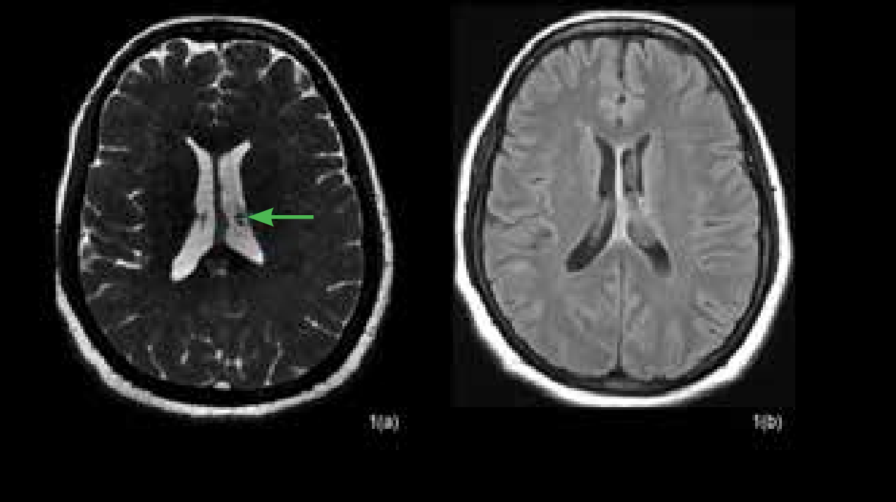
Figure 1a: Axial three-dimensional (3D) constructive interference in steady state (CISS) image reveals a multilobulated T2- weighted hyperintense cystic lesion in the body of the left lateral ventricle (green arrow). A few T2-weighted hypointense foci are also noted within one of the cystic components. Figure 1b: Fluid-attenuated inversion recovery (FLAIR) axial image reveals partial signal suppression within the cysts.
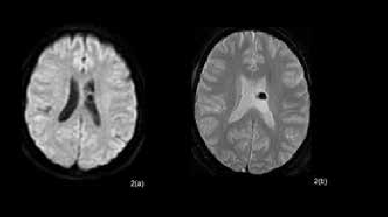
Figure 2a: Axial diffusion-weighted imaging (DWI) image shows absence of diffusion restriction. Figure 2b: Intense susceptibility change is noted on the axial gradient-recalled echo (GRE) image within one of the cystic components (parent cyst) likely representing calcified scolices.
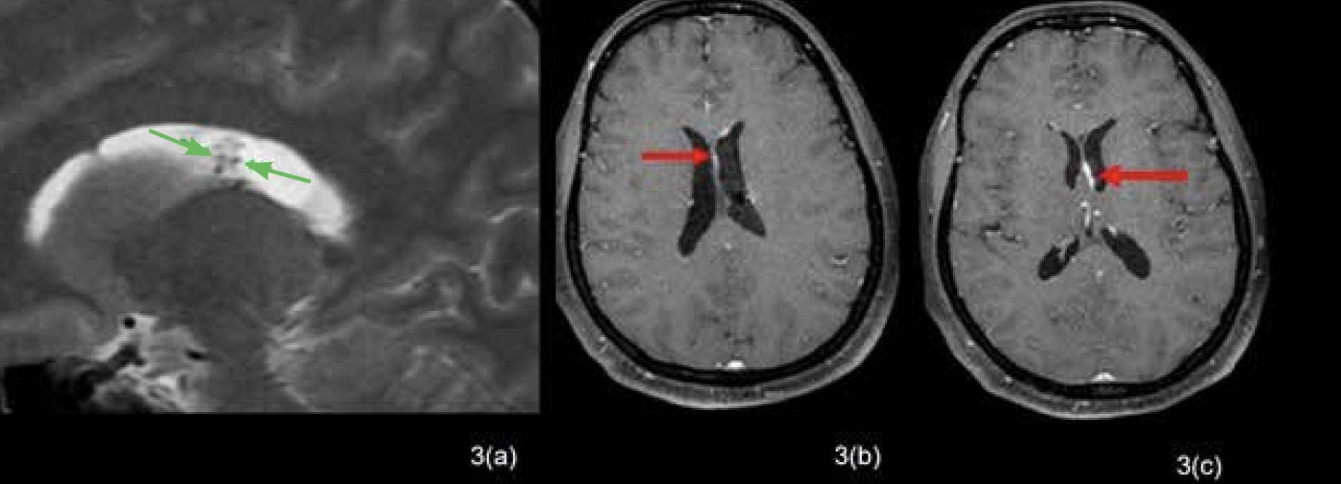
Figure 3a: T2-weighted (T2W) sagittal image also reveals a few T2 hypointense foci within one of the cystic components likely representing scolices within the parent cyst. Figure 3b and 3c: Post-contrast axial T1-weighted (T1W) images show thin, smooth ependymal enhancement along the walls of the left lateral ventricle (red arrows). Findings are suggestive of intraventricular neurocysticercosis with daughter cysts. Signs of ventriculitis are also seen, likely representing an inflammatory response secondary to breach in the wall of the parent cyst.
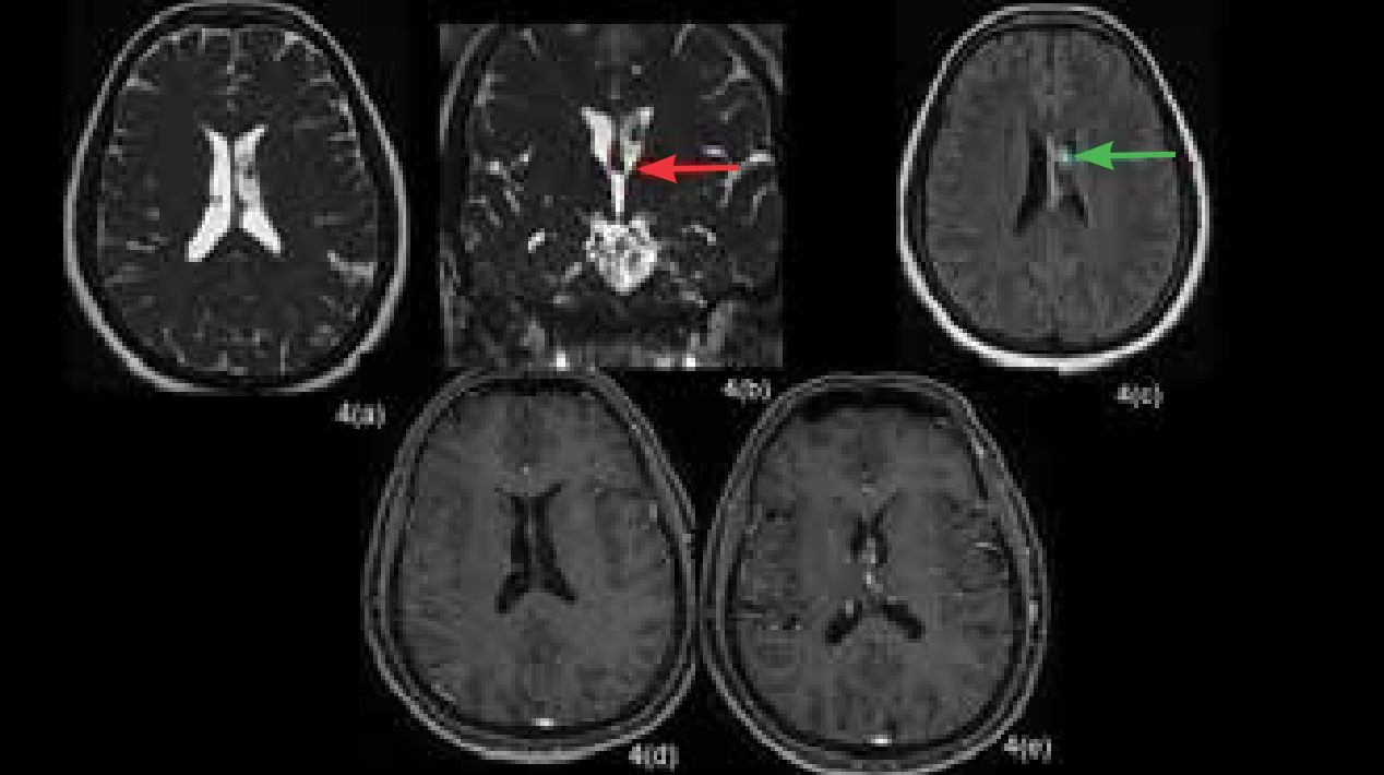
Figure 4: Follow-up imaging with contrast-enhanced magnetic resonance imaging (CEMRI) done at interval of two weeks. Figure 4a: Interval imaging done at 2 weeks reveals anterior displacement of the multilobulated cystic lesion in the anterior aspect of the body of the left lateral ventricle as seen in axial three-dimensional (3D) constructive interference in steady state (CISS) image. Figure 4b: Coronal 3D CISS image also reveals the location of the lesion in the left lateral ventricle, just superior to the left foramen of Monro (red arrow) without causing obstruction. Figure 4c: Axial fluid-attenuated inversion recovery (FLAIR) image also shows the anterior displacement of the cystic lesion as compared to the previous scan. The parent cyst shows hyperintense signal on FLAIR (green arrow). Figure 4d and 4e: Post-contrast T1-weighted (T1W) images reveal significant resolution of the ependymal enhancement.
Conclusion
This case underscores the significance of early MRI diagnosis and prompt medical management in intraventricular NCC, facilitating symptom resolution and preventing complications.
Shashank Raj, Prem Kumar Ganesan, Anchal Singh. Migrating Intraventricular
Neurocysticercosis. MMJ. 2025, March. Vol 1 (5).
References
- Fogang YF, Savadogo AA, Camara M, et al. Managing neurocysticercosis: challenges and solutions. Int J Gen Med. 2015;8:333-344.
- Araujo AL, Rodrigues RS, Marchiori E, et al. Migrating intraventricular cysticercosis: magnetic resonance imaging findings. Arq Neuropsiquiatr. 2008;66:111-113.
- Zee CS, Segall HD, Destian S, et al. MRI of intraventricular cysticercosis: surgical implications. J Comput Assist Tomogr. 1993;17:932-939.
- Ghosh D, Dubey TN, Prabhakar S. Brain parenchymal, subarachnoid racemose, and intraventricular cysticercosis in an Indian man. Postgrad Med J. 1999;75(881):164-166.