 WhatsApp Us (24/7)
 +91 926 888 0303 (24/7)


Abstract:
Systemic onset juvenile idiopathic arthritis (SOJIA) is an autoinflammatory disorder that can present with a wide range of symptoms, often mimicking infectious aetiologies such as tuberculosis and other bacterial infections. As a result, atypical presentations may pose a diagnostic challenge. Here, we report a case of a 4-year-old girl who presented with high-grade fever for two weeks, which was unresponsive to initial antibiotic therapy. Despite extensive investigations, no infection was identified. Imaging revealed bilateral shoulder joint abscesses, which were sterile, accompanied by significantly elevated serum inflammatory markers. Clinical and laboratory findings were consistent with a diagnosis of SOJIA, and corticosteroid and methotrexate therapy were subsequently initiated, to which the child responded.
Key words: Arthritis, Juvenile Idiopathic Arthritis, Shoulder Abscess, Methotrexate.
Introduction
Systemic onset juvenile idiopathic arthritis (SOJIA, formerly called Still's disease or systemic juvenile rheumatoid arthritis) is classified as a category of juvenile idiopathic arthritis (JIA), characterised by arthritis with systemic features such as persistent fever, rash, and prominent visceral involvement, including hepatosplenomegaly, lymphadenopathy, and serositis.1
This case underscores the diagnostic challenges and management strategies for SOJIA presenting with atypical features, such as joint abscesses without any symptoms suggestive of joint involvement,2 which may delay the diagnosis.3 In this case, the child did not have any signs of arthritis, such as joint pain or restricted movements. Bilateral shoulder abscesses were accidentally picked up on computed tomography (CT) of the thorax, which was performed to identify any infectious focus.
Case Report
This 4-year-old female child, born to a non-consanguineous marriage, presented with a high grade fever (up to 104°F) for almost two weeks, which was intermittent in nature and associated with erythematous rashes on the body, which were transient and occurred mainly at the peak of the fever. She took an oral antibiotic (cefixime) for 5 days, with no improvement. She was diagnosed with multisystem inflammatory syndrome in children (MIS-C) 6 months ago and treated with steroids. No similar illnesses were observed in the family. Her immunisations were up to date and there was no history of weight loss (weight: 12.5 kg [3rd-15th centile], height: 96.5 cm [3rd-15th centile]).
On examination, the child was febrile. Other physical findings were normal except for hepatomegaly (liver span: 8.5 cm).
Initial blood investigations revealed neutrophilic leucocytosis (total leukocyte count: 23000, Neutrophil 64%) with a C-reactive protein level of 314 mg/L and an erythrocyte sedimentation rate (ESR) of 37. Chest X-ray, abdominal ultrasound, and echocardiography were normal. An initial diagnosis of infectious cause (enteric fever) was considered and was started on ceftriaxone and azithromycin. Blood and urine cultures were negative. In view of persistent fever spikes, antibiotics were upgraded to meropenem, and repeat blood investigation were conducted, which showed elevated inflammatory markers (D-dimer: 1,553 ng/mL, lactate dehydrogenase (LDH): 496 U/L, ferritin: 798 ng/mL, coronavirus disease (COVID) antibody (IgG): 354 BAU/mL; faecal calprotectin: 150 ug/g), suggested the possibility of an autoimmune disease. Antinuclear antibody (ANA), rheumatoid factor (RF), and human leukocyte antigen-B27 (HLA-B27) were negative.
CT of the thorax was done as a part of evaluation of the unresolving fever and showed a collection in bilateral shoulder joints (Figure 1). Magnetic resonance imaging (MRI) revealed marked subacromial-subdeltoid and sub-coracoid bursitis, tenosynovitis of long head of biceps tendon, mild glenohumeral joint effusion, fluid in the axillary recess, and left axillary and cervical lymphadenopathy (Figure 2).
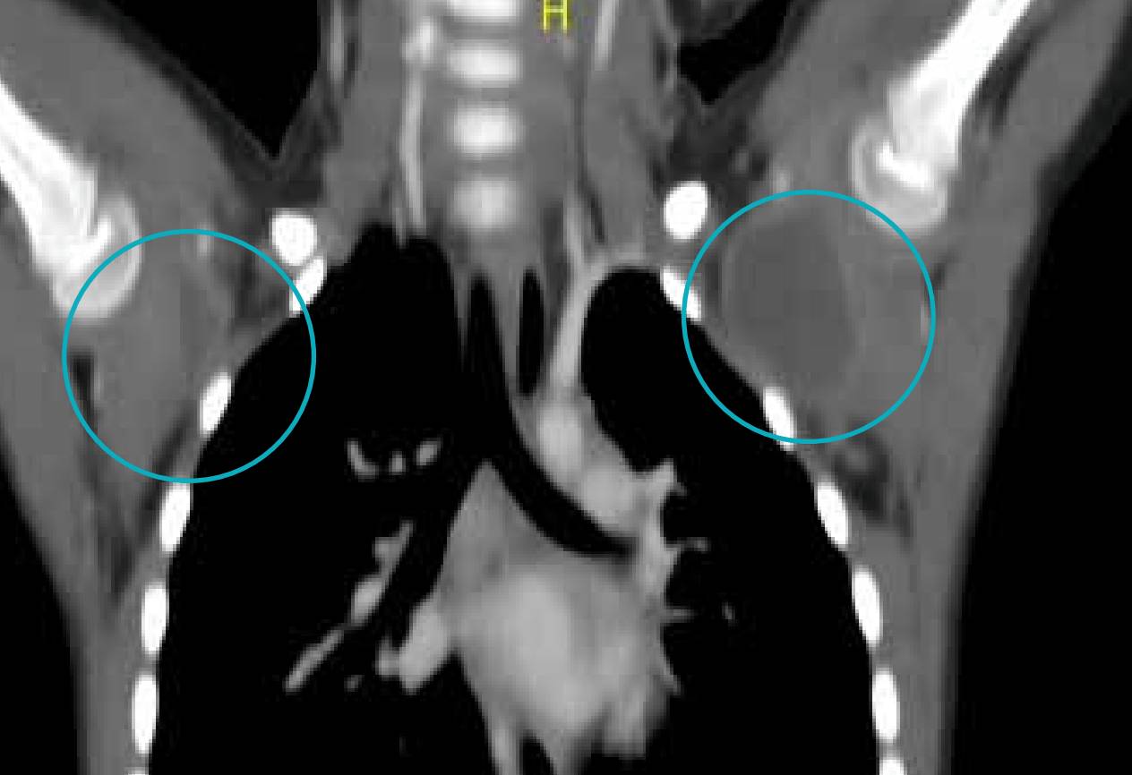
Figure 1: Computed tomography (CT) Thorax showing bilateral shoulder joint abscesses.
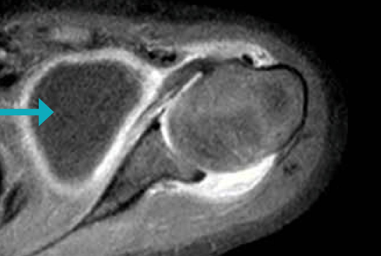
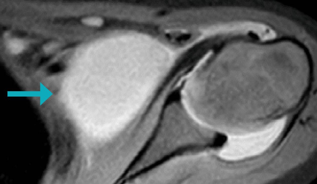
Figure 2: Magnetic resonance imaging (MRI) scan showing marked subacomial-subdeltoid and sub-coracoid bursitis and along with tenosynovitis of long head of the biceps tendon.
USG-guided aspiration of pus was done5; which was negative for Mycobacterium tuberculosis (MTB)/acid-fast bacilli (AFB) stain1 /gram stain/fungal cultures/AFB culture.5 USG-guided biopsy from the left cervical region lymph node of size 1.5 cm was done, suggestive of reactive lymphoid hyperplasia. GeneXpert for MTB was reported as negative.4
The persistent fever, lack of evidence of an infectious process and joint involvement led to considering the possibility of SOJIA.8 She was started on a nonsteroidal anti-inflammatory drug (NSAID), naproxen, and her fever intensity decreased. Subsequent steroid treatment significantly reduced symptoms and the fever resolved. The child was discharged with a diagnosis of systemic JIA. She is currently being treated with methotrexate and a weaning dose of prednisolone.10
Discussion
We report this case to highlight the interesting presentation of SOJIA in a child in the form of fever and bilateral shoulder joint abscess without any other symptoms.3
SOJIA is a subtype of juvenile idiopathic arthritis characterised by systemic symptoms. According to International League of Associations for Rheumatology (ILAR), SOJIA is defined as arthritis in one or more joints, or preceded by fever of >2 weeks that is documented to be daily (Quotidian) for at least 3 days and accompanied by one or more of the following.1
- Evanescent erythematous rash
- Generalised lymph node enlargement
- Hepatomegaly, splenomegaly, or both
- Serositis
Diagnosis is often challenging due to the overlap with infectious diseases. This case underscores the importance of considering SOJIA in differential diagnoses of prolonged fever with elevated inflammatory markers and joint involvement in children.
Conclusion
Early recognition and appropriate management of SOJIA are crucial for improving patient outcomes.7 This case highlights the importance of a multidisciplinary approach in diagnosing and managing complex paediatric cases.9 SOJIA should be considered in children presenting... with persistent fever spikes even without arthralgia.3
Aadinadh M.J, Arvind Bountra. Not All Pus is Bacteria or Tuberculosis: Atypical Presentation of Systemic
Onset Juvenile Idiopathic Arthritis (SOJIA). MMJ. 2025, March. Vol 1 (5).
References
- Kliegman RM, St. Geme JW, Blum NJ, et al. Nelson Textbook of Pediatrics. 22nd Edition. Elsevier; 2024. P. 1471-1482
- Boyarchuk O, Kovalchuk T, Kovalchuk N, et al. Clinical variability of the systemic juvenile idiopathic arthritis course: literature review based on case series. Reumatologia/Rheumatology. 2020;58(6):436-43.
- Shahin MA, Sultan MI, Alam MJ, et al. Tuberculosis is a Mimicker of JIA: A Rare Case Report. Mymensingh Med J. 2016;25(3):575-9.
- Shahin MA, Sultan MI, Alam MJ, et al. Tuberculosis is a Mimicker of JIA: A Rare Case Report. Mymensingh Medical Journal: MMJ. 2016;25(3):575-9.
- Gamalero L, Ferrara G, Giani T, et al. Acute Arthritis in Children: How to Discern between Septic and Non-Septic Arthritis? Children (Basel). 2021;8(10):912.
- Jain D, Aggarwal HK, Rao A, et al. Macrophage activation syndrome in a patient with systemic onset of the juvenile idiopathic arthritis. Reumatologia/Rheumatology. 2016;54(1):42-
- Mirkinson LJ, Katona IM. Systemic onset juvenile idiopathic arthritis: update on pathogenesis and treatment. Expert Rev Clin. Immunol. 2007;3(3):313-21.
- Quartier P. Systemic juvenile idiopathic arthritis/pediatric still’s disease, a syndrome but several clinical forms: Recent therapeutic approaches. J. Clin Med. 2022;11(5):1357.
- Nigrovic PA, Martínez-Bonet M, Thompson SD, et al. Implications of juvenile idiopathic arthritis genetic risk variants for disease pathogenesis and classification. Curr Opin Rheumatol. 2019;31(5):401-10.
- Woo P. Systemic juvenile idiopathic arthritis: diagnosis, management, and outcome. Nat Clin Pract Rheumatol. 2006;2(1):28-34.