 WhatsApp Us (24/7)
 +91 926 888 0303 (24/7)


Abstract:
Lymphoedema is a chronic debilitating condition often perceived as incurable or refractory. It deteriorates the quality of life and can cause severe disability, impacting both the physical and psychosocial health of an individual. With recent advancements in diagnostic and therapeutic tools, its management is evolving. This case report presents a 45-year-old male who reported to our department with a primary complaint of swelling and difficulty while walking. Based upon clinical examination and investigations, the patient was diagnosed with post-traumatic elephantiasis. The patient was successfully treated using a combination of conservative measures, complete decongestive therapy (CDT), lymphatic-venous anastomosis (LVA), and reduction surgeries. The patient’s leg circumference was reduced from 120 cm to 35 cm. Management of lymphatic filariasis has been revolutionised by the introduction of CDT, LVA and super microsurgical vascularised lymph node transfer (VLNT).
Key words: Elephantiasis, Lymphoedema, Complete Decongestive Therapy (CDT), Lymphatic- Venous Anastomosis (LVA).
Introduction
Lymphoedema is defined as a chronic deforming disorder characterised by abnormal interstitial fluid accumulation and stasis in the subcutaneous tissue. Globally, it affects 250 lakh people.1 Primary lymphoedema can be hereditary, caused by inherent abnormal function of the lymphatic channels. Secondary lymphoedema, on the other hand, is attributed to oncological interventions, infections, inflammatory disorders, obesity, posttrauma, or filariasis. Post-traumatic elephantiasis, a severe form of secondary lymphoedema, occurs due to extensive soft tissue damage and lymphatic disruption following trauma. This condition is characterised by persistent, progressive swelling and deformity, often involving the lower limbs. It is aggravated by chronic inflammation, recurrent infections, and fibrosis of the lymphatic channels.² The resulting hypertrophy and hyperkeratosis of the skin can lead to massive enlargement and functional impairment of the affected area, with profound cosmetic, physical, and psychological consequences.
Lymphoedema has two phases: the fluid phase and the solid phase.³ The fluid phase is characterised by pitting oedema, where lymphatic fluid initially accumulates in the affected limb. When left untreated, it progresses to the solid phase, which involves adipocyte stimulation and proliferation, collagen fibre deposition in the extracellular matrix, and lymphatic remodelling.4 In this phase, fibrotic soft tissue changes become apparent.
Irrespective of the pathogenesis, lymphedema is often chronic and leads to long-term physical, social, and psychological challenges.
Case report
A 45-year-old male patient, who had been suffering from elephantiasis of the left lower limb for the past 10 years, presented to our department. He reported a history of trauma to the left groin region that had resulted in disruption of groin lymph nodes. Following this event, he developed oedema of the left leg, which progressively worsened, leading to elephantiasis.
A comprehensive clinical examination revealed striking features. The mid-leg circumference measured an astounding 120 cm, while the mid-thigh circumference was 138 cm. The affected limb weighed approximately 45 kg, contributing significantly to the patient's physical and functional limitations. Peripheral pulses in the left leg were non-palpable due to the extensive oedema. The sheer weight of the affected leg severely restricted its movement, with minimal dorsiflexion and leg extension.
To assess the extent of lymphatic impairment, lymphangiography using indocyanine green dye was performed, which revealed significant lymphatic blockage at the proximal groin region. This blockage caused severe lymphatic stasis, leading to extensive lymphatic fluid accumulation in the leg and foot.
The patient was diagnosed with post-traumatic elephantiasis of the left leg. Before proceeding with surgical intervention, conservative management was initiated, including manual lymphatic drainage and compression bandaging, to facilitate lymphatic flow and prepare the tissues for surgery. Lymphoscintigraphy was performed to identify viable lymphatic vessels, and lymphovenous anastomosis (LVA) was performed at three strategic sites (Figures 1a and 1b).
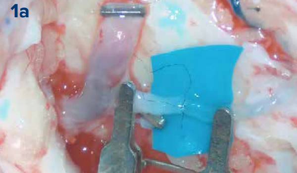
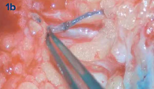
Figure 1a and 1b: Demonstrates lymph-venous anastomosis.
Two weeks post-anastomosis, the patient continued with manual lymphatic drainage and compression therapy. Within a month, the patient’s leg circumference came down to 112 cm, and tissues in all the compartments became notably softer.
A staged reduction approach surgery was then planned to address the persistent tissue hypertrophy in the leg, thigh, and foot. Over the course of three surgeries, combined with ongoing LVA and compression therapy, the patient's leg circumference dramatically decreased from 120 cm to 35 cm (Figures 2a and 2b).
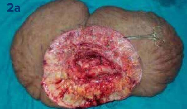
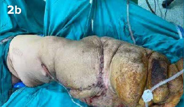
Figure 2a and 2b: Reduction surgery for lymphoedema.
Three months after the first surgery and completion of the three reduction surgeries, the patient experienced significant improvement. The range of motion at the knee and ankle joints increased, enabling him to walk longer distances without much difficulty and to perform routine actives comfortably (Figures 3a and 3b). The patient is now considering rejoining his job, which he had lost due to this condition.
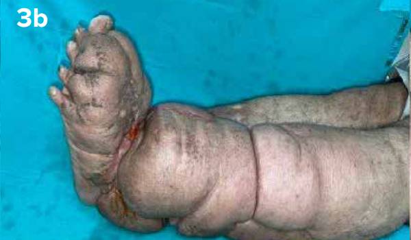
Figure 3a and 3b: Represents range of motion after the reduction surgeries.
Discussion
An accurate diagnosis is mandatory for the effective management of lymphoedema. The diagnosis can be determined by clinical history and physical examination. However, advanced imaging techniques are required for lymphatic mapping to manage lymphatic disorders effectively. The gold standard for diagnosing lymphoedema is lymphoscintigraphy.5 This technique assesses the lymph node drainage speed to evaluate the severity of lymphoedema and identify lymphatic channels.
In recent years, magnetic resonance lymphangiography has emerged as a new method for diagnosing lymphatic diseases. It determines damage to the lymphatic system, enabling the preoperative determination of appropriate vessels for LVA.
Another diagnostic modality is fluorescence lymphangiography which detects the near infrared light emitted by indocyanine green dye. The dye is taken up by the functional lymphatics allowing clinicians to distinguish between functioning and nonfunctioning lymphatics.6
Currently, there is no standardised treatment protocol for lymphoedema. Its management is either conservative, surgical, or a combination of both. Conservative management is the first-line intervention, and includes CDT, which is a two-phase approach. Phase 1 focuses on reducing lymphoedema volume, while phase 2 aims to maintain the reduced volume. CDT involves exercise, daily bandaging, manual drainage therapy, and skin care. Surgical management options include LVA, vascularised lymph node transfer, Charles or modified Charles procedure, and liposuction.7 Surgery is typically considered the last resort, with the ultimate goal of preventing complications and reducing the psychological and physical suffering of the patient. A vast majority of patients require a standardised protocol for treating all stages of lymphoedema, as many are unaware of the condition and the available solutions.
Conclusion
In our case, the patient developed elephantiasis due to trauma that disrupted the groin lymph nodes. The various steps in the management of elephantiasis such as lymphoscintigraphy, manual drainage, compression dressings, and reduction surgeries were as important as the surgery itself. Each step contributed equally to the successful outcome of this case. Lympho-venous anastomosis, along with lymphatic drainage prevented the recurrence of the lymphoedema.
Moreover, recent advances in technology and refinement of surgical techniques have ushered in a new era in the field of reconstructive microsurgery. The management of lymphoedema has been transformed by the introduction of CDT, LVA, and super microsurgical vascularised lymph node transfer (VLNT).8
Our centre is one of its kind in the northern region, focusing on limb preservation. With the right expertise, technology, and comprehensive management, patients with chronic lymphoedema can achieve a better quality of life, breaking the associated stigma through successful management. Therefore, lymphatic disorders, including lymphoedema, should not be neglected.
Pradeep K Singh, Manoj K Johar, Digjeet Kaur. Post-Traumatic Unilateral Elephantiasis: A Case
Report. MMJ. 2025, March. Vol 1 (5).
References
- Wongtrungkapun R. A Case Report of Severe Lymphedema of Lower Limb: Surgical Management. Thai Journal of Surgery. 2017;38(1).
- Agrawal VK, Sashindran VK. Lymphatic filariasis in India: problems, challenges and new initiatives. Medical Journal Armed Forces India. 2006;62(4):359-62.
- Sabesan S, Palaniyandi M, Das PK, et al. Mapping of lymphatic filariasis in India. Annals of Tropical Medicine & Parasitology. 2000;94(6):591-606.
- Kapa DR, Mohamed AJ. Progress and impact of 20 years of a lymphatic filariasis elimination programme in South-East Asia. International Health. 2021;13(Supplement_1):S17-21.
- Cheville AL. Current and future trends in lymphedema management: implications for women's health. Physical medicine and rehabilitation clinics of North America. 2007;18(3):539-53.
- Minasian RA, Samaha Y, Brazio PS. Post-traumatic lymphedema: review of the literature and surgical treatment options. Plast Aesthet Res. 2022;9:18.
- Christopher DL, Annet SK, Aleksandra KA. Scoping literature review of post- traumatic lymphedema: Where are we now? Journal of Plastic, Reconstructive & Aesthetic Surgery. 2022;75(7):2387-2440.
- Dahl VA, Tadisina KK, Xu KY. Limb Salvage and Treatment of Posttraumatic Lymphedema in a Mangled Lower Extremity. Plast Reconstr Surg Glob Open. 2024;12(10):e6209.