 WhatsApp Us (24/7)
 +91 926 888 0303 (24/7)


Abstract:
Meckel's diverticulum is a common congenital anomaly of the gastrointestinal (GI) tract but remains a rare cause of massive GI bleeding, especially in adults. Its presentation can mimic other pathologies, often leading to diagnostic challenges.
We report the case of a 26-year-old male with no comorbidities who presented with a single episode of massive bloody diarrhoea, dizziness, and fainting. On admission, his haemoglobin dropped from 9.9 g/dL to 6.4 g/dL. Initial investigations, including abdominal ultrasound, upper GI endoscopy, and colonoscopy, failed to identify the source of bleeding. Computed tomography (CT) angiography of the abdomen revealed an ileoileal intussusception with a hyperdense lesion at the apex, suspicious for a neuroendocrine or carcinoid tumour.
The patient underwent diagnostic laparoscopy, which confirmed ileoileal intussusception. A mini laparotomy was performed, the affected ileal segment was resected, and ileoileal anastomosis was completed. A prophylactic laparoscopic appendectomy was also conducted. Intraoperatively, the resected ileal segment exhibited a dimpling surface with an intussuscepted mass.
Histopathological examination of the resected segment revealed ectopic pancreatic tissue, leading to a retrospective diagnosis of a bleeding Meckel's diverticulum. This case illustrates the diagnostic challenges of adult intussusception with an atypical lead point. The combination of advanced imaging modalities and surgical intervention was crucial in diagnosing and managing this rare condition.
Key words: Meckel's Diverticulum, Intussusception, Gastrointestinal Bleeding.
Introduction
The diagnosis of adult intussusception is often challenging, requiring a combination of imaging modalities.1 Contrastenhanced computed tomography (CT), with its ability to visualise the characteristic “bowel within a bowel” sign, is the gold standard for detecting intussusception in adults and can also help identify the lead point of the intussusception.1,2 In our case, a young adult male presented with massive gastrointestinal bleeding and was found to have an ileoileal intussusception with a suspicious hyperdense lesion at its apex, suggestive of a neuroendocrine tumour. The subsequent management involved both surgical resection and histopathological examination, which confirmed the presence of pancreatic tissue within the intussuscepted segment, adding a rare twist to the case. Since the pancreatic tissue was found at the location consistent with Meckel’s diverticulum, a retrospective diagnosis of inverted Meckel’s diverticulum was made.
This case underscores the importance of a thorough diagnostic workup in patients presenting with unexplained gastrointestinal bleeding and highlights the role of advanced imaging techniques in identifying rare but potentially serious causes of adult intussusception. Additionally, it emphasises the significance of considering a broad differential diagnosis, including neuroendocrine tumours, in such presentations.
Case report
A 26-year-old male with no comorbidities was admitted to the hospital following a single episode of massive bloody diarrhoea, accompanied by dizziness and a fainting episode. After admission, the patient experienced a single episode of fever, but the fever profile results were normal. An abdominal ultrasound showed no abnormalities. At the time of admission, the patient’s haemoglobin was 9.9 g/dL, which dropped to 6.4 g/dL after the diarrhoea episode. The patient was transferred to a tertiary care centre where, after haemoglobin correction, both upper GI endoscopy and colonoscopy were performed, revealing no obvious source of bleeding.
A CT angiography of the abdomen was conducted, revealing an intussusception (bowel within bowel appearance) (Figures 1 and 2) with a hyperdense lesion at the apex, suspicious for a neuroendocrine tumour or carcinoid tumour (Figure 3).
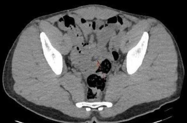
Figure 1: Bowel within bowel appearance on plain computed tomography (CT) image.
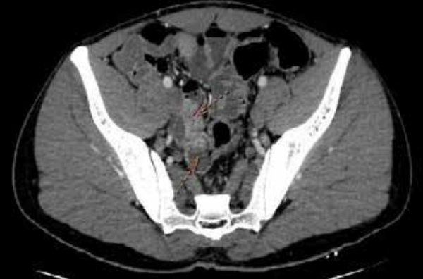
Figure 2: Bowel within bowel appearance on computed tomography (CT) angiography Image.
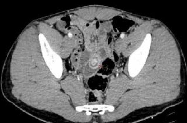
Figure 3: Hyperdense mass shown by the arrow at apex, suspicious for the neuroendocrine tumour or carcinoid tumour.
The patient was scheduled for surgery, and diagnostic laparoscopy confirmed ileoileal intussusception (Figure 4). Given the CT findings suggestive of a neuroendocrine tumour, a prophylactic laparoscopic appendectomy was performed. A mini laparotomy followed, with the affected bowel segment brought out (Figure 5). The segment was resected, and an ileoileal anastomosis was completed. The resected segment exhibited dimpling on the surface and an intussuscepted mass (Figures 6 and 7).
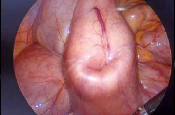
Figure 4: Laparoscopic view of intussusception with feeding vessel.
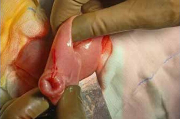
Figure 5: Bowel loop with intussuscepted segment.
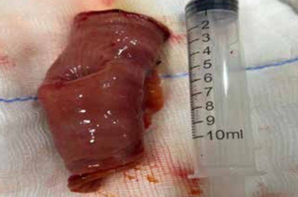
Figure 6: Dimpling on the surface of the resected bowel segment.
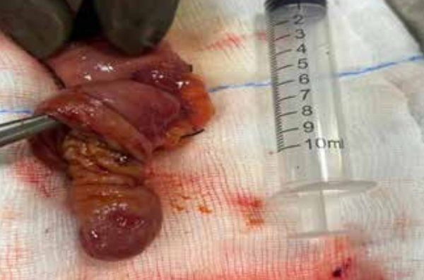
Figure 7: Intussuscepted mass with serosal erosion.
Post-operative recovery was uneventful, and the patient got discharged on post-operative day 5. The histopathological examination of the intussuscepted mass revealed the presence of pancreatic tissue (Figure 8), so a retrospective diagnosis of Meckel's diverticulum was made.
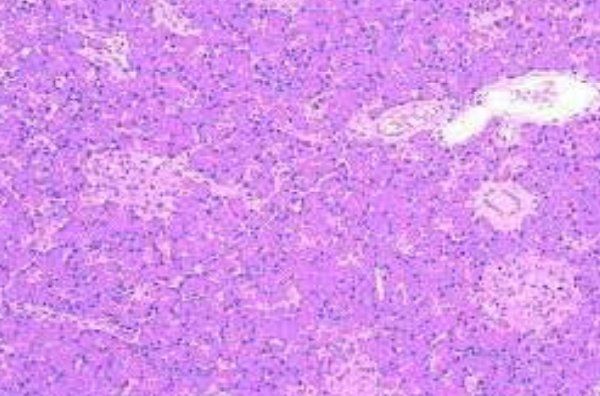
Figure 8: Intussuscepted mass with serosal erosion.
Discussion
Intussusception in adults
Intussusception, more common in children, is a rare cause of intestinal obstruction in adults, often presenting with nonspecific symptoms such as abdominal pain, nausea, and vomiting. In adults, intussusception is frequently secondary to a pathological lead point, such as a benign or malignant tumour.3 This case is unusual as the intussusception involved the ileum, which is less commonly affected compared to the colon in adult patients. The hyperdense lesion seen on CT angiography raised concern for a neuroendocrine tumour or carcinoid tumour, a common lead point for such a presentation in adults. The suspicion of a neuroendocrine tumour highlights the importance of advanced imaging techniques, such as CT and magnetic resonance imaging (MRI), in identifying atypical causes of intussusception and guiding clinical decision-making.
Neuroendocrine tumours and diagnostic challenges
Neuroendocrine tumours (NETs), including carcinoid tumours, are rare in the small intestine but are known to cause intussusception by acting as a lead point.4 These tumours often present with vague symptoms, including abdominal pain, diarrhoea, and weight loss, making early diagnosis difficult.3,5 The radiological features of a hyperdense lesion, as seen in this patient’s CT angiography, are suggestive of a NET or carcinoid tumour.6 Although the patient did not exhibit any clear clinical signs of a neuroendocrine tumour, the imaging findings were significant enough to warrant further surgical investigation.
Surgical management and outcomes
Given the suspicion of a NET, a prophylactic laparoscopic appendectomy was performed. The subsequent laparotomy, resection of the affected ileal segment, and ileoileal anastomosis were appropriate surgical interventions, ensuring the resolution of the intussusception and preventing recurrence. While the presence of pancreatic tissue did not alter the immediate surgical strategy, its discovery after resection emphasises the need for thorough histopathological examination to confirm the nature of any suspected masses in the bowel.
Conclusion
Although intussusception in adults due to inverted Meckel’s diverticulum is rare, awareness of this entity is essential. This case illustrates the diagnostic challenges of adult intussusception with an atypical lead point. The combination of advanced imaging modalities and surgical intervention was crucial in diagnosing and managing this rare condition. It highlights the need for a high degree of suspicion when encountering unusual radiological findings, as well as the importance of histopathological analysis in uncovering unexpected aetiologies.
Manmohan M Kamat, Sharvari Gorkar, Sakshi Parmar, Sai Sree, Rohan KJ. Rare Case of Inverted
Meckel's Diverticulum Causing Intussusception in Adults. MMJ. 2025, March. Vol 1 (5).
References
- Amin MU, Siddiqui MK, Mahmood R. Inverted Meckel’s diverticulum causing intussusception in an adult. J Coll Physicians Surg Pak. 2008;18(9):574-5.
- Kamel M, Barsoum H, Talaat S, et al. Inverted Meckel's diverticulum associated with heterotopic pancreatic tissue causing intestinal obstruction: a case report. Bull Natl Res Cent. 2021;45:1-4.
- Lee S, Cho SW. Adult Intussusception Caused by Inverted Meckel's Diverticulum Containing Mesenteric Heterotopic Pancreas and Smooth Muscle Bundles. J Pathol Transl Med. 2017;51(1):96-98.
- Baez-Giangreco A, Afzal M, Shakaki MA. Intussuscepted inverted meckel diverticulum: a case report. Saudi J Gastroenterol. 1997;3:99–102.
- Snyder LC. Meckel's diverticulum. In: Grosfeld JL, O’Neill AJ, Fonkalsrud WE, Coran GA, editors. Pediatric surgery. 6th Edition. Philadelphia: Mosby; 2006. pp. 865-903.
- Banli O, Karakoyun R, Altun H. Ileo-ileal intussusception due to inverted Meckel's diverticulum. Acta Chir Belg. 2009;109:516– 518.