 WhatsApp Us (24/7)
 +91 926 888 0303 (24/7)


Abstract:
Olecranon fractures are common fractures that are usually caused by direct trauma. It can result in complications such as infection, non-union, and bone loss which can significantly impact patient healing and recovery. This case report discusses the successful management of a 27-year-old male patient who presented with a postoperative infected non-union of the olecranon with associated osteoarticular bone loss. The patient was involved in an accident in which he sustained a shaft fracture of the right humerus, a fracture of the medial epicondyle of the right humerus, and a fracture of the right olecranon. He underwent open reduction and internal fixation (ORIF) surgery with plating of both the humerus, medial epicondyle, and tension band for the olecranon. However, complications arose, including infection and subsequent non-union, leading to a discharging sinus and significant bone loss from the olecranon. This was treated with dressing and antibiotics. He presented to our out-patient department (OPD) 6 months after his first surgery with two problems: postoperative osteomyelitis with non-union with osteochondral bone loss with failure of fixation of olecraonon and non-union of the humeral shaft fracture.
The Masquelet technique was employed in a staged manner to treat this complex olecranon fracture. The first stage involved thorough debridement, implant removal, anatomical plate fixation, and placement of bone cement. The second stage, performed after two months, involved removal of the bone cement and autologous bone grafting. Olecranon bone defect recostruction done using a tricortical iliac crest graft where the inner table of the iliac crest was contoured to mimic the articular surface of the olecranon. Three months postgrafting and fixation, the patient achieved union with a functional range of motion (0-130 degrees) and no signs of infection. This report highlights the effectiveness of the Masquelet technique in managing complex post-operative complications in orthopaedic trauma even in intra-articular fracture.
Key words: Infected Non-Union of Olecranon, Masquelet Technique, Osteoarticular Bone Loss.
Introduction
Olecranon fractures are common injuries often resulting from direct trauma. The standard treatment involves fixation using tension band wiring (TBW) and K-wires. However, complications such as infection, non-union, and bone loss can significantly impact patient outcomes.1,2 The management of infected nonunion with bone loss presents a unique challenge, requiring meticulous surgical planning and execution.2 Various forms of treatment have been de-scribed for the management of traumatic bone loss of olecranon like cancellous grafting, synthetic bone graft substitute, and prosthesis but in the case of infected non-union with chronic osteomyelitis with bone loss is a unique challenge.3,4
The masquelet technique, also known as the induced membrane technique, was first presented by a French orthopaedic surgeon, Dr. Alain C. Masquelet in 1986.2 He developed a 2-stage surgical technique for the treatment of segmental bone defects, particularly in the case of infections, open fractures, and trauma with significant bone loss.2
This case report details the management of a 27-year-old male with a post-operative infect-ed non-union of the olecranon, compounded by articular cartilage and bone loss.
Case report
Patient demographics and history: A 27-year-old male presented 6 months post-operatively with a discharging sinus at the site of an olecranon fracture that had initially been treated with TBW and K-wire fixation. The patient had an infection following the primary surgery, which was managed conservatively at that time. Over the subsequent 6 months, the patient developed non-union with significant osteoarticular bone loss (Figure 1).
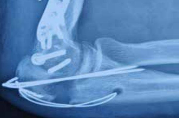
Figure 1: Infected non-union with osteoarticular bone loss.
Initial presentation: On examination, the patient exhibited signs of infection, including a discharging sinus, and there was clinical and radiological evidence of non-union with associ-ated bone loss at the olecranon. There was also a non-union of the humeral shaft fracture.
Investigations: Following investigations were performed:
- Radiographs: Demonstrated non-union with significant bone and cartilage loss.
- Blood tests: Elevated inflammatory markers indicating ongoing infection.
- Microbiological cultures: Positive for bacterial growth, confirming infection.
Treatment
Stage 1:
Masquelet technique: Initial surgery
Procedure:
- The patient underwent thorough debridement to remove all infected and necrotic tis-sue.
- The previous TBW and K-wire implants were removed.
- Anatomical plate fixation was performed to stabilise the fracture site.
- Bone cement impregnated with antibiotics (vancomycin with gentamicin) was placed at the defect site to fill the bone, and help avoid and manage infection.
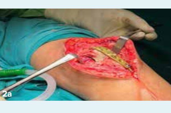
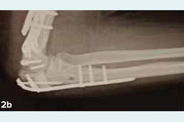
Figure 2: Stage 1 intra-operative clinical image; 2b: post-operative X-ray
Post-operative care: The patient was closely monitored, with regular wound checks and administration of targeted intravenous antibiotics based on culture sensitivity (Figure 2).
Stage 2:
Masquelet technique: Initial surgery
Procedure:
- Two months after the initial surgery, the bone cement was removed.
- Autologous bone grafting was performed to fill the defect.
- A similar size and shape bone graft taken from iliac crust (Figures 3 and 4).
- He also underwent plating with bone grafting for non-union of humerus fracture.
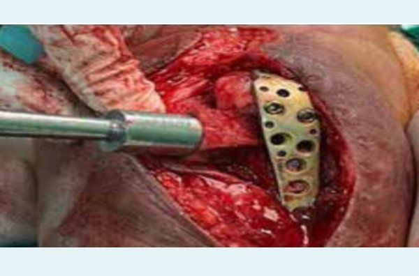
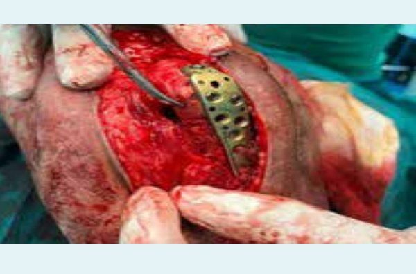
Figure 3 and 4: Intraoperative images of bone grafting.
Post-operative care: Post-operative X-ray has been shown in Figure 5. Continued monitoring for signs of infection and promoting fracture healing through physiotherapy.
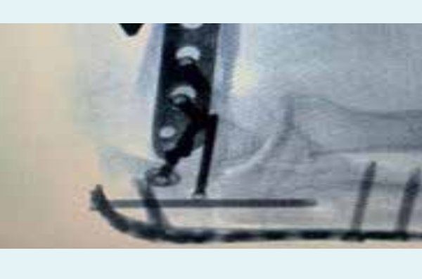
Figure 5: Post-operative X-ray (stage 2).
Outcome and follow-up: Two months post-surgery, signs of union were seen (Figure 6). Three months after the second surgery, radiographs confirmed the successful union of the olecra-non. The patient demonstrated a functional range of motion, with full extension and flexion to 130 degrees. Importantly, there were no clinical signs of infection, and the patient re-turned to daily activities with minimal discomfort.
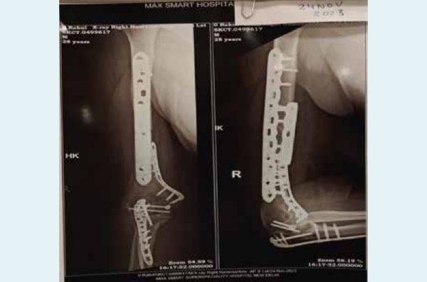
Figure 6: Two months post-surgery showing signs of union.
Discussion
The Masquelet technique is particularly advantageous in managing complex cases of infected non-union with bone loss, as seen in this patient.2 The staged approach allows for infection control and the creation of a biologically active membrane that supports subsequent bone graft incorporation and healing.1,3 In this case, the anatomical plate fixation provided the necessary stability, and the use of bone cement in the first stage effectively managed the infection.
Fibrocartilage formation is a common concern after intraarticular fixation, especially when there is significant cartilage damage. In this case, the combination of staged surgery and bone grafting mitigated the risk of fibrocartilage dominating the repair site, leading to a more favourable functional outcome. This case underscores the importance of a thorough and staged approach in managing post-operative complications, particularly in the presence of infection and bone loss. The successful outcome highlights the Masquelet technique as a viable option in similar cases.
Consent
The patient has provided informed consent for the publication of this case report, acknowledging their understanding of the content and the potential implications of sharing their medical information.
Competing interests
The author(s) declare that they have no competing interests
Conclusion
The Masquelet technique offers a robust solution for treating infected non-unions with significant bone loss.2 This case demonstrates the technique's effectiveness in achieving union and restoring function in a young patient with an olecranon non-union complicated by infection and bone loss. With careful surgical planning and execution, even challenging cases can achieve successful outcomes.
Harshavardhan Hegde, Akshay Kumar Saxena, Ajay Dhanopeya. Management of Post-Operative Infected
Non-Union with Osteoarticular Bone Loss of the Olecranon in a 27-Year-Old Male Using the Masquelet
Technique. MMJ. 2025, March. Vol 1 (5).
References
- Masquelet AC, Begue T. The concept of induced membrane for reconstruction of long bone defects. Orthopedic Clinics. 2010;41(1):27-37.
- Giannoudis PV, Faour O, Goff T, et al. Masquelet technique for the treatment of bone defects: tips-tricks and future directions. Injury. 2011;42(6):591-598.
- Claes L, Recknagel S, Ignatius A. Fracture healing under healthy and inflammatory conditions. Nat Rev Rheumatol. 2012;8(3):133-143.
- Morris R, Hossain M, Evans A, et al. Induced membrane technique for treating tibial defects gives mixed results. The bone & Joint Journal. 2017;99(5):680-685.