 WhatsApp Us (24/7)
 +91 926 888 0303 (24/7)


Abstract:
Neurosarcoidosis is a rare manifestation of systemic sarcoidosis, with involvement of the pituitary– hypothalamic axis being particularly uncommon and often posing a diagnostic challenge due to its close resemblance to other sellar and suprasellar lesions, especially pituitary adenomas. We describe a case of a middle-aged patient who presented with headache, nausea, fever, anorexia, and hypothyroidism. Magnetic resonance imaging (MRI) revealed a large, lobulated sellar and suprasellar lesion that appeared isointense on both T1- and T2-weighted images with heterogeneous enhancement. Notably, there was interval development of significant infundibular stalk thickening measuring 8.5 mm, while the posterior pituitary bright spot was preserved. Further evaluation with multimodality imaging, including positron emission tomography–computed tomography (PET–CT) and ultrasound, demonstrated metabolically active cervical lymphadenopathy, hepatic involvement, and pulmonary ground-glass opacities. These systemic findings, in conjunction with histopathological confirmation, established the diagnosis of neurosarcoidosis. This case underscores the importance of a comprehensive multimodality imaging approach in distinguishing neurosarcoidosis from primary pituitary neoplasms. Recognition of key imaging features such as infundibular stalk thickening, along with evidence of systemic granulomatous disease, is essential for accurate and timely diagnosis, facilitating appropriate management and preventing misdiagnosis.
Key words: Neurosarcoidosis, Pituitary Gland, Infundibulum, Magnetic Resonance Imaging.
Introduction
Sarcoidosis is an idiopathic, multisystem granulomatous disorder characterised by non-caseating granulomas that most commonly affect the lungs and lymphatic system. 1 Central nervous system (CNS) involvement, termed neurosarcoidosis, is histologically present in up to 25% of systemic cases in postmortem studies, though it is clinically symptomatic in only about 5% of patients. 1 Isolated neurosarcoidosis without systemic disease is extremely rare, accounting for less than 1% of cases. 1,2
Imaging evidence of CNS involvement is detected in nearly 10% of systemic cases. 1 Magnetic resonance imaging (MRI) is the modality of choice for assessing parenchymal, leptomeningeal, and hypothalamic– pituitary involvement. 1,3 Pituitary or infundibular involvement may manifest as cystic or solid enhancing lesions or thickening of the pituitary stalk, often mimicking neoplastic conditions like pituitary adenoma, particularly in the sellar and suprasellar regions. 1,4
Case Report
Case presentation
A middle-aged patient presented with a history of headache, nausea, fever, loss of appetite, and endocrine dysfunction (hypothyroidism). 1,2
Imaging findings
MRI of the brain revealed a large, oval, lobulated lesion centred in the sellar and suprasellar region. The lesion appeared isointense on T1- and T2-weighted images and demonstrated inhomogeneous post-contrast enhancement (Figure 1A–D). 1 The lesion abutted the optic chiasm with minimal associated oedema and showed minor extension into the right cavernous sinus.3,4 A key finding was the interval development of infundibular stalk thickening, measuring 8.5 mm (Figure 1B), which was not present on a prior MRI dated two months earlier (Figure 2A and B). 1 The posterior pituitary bright spot was normal in location and signal on T1-weighted images (Figure 1A). 1,4
MRI findings:
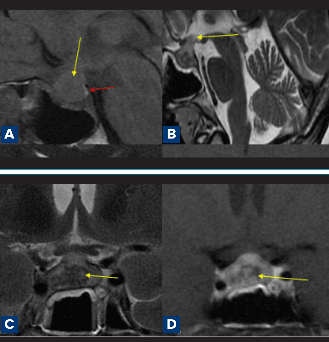
Figure 1A–D: Brain magnetic resonance imaging (MRI) demonstrates a large, oval, lobulated lesion centred in the sellar and suprasellar region, appearing isointense on both T1- weighted and T2-weighted sequences, with inhomogeneous post–contrast enhancement: A. Red arrow - Preserved posterior pituitary bright spot in its normal anatomical location on T1- weighted imaging; B. Marked thickening of the infundibular stalk, measuring approximately 8.5 mm; C. Coronal post-contrast image demonstrating heterogeneous enhancement of the sellar–suprasellar lesion with extension towards the optic chiasm; D. Sagittal post-contrast image showing superior extension of the lesion with mild mass effect on the optic apparatus.
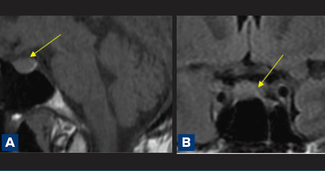
Figure 2A and B: Comparison with imaging performed two months earlier demonstrates a mild superior convexity of the pituitary gland. The gland appears normal in size and signal intensity, with no evidence of stalk thickening at that time.
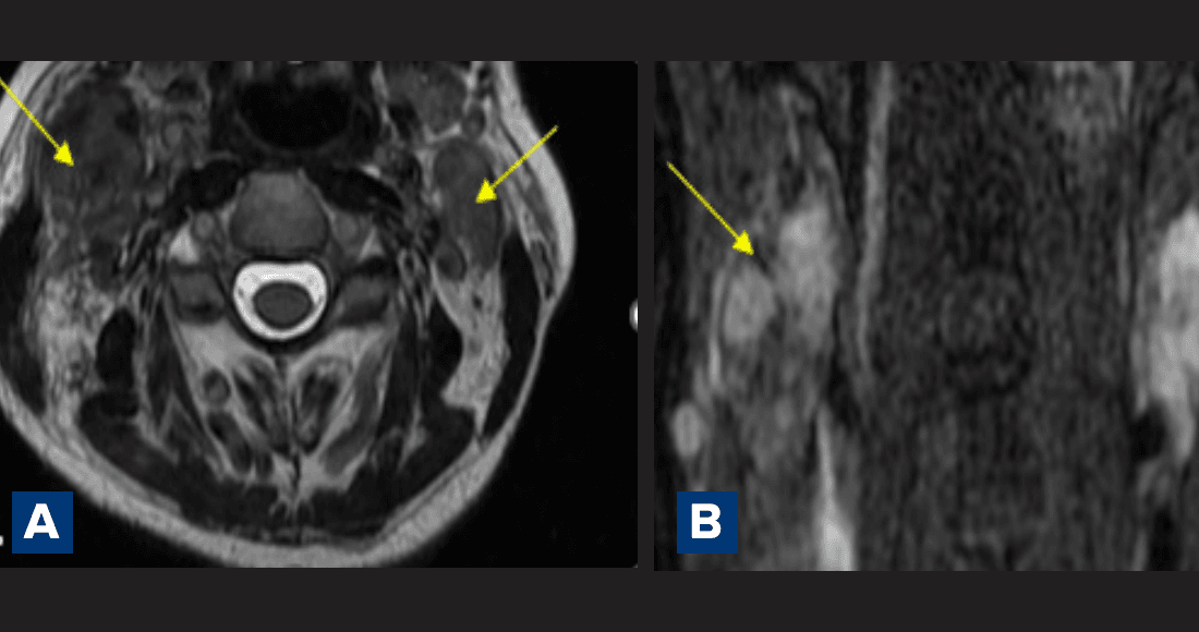
Figure 3A and B : Imaging reveals multiple enlarged, partly necrotic cervical lymph nodes
Cervical screening: Further screening revealed multiple enlarged, partly necrotic cervical lymph nodes on MRI (Figure 3A and B).2
CT/PET–CT findings: Positron emission tomography–computed tomography (PET–CT) confirmed metabolically active cervical lymphadenopathy (levels II–V), peripherally enhancing hepatic lesions in segments II, III, VII, and VIII, and pulmonary abnormalities including interlobular septal thickening and ground-glass opacities (Figures 4 and 5).
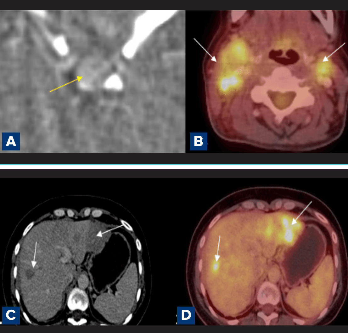
Figure 4: A. Computed tomography (CT) imagining showing an inhomogeneously enhancing lesion replacing the pituitary gland, with minimal suprasellar extension; B. Positron emission tomography–computed tomography (PET–CT) neck showing metabolically active conglomerate cervical lymph nodes, involving nodal levels II–V on the right and level II on the left; C, D. PET–CT liver showing multiple peripherally enhancing, metabolically active lesions involving hepatic segments II, III, VII, and VIII.
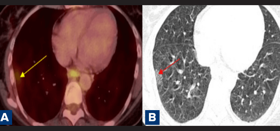
Figure 5A and B: Positron emission tomography–computed tomography (PET–CT) lungs demonstrating patchy ground-glass attenuation with interlobular septal thickening in the superior segment of the right lower lobe.
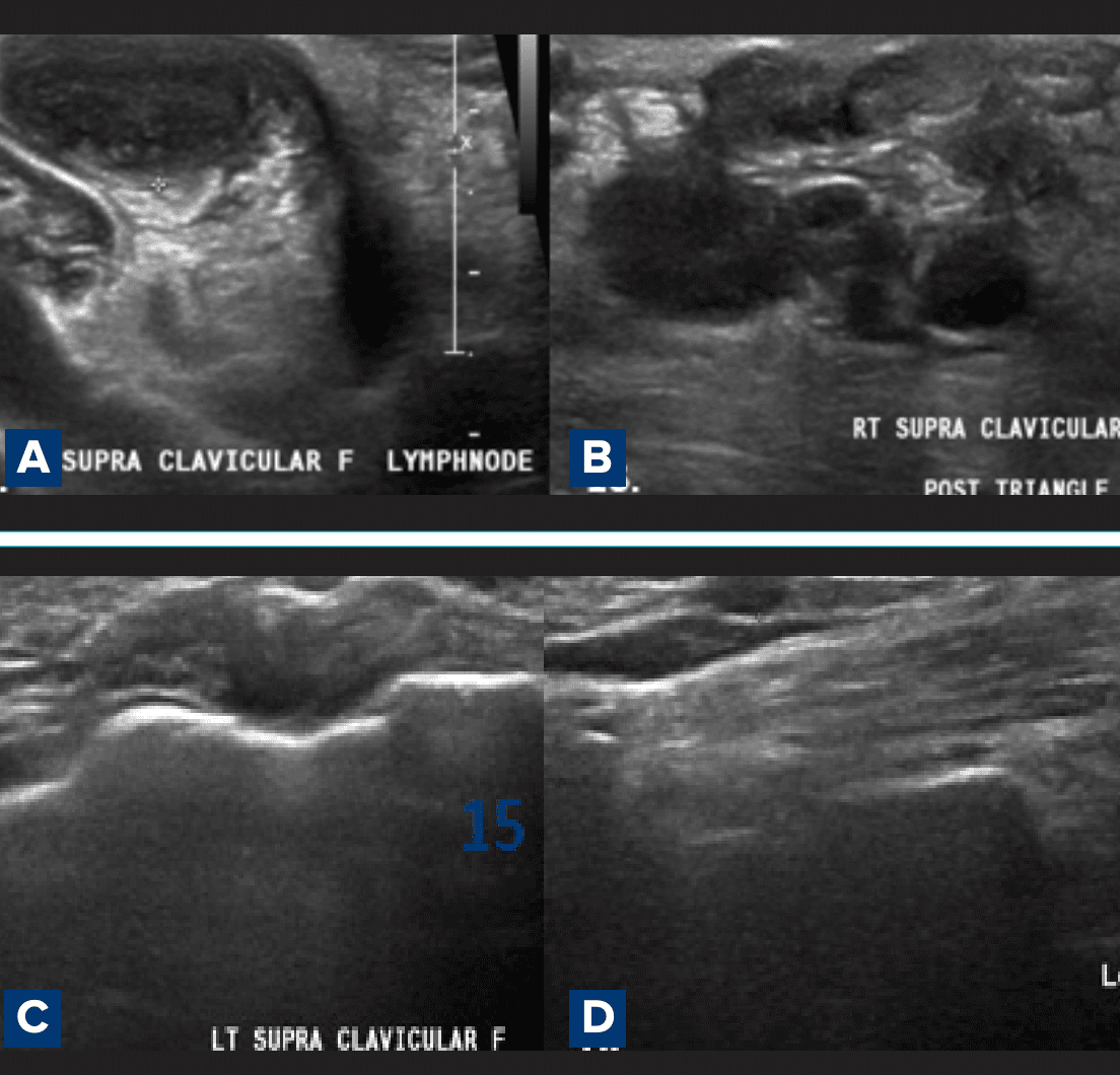
Figure 6A–D: Ultrasound of the neck corroborates the presence of multiple conglomerate lymph nodes in the right posterior triangle and supraclavicular region, with some showing partial central necrosis.
USG neck findings: Ultrasound of the neck corroborated these findings, showing conglomerate nodes with partial central necrosis (Figure 6A–D). 3 These multimodality findings, combined with systemic evaluation and histopathological confirmation, were consistent with neurosarcoidosis involving the pituitary gland.
Discussion
Neurosarcoidosis of the hypothalamic-pituitary axis is rare and often diagnostically challenging. 1,2 In this case, the preservation of the posterior pituitary "bright spot" suggested sparing of the neurohypophysis, while the rapid interval thickening of the infundibular stalk served as a primary clue for a granulomatous process rather than a typical pituitary adenoma. 2 The differential diagnosis includes lymphocytic hypophysitis, lymphoma, metastasis, and germinoma. 2
A multimodality approach was pivotal. 1,3 While MRI remains the gold standard for detailed CNS evaluation, PET–CT and ultrasound were essential for assessing the systemic disease burden and identifying accessible targets for tissue sampling. 1,4 This case highlights that integrating characteristic imaging patterns across modalities facilitates the differentiation of neurosarcoidosis from neoplastic conditions, enabling the timely initiation of systemic therapy. 3,4
Acknowledgements
I, Dr. Mohit H. Tamakuwala, would like to acknowledge Dr. Deepak Patkar (H.O.D, Department of Radiology) and Dr. Mitusha Verma (Consultant Radiologist) at Nanavati Max Super Speciality Hospital, Mumbai, for their guidance. We also thank the patient and family for their consent.
Conclusion
Neurosarcoidosis is a critical differential diagnosis for infiltrative sellar–suprasellar lesions, especially when accompanied by pituitary stalk thickening.1,3 This case underscores the importance of a comprehensive imaging strategy — utilising MRI, PET–CT, CT, and ultrasound — to establish a diagnosis of systemic sarcoidosis and avoid unnecessary surgical intervention for suspected primary pituitary neoplasms.3,4
Mohit Tamakuwala, Mitusha verma, Deepak Patkar. Beyond Adenoma: Imaging Clues to Pituitary
Involvement in Systemic Sarcoidosis. MMJ. 2026, June. Vol 3 (2).
References
- Smith JK, Matheus MG, Castillo M. Imaging manifestations of neurosarcoidosis. AJR Am J Roentgenol. 2004;182(2):289–95.
- Samuelsson K, Stiernstedt G, Söderman M. Neurosarcoidosis can occur without any other signs of sarcoidosis. Lakartidningen. 1992;89(38):3052–5.
- Ginat DT, Dhillon G, Almast J. Magnetic resonance imaging of neurosarcoidosis. J Clin Imaging Sci. 2011;1:15.
- Arle JE, Judkins AR, Kotapka MJ. Neurosarcoidosis presenting in the pituitary gland with normal endocrine studies. Skull Base Surg. 1999;9(2):149–53.
- Al-Badri SG, Duhan A, Al-Taie RH, et al. Hypothalamic Neurosarcoidosis With Multisystem Involvement: Case Report and Literature Review. Clin Case Rep. 2025;13(10):e71106.