 WhatsApp Us (24/7)
 +91 926 888 0303 (24/7)


Abstract: An uncommon class of illnesses known as mastocytosis is defined by aberrant mast cells in different tissues or organs. It encompasses mast cell sarcoma, systemic mastocytosis, and cutaneous mastocytosis. For those without advanced manifestations of the disease, the diagnosis of Systemic Mastocytosis can be protracted due to the high rate of symptom overlap with other illnesses. A 56-year-old female patient was brought to our department for a diagnosis because of her history of eosinophilia, chronic colitis, and skin allergies. The patient's eosinophilia remained in spite of treatment. The patient's blood count revealed persistent eosinophilia even after five months. An examination of the bone marrow aspirate showed a hypercellular bone marrow with a noticeable eosinophilic prominence. To our astonishment, the trephine biopsy showed sheets and nodules of mast cells, expressing strong positivity for CD117 immunostain. A review of the colonic biopsy also showed a significant mast cell presence as highlighted by CD117 immunostain. A definitive diagnosis of systemic mastocytosis was reached, indicating that, in situations of eosinophilia, a low index of suspicion and meticulous morphological examination are necessary to prevent delays in the diagnosis of mastocytosis, particularly in cases where there is no cutaneous involvement.
Key words: Systemic Mastocytosis, Eosinophilia, Mast Cells
Introduction
Mastocytosis is one of the greatest masquerades with varied symptomatology, thus, imposing a big diagnostic challenge. This disease entity comprises a rare group of heterogeneous diseases characterized by the accumulation of abnormal mast cells in various organs or tissues and includes cutaneous mastocytosis (CM), systemic mastocytosis (SM) with its various clinic-pathological subtypes, and mast cell sarcoma.1 Systemic mastocytosis includes bone marrow mastocytosis (BMM), Indolent systemic mastocytosis (ISM), Smouldering systemic mastocytosis (SSM), Aggressive systemic mastocytosis (ASM), Systemic mastocytosis with an associated hematological neoplasm and mast cell leukemia. Despite advancements in diagnosis of this rare disorder, the diagnostic journey for SM patients is often long, especially for those who do not have an advanced form of the disease for which diagnosis can take years. Even in cases of advanced SM, diagnosis can take up to 2 years as the presenting symptoms have a considerable overlap with other diseases. Herein we report a case of a 56-year-old female who was referred to our department with a long-standing history of skin allergy and chronic colitis, and persistent eosinophilia, to exclude an eosinophilic myeloproliferative neoplasm that was subsequently diagnosed as Systemic Mastocytosis.
History
Bone marrow samples (aspirate and biopsy) was received for a 56-year-old female to evaluate persistent eosinophilia, despite treatment. A clinical diagnosis of hyper-eosinophilia/eosinophilic myeloproliferative neoplasm had been entertained. Interestingly, the lady had presented nearly six months earlier with significant gastro-intestinal symptoms, having multiple episodes of loose stools for last three years with bleeding per rectum for two weeks associated with pain in abdomen and intermittent vomiting. She also had a history of skin allergy for 8-9 years and weight loss of 7 to 8 kgs. Per-rectal examination was normal, however, colposcopy was suggestive of colitis with rectal sparing. Routine investigations revealed a C-Reactive Protein (CRP) of 7.18 mg/L, negative tissue trans-glutaminase antibody (IgA) with complete blood count (CBC) showing hemoglobin of 11g/dL, raised total leukocyte count (TLC) of 11,200/uL, platelet count of 2,87,000/uL and absolute eosinophilia (eosinophils-15%; absolute eosinophil count = 1680/☐l). Urine routine examination and kidney function test were normal. Liver function tests showed increased alkaline phosphatase (135 U/L ) and mild hypoalbuminemia (albumin-3.7gm/dL). Urine and blood culture showed no growth. Colonoscopic biopsy was done which showed chronic active colitis with marked prominence of eosinophils (>100/hpf) and no evidence of granuloma or malignancy (Figures 1 A and B). GeneXpert® Mycobacterium complex PCR and CMV DNA PCR on colonic biopsy were negative. Biofire® GI PCR confirmed the presence of Giardia Lambia. Based on this, the patient was treated with intravenous(IV) proton pump inhibitors (PPIs), antibiotics, IV fluids and other supportive measures.
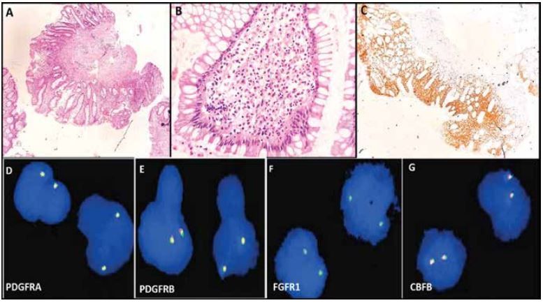
Figure 1: A. H & E stained sections revealed fragments of colonic mucosa with mild distortion of glandular epithelium with surface ulceration and granulation tissue (100X). B. Lamina shows dense inflammatory infiltrate consisting of sheets of eosinophils (>100/hpf) along with neutrophils (H&E;400X) C. Immunohistochemistry for CD117 highlighted clusters of mast cells in lamina propria(100X) D. PDGFRA gene (4q12) break apart Probe, BioMolds Pvt. Ltd -Negative E. PDGFRB gene (5q32) break apart Probe, BioMolds Pvt. Ltd- Neative F. Zytolight Spec FGFR1 dual colour breakapart probe- Negative G. Zytolight SPEC CBFB dual colour breakapart probe- Negative
Differential diagnosis and follow-up
The patient showed good response to the medication, but the eosinophilia remained. Six months later, a CBC revealed a platelet count of 3,22,000/uL, a TLC of 9340/uL, and hemoglobin of 13.5 gm/dL. Differential leukocyte count (DLC) showed: Neutrophils 55%, lymphocytes 27%, monocytes 06% and eosinophils 14%. Differential diagnoses included gastrointestinal conditions such as eosinophilic enteropathy/ inflammatory bowel disease; allergic conditions; drug reactions hyper-eosinophilic syndrome or underlying any other hematological neoplasm. For additional assessment, a bone marrow aspirate and biopsy were conducted on the patient after a re-evaluation.
Diagnostic work-up
Bone marrow aspirate examination showed a hyper-cellular bone marrow with granulocytic hyperplasia, marked eosinophilic prominence and 4% blasts (Figure 2 A-C). Only a few mast cells were noted. Myelogram showed: blasts 04%, myelocytes 14%, metamyelocytes 04%, neutrophils 40%, eosinophils 20%, lymphocytes 06%, mast cells 02% and erythroid precursors 10%.
Bone marrow biopsy was markedly hypercellular (~80-90% cellularity) and to our surprise, was remarkable for presence of nodular aggregates and sheets of mast cells (as highlighted by CD117 immunostain; Figure 2 K, L) with many of them having spindled morphology (Figure 2 F, G). In focal areas central core of lymphoid cells with encircled mast cells were also seen giving it a characteristic ‘’coin lesions”. In all the lesions areas, there was profound prominence of eosinophils and its precursors (Figure 2 D-G). Focal perivascular streaming of spindled mast cells, focal increased osteoclastic and osteoblastic activity were seen. Preserved areas of trilineage hematopoiesis were also identified comprising predominantly of granulocytic cells showing adequate sequential maturation and abundant megakaryocytes with dysplasia. CD34 immunostain showed ~4% blasts with no blast clusters. Immunohistochemistry for CD2, CD3 and CD20 highlighted scattered B and T cells. CD68 immunostain was positive on histiocytes and few mast cells. CD2 immunostain was negative on mast cells (Figure 2 H-L). CD30 immunostain also did not highlight any cell in the sections examined.
Review of colonic biopsy was done which also revealed clusters of CD117 positive mast cells as well as many eosinophils in the lamina propria (Figure 1C). FISH eosinophilic panel (for PDGFRA, PDGFRB, FGFR1 and CBFB gene rearrangement) was negative (Figure 1D-G). A final diagnosis of “Systemic Mastocytosis” was made, however the patient was lost to follow up after the diagnosis.
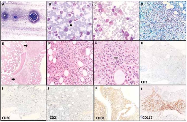
Figure 1: A. Hypercellular bone marrow aspirate (Wright Geimsa ; Scanner) B. Mast cells (Arrowhead; 1000X) C. Prominence of eosinophils in aspirate smear(Wright Geimsa ; 1000X) D. Hypercellular bone marrow biopsy showing prominence of eosinophils highlighted by Geimsa stain on trephine biopsy examined (H &E; 400X) E. Trephine biopsy showing sheets and nodules of mast cell appearing as “coin” lesions ( H & E; 200X) F. & G. Atypical spindled morphology of mast cells (thick arrow) along with eosinophilia (thin arrow; H& E 400X) H. CD3 immunostain highlighted scattered T cells I. Few scattered B cells seen as evidenced by CD20 immunostain however no definite lymphoid aggregate noted. J. CD2 immunostain was negative on mast cells however, highlighted T cells as similar as CD3 immunostain. K. CD68 immunostain positive on some mast cells along with highlighting histiocytes in the background. L. Strong positivity of CD117 immunostain seen highlighting mast cells
Discussion
Many of the symptoms of systemic mastocytosis are non-specific in nature. To enable a suitable investigation, a low index of suspicion is necessary.2 Years lost before diagnosis are particularly significant in cases when there is no cutaneous involvement, especially in patients with an aggressive form of the illness.3,4 According to current WHO criteria1 , one major and one minor or three minor criteria should be present, to diagnose the disorder.
Major
- Multifocal dense infiltrates of mast cells (≥ 15 mast cells in aggregates) detected in sections of bone marrow and/or other extracutaneous organ(s)
Minor
- Atypical mast cell morphology, including spindle shape or immature morphology, present in > 25% of all mast cells on bone marrow smears or in other extracutaneous organ(s)
- Mast cells aberrantly express one or more of the following antigens: CD2, CD25, CD30
- KIT p.D816V mutation or other activating KIT mutationb detected in peripheral blood, bone marrow, or other extracutaneous organ(s)
- Baseline serum tryptase concentration of > 20 ng/mL in the absence of an associated myeloid neoplasm; in the case of a known HαT, the tryptase level could be adjusted
The classification of systemic mastocytosis (SM) incorporates molecular and clinical features (B and C findings) along with morphological, immunohistochemical and bicochemical parameters. In our patient, one major criterion (multifocal dense infiltrates of mast cells in bone marrow sections, in bone marrow and gastric biopsy) and one minor criterion (greater than 25% of mast cells being spindle shaped ) were present. Together with SM criteria fulfilled and atleast one C findings (gastrointestinal symptoms: Malabsorption with hypoalbuminaemia and weight loss), our case best fitted with Aggressive Systemic Mastocytosis (ASM).1
CONCLUSION:
This case stresses the need of careful morphological examination and to investigate for mastocytosis in patients especially in the presence of longstanding skin allergy and prominent eosinophilia, even with the co-existence of parasitic infection and allergic sensitization. In our case, the bone marrow aspiration showed only eosinophilic prominence with only a few mast cells however trephine biopsy examination clinched the diagnosis.
Acknowledgments:
None
Funding source:
None
Conflict of interest:
None
References
- Hochhaus A etal. Mastocytosis. In: WHO Classification of Tumours Editorial Board. Haematolymphoid tumours [Internet]. Lyon (France): International Agency for Research on Cancer; 2024 (WHO classification of tumours series, 5th ed.; vol. 11). Available from: https://tumourclassification.iarc.who.int.
- Nallamilli S, O’Neill A, Wilson A, Sekhar M, Lambert J. Systemic mastocytosis: variable manifestations can lead to a challenging diagnostic process. BMJ Case Reports CP. 2019 Aug 1;12(8):e229967.
- Butterfield JH. Systemic mastocytosis: clinical manifestations and differential diagnosis. Immunology and Allergy Clinics. 2006 Aug 1;26(3):487-513. doi: 10.1016/j.iac.2006.05.006.
- Rama TA, Martins D, Gomes N, Pinheiro J, Nogueira A, Delgado L et al. Case report: Mastocytosis: The long road to diagnosis. Frontiers in Immunology. 2021 Feb 12;12:635909. doi: 10.3389/ fimmu.2021.635909. PMID: 33643319; PMCID: PMC7907169.