 WhatsApp Us (24/7)
 +91 926 888 0303 (24/7)


Abstract:
Fat necrosis is a non-malignant breast condition that can closely mimic cancer recurrence on imaging, especially in individuals with a prior history of breast cancer and reconstructive procedures. We report the case of a 34-year-old woman with a history of left breast cancer who developed a new palpable breast mass after undergoing mastectomy and reconstruction. Multimodal imaging — including mammography, ultrasound, magnetic resonance imaging (MRI) and positron emission tomography (PET)/computed tomography (CT) — demonstrated features that raised concern for possible malignancy. A definitive diagnosis of fat necrosis was confirmed through histopathological analysis of a biopsy sample.
This case underscores the importance of correlating imaging findings with tissue diagnosis to accurately differentiate benign post-surgical changes from true cancer recurrence, thereby avoiding unnecessary treatments or procedures.
Key words: Fat Necrosis, Recurrent Breast Carcinoma, Breast Imaging, Post-Surgical Changes.
Introduction
Fat necrosis is a benign, non-suppurative inflammatory process of adipose tissue. Accurate recognition is important, as its imaging characteristics can closely resemble those of breast carcinoma.1
Fat necrosis of the breast most commonly occurs following trauma, surgery, or radiotherapy. Less common causes include anticoagulation, infection, or idiopathic origin. Other rare causes include polyarteritis nodosa, WeberChristian disease, and granulomatous angiopanniculitis. In some patients, the cause of fat necrosis remains unknown.1-3
The imaging manifestations of fat necrosis on mammography, ultrasonography, and magnetic resonance imaging (MRI) vary depending on the stage of evolution, which has important clinical implications as certain appearances can be virtually indistinguishable from malignancy, often necessitating biopsy for definitive diagnosis.1-4
Furthermore, fluorodeoxyglucose (FDG) positron emission tomography (PET)/computed tomography (CT) can misinterpret fat necrosis as malignancy due to its metabolic activity, resulting in false-positive findings and physicians must be aware of this as a potential pitfall.5,6
In this report, we present a case of a 34-year-old female with a history of breast cancer who underwent mastectomy and reconstruction surgery and subsequently presented with a breast lump, raising concern for recurrence. In this case, ultrasound, mammography, and PET/CT demonstrated features that were suspicious of malignancy.
Case Report
A 34-year-old female presented with a new, painless lump in the left breast. She had a history of left breast cancer (invasive ductal carcinoma [IDC], grade 2 with extensive ductal carcinoma in situ [DCIS], hormone receptor [HR] negative, and human epidermal growth factor receptor 2 [HER -2] positive) — eight months prior to this presentation. She had undergone left mastectomy and sentinel lymph node biopsy (SLNB) and was managed with chemotherapy (paclitaxel) and HER2-targeted therapy (trastuzumab). She subsequently underwent deep inferior epigastric perforator (DIEP) breast reconstruction.
On examination, there was irregular thickening at 12 to 1 o’clock. No palpable axillary lymph nodes were noted.
She underwent several imaging investigations, including mammography, ultrasound, MRI and PET/CT examinations, which are described below (Figure 1–4).
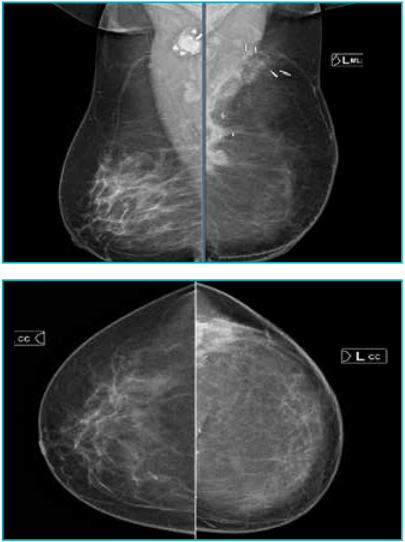
Figure 1: Mediolateral oblique (MLO) and craniocaudal mammographic images of both breasts demonstrate a reconstructed left breast with irregular isodense areas and dystrophic calcifications seen superolaterally along the flap. Surgical clips are noted.
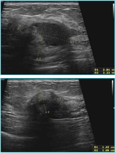
Figure 2: Ultrasound images show multiple heteroechoic areas, some demonstrating posterior shadowing, along the lateral and superior margin of the flap in the reconstructed left breast. The largest lesion measures approximately 2.8 cm x 2.2 cm.
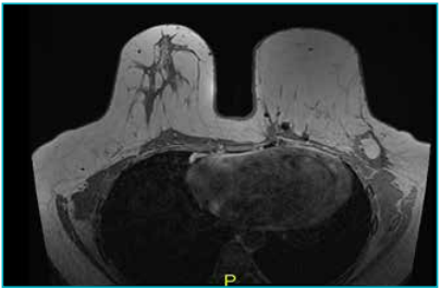
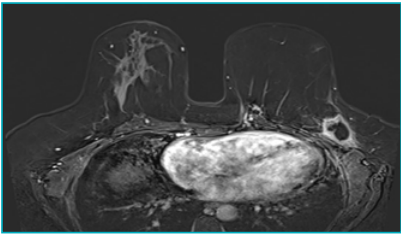
Figure 3: MRI images showing multiple coalescent lesions with central fat signal intensity and a thick, irregular peripheral rim enhancement seen along the posterolateral margin of the flap in the reconstructed left breast, the largest measuring approximately 30 mm x 27 mm. Mild surrounding parenchymal oedema pattern, skin thickening, and architectural distortion are also noted.
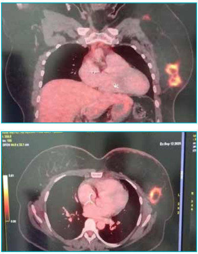
Figure 4: PET/CT images show metabolically active, multiple coalescent and discrete nodular enhancing soft-tissue lesions along the lateral and superior margins of the flap, with areas of fat density within the reconstructed left breast. The largest lesion measures approximately 3.1 cm x 2.5 cm, suggestive of inflammation versus tumour recurrence. Clinical and histopathological evaluation was advised. Low-grade metabolically active and inactive, left level I axillary lymph nodes are also seen, the largest measuring approximately 9 mm x 7 mm, likely inflammatory in nature. No other significant metabolically active lesion was identified in the body surveyed.
Core needle biopsy of the left breast lesion was performed. Histopathology findings revealed fat necrosis. Core needle biopsy of the left axillary node revealed benign lymphoid tissue.
Discussion
Fat necrosis often presents as a palpable mass and can raise suspicion for breast carcinoma, especially in patients with a history of breast cancer or surgery.7 Our patient had a history of mastectomy and reconstructive surgery following breast cancer diagnosis and presented with a palpable mass, which raised suspicions for recurrence.
Ultrasound findings in fat necrosis are variable and depend on the stage of the lesion. In the early phase, fat necrosis may appear as a hypoechoic or complex cystic mass with indistinct margins and posterior acoustic shadowing. These features have been reported in association with breast cancer and were also observed in our case.8
On mammography, fat necrosis can present with a wide spectrum of appearances, ranging from benign-appearing oil cysts with or without rim calcifications to suspicious spiculated masses or focal asymmetries. In the early stages, the small foci of calcification in the wall of an oil cyst appear similar to fine microcalcifications and need differentiation from the disease process.4
In our patient, the mammographic findings included a high-density, irregular mass with architectural distortion — features that closely mimic invasive carcinoma. This underscores the well-documented potential of fat necrosis to imitate malignancy on conventional imaging, particularly when classical features such as rim calcifications or fat lucencies are absent.
Fat necrosis produces a wide spectrum of findings on MRI. The MRI appearance of fat necrosis may be indistinguishable from that of malignancy and can mimic tumour recurrence after breast conservation therapy.4
MRI can offer valuable diagnostic information in complex or equivocal cases, particularly when conventional imaging raises suspicion for malignancy. In our case, MRI of the left breast revealed a reconstructed flap with multiple coalescent lesions along the posterolateral margin. These lesions demonstrated central fat signal intensity with thick, irregular peripheral rim enhancement, associated with mild surrounding parenchymal oedema, skin thickening, and architectural distortion. These findings closely mimicked a malignant process, particularly due to the irregular rim enhancement and surrounding tissue changes. However, the presence of a central fat signal was an important clue pointing toward a benign aetiology, such as fat necrosis.
PET/ CT is commonly employed in the surveillance of breast cancer patients, particularly post-surgical or post-reconstructive cases, to evaluate for recurrence or metastatic disease. However, fat necrosis can exhibit increased FDG uptake, especially when associated with active inflammation, further complicating differentiation from malignancy.6
In our case, PET/CT showed metabolically active nodular soft tissue lesions along the lateral and superior margins of the reconstructed left breast flap, raising the possibility of either an inflammatory process or recurrent malignancy. The FDG avidity, combined with the morphological features on mammography and ultrasound, heightened clinical concern for tumour recurrence, especially in a post-surgical breast where architectural distortion is common, therefore necessitating a histological diagnosis.
Conclusion
Fat necrosis of the breast is a benign condition that can mimic malignancy across all imaging modalities, especially in post-surgical or reconstructed breasts. This case underscores the need for a broad differential diagnosis, clinical correlation, and, when imaging is inconclusive, timely histopathologic confirmation to guide appropriate management and avoid unnecessary interventions.
Flora A. Lwakatare, Eeta Jain, Sylvia Nsato, Richa Bansal, Bharat Aggarwal. Imaging Pitfalls: A Case of Fat Necrosis Masquerading as Recurrent Breast Carcinoma. MMJ. 2025, December. Vol 2 (4).
References
-
Kerridge WD, Kryvenko ON, Thompson A, et al. Fat necrosis of the breast: a pictorial review of the mammographic, ultrasound, CT, and MRI findings with histopathologic correlation. Radiol Res Pract. 2015(1):613139.
-
Bilgen IG, Ustun EE, Memis A. Fat necrosis of the breast: clinical, mammographic and sonographic features. Eur J Radiol. 2001;39(2):92–9.
-
Tan PH, Lai LM, Carrington EV, et al. Fat necrosis of the breast—a review. Breast. 2006;15(3):313–8.
-
Majithia J, Haria P, Popat P, et al. Fat necrosis: A consultant’s conundrum. Front Oncol. 2023;12:926396.
-
Akkas BE, Vural GU. Fat necrosis may mimic local recurrence of breast cancer in FDG PET/CT. Rev Esp Med Nucl Imagen Mol. 2013;32(2):105–6.
-
Adejolu M, Huo L, Rohren E, et al. False-positive lesions mimicking breast cancer on FDG PET and PET/CT. AJR Am J Roentgenol. 2012;198(3):W304–14.
-
Al-Khalili R, Alzeer A, Nguyen GK, et al. Palpable lumps after mastectomy: radiologic-pathologic review of benign and malignant masses. Radiographics. 2021;41(4):967–89.
-
Sharma R, Thulkar S, Parshad R, et al. Mammographic and sonographic features of fat necrosis of the breast: a pictorial essay. Indian J Radiol Imaging. 2015;25(3):282–6.