 WhatsApp Us (24/7)
 +91 926 888 0303 (24/7)


Abstract:
Intestinal malrotation is a rare congenital anomaly resulting from abnormal rotation and fixation of the midgut during embryogenesis. While commonly diagnosed in infancy, adult presentation is rare and often incidental. This report describes an incidentally discovered complete gut malrotation in an elderly patient who was evaluated for bilateral ureteric calculi. A 68-year-old male with a history of hypertension and diabetes, underwent a non-contrast computed tomography of the kidney, ureters, and bladder (CT KUB) to investigate flank pain. Imaging revealed bilateral moderate hydroureteronephrosis secondary to ureteric calculi with an incidental finding of complete intestinal malrotation. The duodenojejunal (DJ) flexure and jejunal loops were located on the right side, while the cecum, ileocecal junction, and ascending colon were positioned on the left. The relationship between the superior mesenteric artery (SMA) and the superior mesenteric vein (SMV) was reversed, confirming the diagnosis of malrotation. There was no evidence of bowel obstruction or volvulus. Although rare in adults, intestinal malrotation may be incidentally identified during imaging for unrelated clinical conditions. Recognising key CT features, including reversed SMA–SMV orientation and abnormal bowel positioning, is essential for accurate diagnosis and appropriate surgical planning.
Key words: Intestinal Malrotation, CT Abdomen, SMA–SMV Relationship, Intestinal Malrotation, Radiology.
Introduction
Intestinal malrotation arises from incomplete rotation of the midgut around the superior mesenteric artery (SMA) during embryonic development. Although its estimated incidence is approximately 1 in 500 live births, detection in adulthood is exceedingly rare and typically occurs incidentally or during assessment for non-specific abdominal complaints.1,2 Adult presentations of malrotation present diagnostic challenges, and radiologists must be aware of its characteristic imaging features to prevent misdiagnosis.3
Case Report
A 68-year-old male with a known history of hypertension and type II diabetes mellitus presented for routine followup. He reported mild flank discomfort but denied vomiting, abdominal pain, or bowel disturbance. Non-contrast computed tomography of the kidney, ureters, and bladder (CT KUB) was performed to evaluate for renal calculi.
The scan incidentally revealed features consistent with complete intestinal malrotation (Figures 1–3). The duodenojejunal (DJ) flexure and jejunal loops were located on the right side, while the cecum, ileocecal junction, and ascending colon were positioned on the left (Figures 1 and 2). The relationship between the superior mesenteric artery (SMA) and superior mesenteric vein (SMV) was reversed, with the SMA situated to the right of the SMV (Figure 3). No evidence of obstruction, volvulus, or ischaemia was identified.
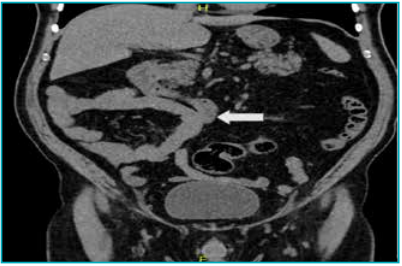
Figure 1: Plain coronal computed tomography (CT) image showing complete malrotation of the bowel. Jejunal loops are located on the right side of the abdomen (White arrow), while both the ascending and descending colon lie on the left.
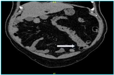
Figure 2: Plain coronal computed tomography (CT) image demonstrating a malpositioned cecum in the left iliac fossa with the ileocecal junction and ascending colon seen in the left half of the abdomen (white arrow).
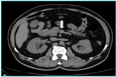
Figure 3: Axial computed tomography (CT) image showing reversed superior mesenteric artery–superior mesenteric vein (SMA–SMV) relationship with the superior mesenteric artery lying to the right of the vein (white arrow).
Further evaluation in imaging revealed both kidneys to be normal in size and position, with bilateral mild perinephric fat stranding and moderate hydroureteronephrosis (Figure 4). A 6 × 4 mm calculus was seen in the right upper ureter at the level of L4 (Figures 5 and 6A), and an 8.6 × 6.6 mm calculus with a density of 1000 Hounsfield units (HU) was located in the left lower ureter just proximal to the ureterovesical junction (Figure 6B).
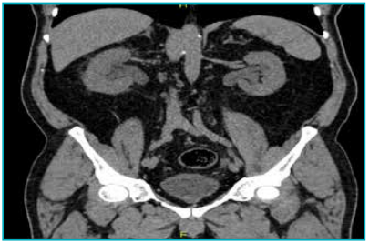
Figure 4: Coronal computed tomography of kidney, ureters, and bladder (CT KUB) image showing bilateral moderate hydroureteronephrosis with mild perinephric fat stranding.
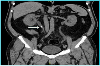
Figure 5: Coronal computed tomography of kidney, ureters, and bladder (CT KUB) image demonstrating a 6 × 4 mm ureteric calculus in the right upper ureter at the level of the upper border of L4 vertebral body (white arrow) with associated perinephric fat stranding.
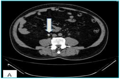
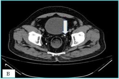
Figure 6: A. Axial computed tomography of kidney, ureters, and bladder (CT KUB) image showing a 6 × 4 mm calculus in the right upper ureter (White arrow), which was seen at the level of the upper border of L4. B. Axial CT KUB image showing an 8.6 × 6.6 mm calculus (1000 HU) in the left lower ureter just proximal to the ureterovesical junction (white arrow).
Imaging findings
Non-contrast CT KUB revealed characteristic features of complete intestinal malrotation:
-
Jejunal loops located on the right side of the abdomen (Figure 1)
-
Cecum and ascending colon lying in the left abdomen (Figure 2)
-
Reversed SMA–SMV relationship (SMA on the right side of SMV) (Figure 3)
-
No evidence of midgut volvulus, Ladd’s bands, or bowel obstruction
-
Bilateral moderate hydroureteronephrosis with ureteric calculi (Figures 4–6)
-
Additional findings included hepatic steatosis
Discussion
Adult intestinal malrotation is a rare congenital anomaly caused by incomplete rotation and fixation of the midgut during embryologic development, with an estimated incidence of less than 0.2% in adults.4,5 Most adult cases remain asymptomatic and are often identified incidentally during imaging for unrelated conditions.
In this case, the anomaly was incidentally detected on a non-contrast CT KUB, performed to evaluate ureteric calculi. Although contrast-enhanced CT or CT angiography is considered the standard imaging modality for assessing mesenteric vascular orientation and related complications,6,7 careful evaluation of bowel and vascular landmarks on plain CT can still reveal characteristic features of malrotation.
The hallmark findings include failure of the DJ flexure to cross the midline, small bowel loops predominantly on the right, colon positioned on the left, and reversal of the SMA–SMV relationship.8,9 In the present case, all these features were appreciable despite the absence of intravenous contrast, underscoring the importance of maintaining a high index of suspicion even on routine KUB scans.
CT remains the imaging modality of choice for diagnosing intestinal malrotation, as it enables identification of both abnormal vascular orientation and potential complications such as volvulus or ischaemia.9 However, in asymptomatic or incidentally discovered cases, as in this patient, conservative management with documentation is typically appropriate.10
This case emphasises that meticulous assessment of noncontrast CT images can occasionally reveal significant congenital anomalies. Awareness of these findings prevents misinterpretation as postsurgical changes or internal hernia, ensuring accurate reporting and clinical correlation.
Teaching Points
-
Adult intestinal malrotation is a rare but noteworthy incidental finding on CT.
-
Recognition of reversed SMA–SMV relationship and abnormal bowel positioning is diagnostic.
-
Awareness of this anomaly prevents misinterpretation as postsurgical anatomy or internal hernia.
-
Asymptomatic incidental malrotation typically requires no surgical intervention but should be documented clearly
Declarations
-
Ethics approval and consent to participate: Not required; this report describes anonymised radiological data.
-
Consent for publication: Not applicable; no identifiable patient information is included.
-
Competing interests: The authors declare no competing interests.
-
Funding: None.
-
Authors’ contributions: Dr. Sylvia Bedas Nsato conceived the case, performed imaging analysis, conducted literature review, and drafted the manuscript. Supervisors and co-authors contributed to imaging interpretation, critical revision, and manuscript approval. All authors reviewed and approved the final manuscript.
Conclusion
This case highlights an incidental detection of intestinal malrotation on a non-contrast CT KUB, illustrating that careful scrutiny of even limited-field, non-enhanced scans can reveal important congenital anomalies. While contrast-enhanced CT or CT angiography remains the gold standard for vascular assessment, awareness of the characteristic anatomic landmarks allows radiologists to recognize malrotation in unexpected contexts and guide appropriate management.
Sylvia Bedas Nsato, Karthikeya Jain, Flora Lwakatare, Vandana, Amit Kumar Sahu. Complete Gut Malrotation Incidentally Discovered in an Adult: A Radiological Case Report. MMJ. 2025, December. Vol 2 (4).
References
-
Nehra D, Goldstein AM. Intestinal malrotation: varied clinical presentation from infancy through adulthood. Surgery. 2011;149(3):386–93.
-
Von Flüe M, Herzog U, Ackermann C, et al. Acute and chronic presentation of intestinal nonrotation in adults. Dis Colon Rectum. 1994;37(2):192–8.
-
Wang C. Anomalies of intestinal rotation in adolescents and adults. Surgery. 1963;54:839–55.
-
Dietz DW, Walsh MR, Grundfest-Broniatowski S, et al. Intestinal malrotation: a rare but important cause of bowel obstruction in adults. Dis Colon Rectum. 2002;45(10):1381–6.
-
Gamblin TC, Stephens RE Jr, Johnson RK, et al. Adult malrotation: a case report and review of the literature. Curr Surg. 2003;60(5):517–20.
-
Butterworth WA, Butterworth JW. An adult presentation of midgut volvulus secondary to intestinal malrotation: a case report and literature review. Int J Surg Case Rep. 2018;50:46–9.
-
Emanuwa OF, Ayantunde AA, Davies TW. Midgut malrotation first presenting as acute bowel obstruction in adulthood: a case report and literature review. World J Emerg Surg. 2011;6(1):22.
-
Pickhardt PJ, Bhalla S. Intestinal malrotation in adolescents and adults: spectrum of clinical and imaging features. Am J Roentgenol. 2002;179(6):1429–35.
-
Strouse PJ. Disorders of intestinal rotation and fixation (“malrotation”). Pediatr Radiol. 2004;34(11):837–51.
-
Xiong Z, Shen Y, Morelli JN, et al. CT facilitates improved diagnosis of adult intestinal malrotation: a 7-year retrospective study based on 332 cases. Insights Imaging. 2021;12(1):58