 WhatsApp Us (24/7)
 +91 926 888 0303 (24/7)


Abstract:
Squamous cell carcinoma (SCC) of the lung most often arises in the central bronchi and is strongly associated with tobacco exposure. However, peripheral squamous cell carcinoma (pSCC) is increasingly recognised as a distinct variant, often radiologically resembling adenocarcinoma and posing diagnostic difficulties, particularly in smokers with emphysema. A 76-year-old chronic smoker presented with persistent cough and weight loss. Chest X-ray revealed an ill-defined opacity in the right perihilar region, with a few thin atelectatic linear bands in the lower zones. Contrast-enhanced computed tomography (CT) of the chest showed an irregular, spiculated soft-tissue mass measuring approximately 4.4 × 3.7 cm in the superior segment of the right lower lobe, with multiple smaller spiculated nodules in the right upper and middle lobes on a background of centrilobular emphysema. Mildly enlarged right pre-paratracheal and subcarinal lymph nodes were also noted. CT-guided biopsy revealed SCC. Histopathology showed well-differentiated SCC with keratin pearls and intercellular bridges. Immunohistochemistry was positive for p40 and cytokeratin 5/6 (CK5/6) and negative for thyroid transcription factor-1 (TTF-1) and Napsin A. Peripheral squamous cell carcinoma, though less common than central SCC, should be considered in smokers presenting with spiculated peripheral pulmonary nodules. These lesions can mimic adenocarcinoma radiologically, may invade the pleura, and often require CT-guided biopsy or surgical resection for definitive diagnosis. Awareness of this variant is important to avoid misclassification and guide appropriate management.
Key words: Peripheral Lung Carcinoma, Squamous Cell Carcinoma, Smoking, CT Chest, Radiology, Case Report.
Introduction
Lung cancer remains the leading cause of cancer-related mortality worldwide. Squamous cell carcinoma (SCC) traditionally arises from the central bronchi and is strongly linked to long-term smoking.1 However, an increasing number of SCCs are being identified peripherally within the lung parenchyma, a phenomenon attributed to changes in smoking habits and improved imaging detection.1-3 Peripheral squamous cell carcinoma (pSCC) can closely mimic adenocarcinoma on imaging due to its peripheral location and spiculated appearance.4,5 Recognition of this subtype is important, as it exhibits different histopathological characteristics, biological behaviour, and therapeutic implications.6,7
Case Report
A 76-year-old male chronic smoker presented with chronic cough, dyspnoea, and unintentional weight loss. Chest X-ray (posteroanterior view) revealed an ill-defined opacity in the right perihilar region, with thin atelectatic bands in both lower zones (Figure 1). Contrast-enhanced computed tomography (CT) of the chest showed an irregular, spiculated soft-tissue mass measuring 4.4 × 3.7 cm in the superior segment of the right lower lobe, along with multiple smaller spiculated nodules in the right upper and middle lobes on a background of centrilobular emphysema (Figure 2A–C). Mild right preparatracheal and subcarinal lymphadenopathy was noted, with no evidence of pleural effusion or chest wall invasion.
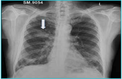
Figure 1: Chest X-ray (anteroposterior view) showing an illdefined opacity in the right perihilar region, with thin atelectatic linear bands in the lower zones of the right lung (white arrow).
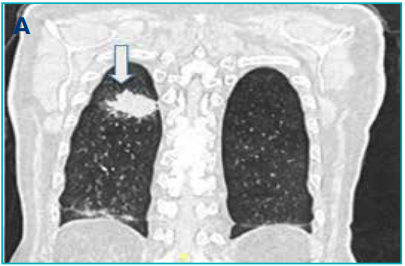
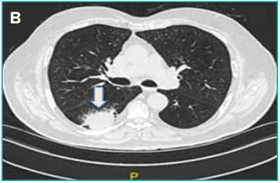
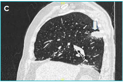
Figure 2A–C: High-resolution computed tomography (HRCT) images — coronal, axial (A and B), and sagittal (C) views — showing a spiculated soft-tissue mass in the superior segment of the right lower lobe, with background centrilobular emphysema (white arrows).
Histopathology findings:
CT-guided biopsy revealed SCC characterised by keratin pearls and intercellular bridges. The degree of differentiation was consistent with well to moderately well-differentiated SCC. Immunohistochemistry showed positivity for p40 and cytokeratin 5/6 (CK5/6) and negativity for thyroid transcription factor-1 (TTF-1) and Napsin A, confirming squamous differentiation
Discussion
pSCC is an uncommon subtype of pulmonary SCC that arises from smaller bronchi or alveolar epithelium, unlike the more typical central SCC.3,4 Although most SCCs are centrally located, peripheral SCC should be considered in smokers presenting with spiculated peripheral pulmonary lesions, as failure to recognise this variant may delay accurate diagnosis and management. Radiologically, pSCC appears as a spiculated or lobulated peripheral mass, occasionally with cavitation or adjacent emphysema.5,6 Distinguishing pSCC from adenocarcinoma based solely on imaging is challenging.7 Histopathologically, pSCC demonstrates keratinisation and intercellular bridges, with immunohistochemistry (p40+, CK5/6+, TTF-1−, Napsin A−) being crucial for confirming squamous differentiation.8 Several studies and case series, including the one by Watanabe et al., have characterised the clinicopathologic and radiologic features of pSCC,3,4 highlighting the importance of awareness and accurate diagnosis to guide management and prognostication.
Declarations
-
Ethics approval and consent to participate: Not required, as this report describes anonymised radiological data.
-
Consent for publication: Not applicable, as no identifiable patient information is included.
-
Competing interests: The authors declare no competing interests.
-
Funding: None.
-
Authors’ contributions: Dr. Sylvia Bedas Nsato conceived the case, performed imaging analysis, conducted the literature review, and drafted the manuscript. Supervisors and co-authors contributed to imaging interpretation, critical revision, and manuscript approval. All authors reviewed and approved the final manuscript.
Conclusion
pSCC is a less frequent but increasingly recognised variant of pulmonary SCC. It should be actively considered in the differential diagnosis of spiculated peripheral lung masses in smokers, as imaging alone is non-specific. Histopathological and immunohistochemical confirmation are essential for accurate diagnosis, appropriate treatment planning, and prognostication.
Sylvia Bedas Nsato, Eeta Jain, Flora A Lwakatare, Richa Bansal, Bharat Aggarwal. Peripheral Squamous Cell Carcinoma of the Lung in a Smoker: A Radiological Case Report and Literature Review. MMJ. 2025, December. Vol 2 (4).
References
-
Nicholson AG, Tsao MS, Beasley MB, et al. The 2021 WHO classification of lung tumours: impact of advances sinc
-
Travis WD, Brambilla E, Noguchi M, et al. Diagnosis of lung cancer in small biopsies and cytology: implications of the 2011 International Association for the Study of Lung Cancer/ American Thoracic Society/European Respiratory Society classification. Arch Pathol Lab Med. 2013;137(5):668–84
-
Sung YE, Cho U, Lee KY. Peripheral type squamous cell carcinoma of the lung: clinicopathologic characteristics in comparison to the central type. J Pathol Transl Med. 2020;54(4):290–9.
-
Watanabe Y, Tsuta K, Kusumoto M, et al. Clinicopathologic features and computed tomographic findings of 52 surgically resected adenosquamous carcinomas of the lung. Ann Thorac Surg. 2014;97(1):245–51.
-
Ciofiac CM, Mămuleanu M, Florescu LM, et al. CT imaging patterns in major histological types of lung cancer. Life (Basel). 2024;14(4):462.
-
Yue JY, Chen J, Zhou FM, et al. CT-pathologic correlation in lung adenocarcinoma and squamous cell carcinoma. Medicine (Baltimore). 2018;97(50):e13362.
-
Bishop JA, Teruya-Feldstein J, Westra WH, et al. p40 (ΔNp63) is superior to p63 for the diagnosis of pulmonary squamous cell carcinoma. Mod Pathol. 2012;25(3):405–15.
-
Guo R, Tian Y, Zhang N, et al. Use of dual-marker staining to differentiate between lung squamous cell carcinoma and adenocarcinoma. J Int Med Res. 2020;48(4):300060519893867.