 WhatsApp Us (24/7)
 +91 926 888 0303 (24/7)


Abstract:
Back pain is a frequent complaint among young individuals and is often attributed to benign musculoskeletal causes. However, infectious aetiologies such as paravertebral abscess, though rare in this age group, must be considered, as delayed diagnosis can result in severe complications, including neurological deficits or sepsis. We report the case of a 13-year-old previously healthy female presenting with a two-day history of progressive lower back pain, intermittent fever, and mild restriction of movement. Initial evaluation suggested mechanical back strain, but the persistence of pain and elevated inflammatory markers, such as erythrocyte sedimentation rate (ESR) and C-reactive protein (CRP), warranted further investigation. Magnetic resonance imaging (MRI) of the spine revealed multiple abscesses from C7–T4 and a small collection from T10–L1 levels, with surrounding soft-tissue inflammation. Blood cultures and abscess aspirate confirmed Staphylococcus aureus infection. The patient was treated with intravenous antibiotics followed by a course of oral therapy. Significant clinical improvement was observed, which prevented surgical drainage of the abscess, and follow-up imaging showed significant resolution. This case emphasises the diagnostic challenge of differentiating spinal infections from mechanical back pain in young patients. The absence of classic risk factors or systemic toxicity often contributes to a diagnostic delay. MRI remains the gold standard for early detection of spinal infections. Paravertebral abscess, though uncommon in young individuals, should be included in the differential diagnosis of persistent or atypical back pain. Early recognition and appropriate management are crucial to prevent morbidity and ensure favourable outcomes. A high index of suspicion, timely imaging, and multidisciplinary management are essential for optimal care.
Key words: Back Pain, Paravertebral Abscess, Spinal Infection, MRI, MRSA, Spinal Abscess.
Introduction
Paravertebral abscesses — collections of pus in the soft tissues adjacent to the vertebrae — are rare but potentially life-threatening conditions in the paediatric population.1 These abscesses can result from haematogenous spread, contiguous infections, or post-surgical complications. When symptoms of back pain persist or worsen, particularly in the absence of clear trauma, serious underlying pathologies should be considered. In young children, the presentation of paravertebral abscesses is often non-specific and can be easily overlooked. Symptoms such as back pain, fever, irritability, and decreased mobility may be mistaken for more common musculoskeletal complaints or viral infections.2 As a result, the diagnosis is frequently delayed, which can lead to severe complications, including vertebral osteomyelitis, spinal deformities, or even sepsis. Despite its rarity, the condition should be considered in any child with unexplained or progressive back pain, particularly when accompanied by systemic signs such as fever or lethargy. Imaging, especially magnetic resonance imaging (MRI), plays a crucial role in confirming the diagnosis.3 Early recognition and prompt treatment, including surgical drainage and antibiotic therapy, are essential for improving outcomes and preventing long-term sequelae.
Case Report
A 13-year-old female presented to Max Super Speciality Hospital, Nagpur, with complaints of shortness of breath, fever, back pain, and lower limb pain for two days. She was referred from an outside hospital where she had been admitted one day earlier for the same complaints. She was initiated on non-invasive ventilation (NIV) support and referred.
Her significant outside reports wereN-terminal proB-type natriuretic peptide (NT-proBNP): 6806 ng/L, D-dimer: 3.3 mg/L. Clinical evaluation showed her to be febrile with bilateral crepitations in the infra-axillary and subscapular areas, requiring FiO2 of 50% on NIV support. The initial laboratory investigations revealed C-reactive protein (CRP), 323 mg/L and procalcitonin of 22 ng/mL, with a normal total leukocyte count.
On imaging, computed tomography pulmonary angiography (CTPA) showed a few subpleural groundglass nodular opacities in both lobes, indicating a septic emboli-like picture, with no evidence of pulmonary oedema or embolism. Echocardiography showed a normal ejection fraction with no evidence of right atrial or right ventricular dilatation. The flu panel was negative. Thus, workup for pulmonary embolism and viral pneumonia as a cause of acute respiratory distress with hypoxia was insignificant.
The patient was therefore started on potent antibiotics — meropenem and teicoplanin — in view of raised procalcitonin and CRP. The patient gradually responded to the line of treatment, with improvement in O2 saturation and CRP levels. She was gradually weaned off NIV support and shifted to nasal prongs. Blood culture revealed growth of methicillin-resistant Staphylococcus aureus (MRSA). The source of MRSA sepsis was still undiagnosed.
The patient complained of persistent back pain with mild bilateral lower limb weakness and myalgia (power 4/5). Therefore, creatine phosphokinase (CPK) levels were sent in view of possible viral myositis, which came out to be 515 U/L, indicating that it was not significant. Detailed history and evaluation of the back pain revealed that all the other presenting complaints had initially started with back pain. MRI of the whole spine with contrast (Figure 1) revealed multiple abscesses in the prevertebral region from C7–T4 and a small collection from the T10–L1 level (Figure 2). Multiple multiloculated, peripherally enhancing collections are scattered throughout the intermuscular planes and intramuscular tissues of the back, including the splenius cervicis and erector spinae muscles on either side, with pleural-based collections in the region of the bilateral upper lung fields along the mediastinal and pleural surfaces. Further imageguided aspiration of the abscess showed no evidence of tuberculosis (TB) but did demonstrate growth of Staphylococcus aureus.
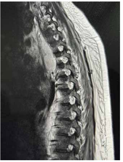
Figure 1: T2-weighted magnetic resonance imaging (MRI) images showing paravertebral abscess.
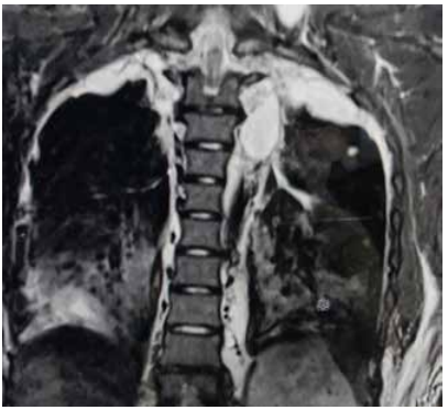
Figure 2: T2-weighted magnetic resonance imaging (MRI) coronal images showing multiloculated enhancing abscesses along the intermuscular and intramuscular planes, along the mediastinal and pleural surfaces.
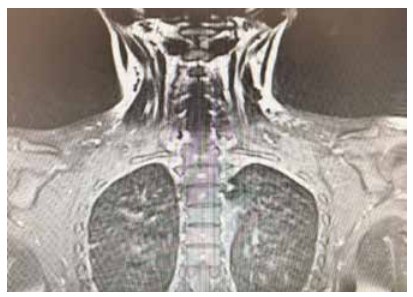
Figure 3: Follow-up magnetic resonance imaging (MRI) showing significant resolution of the abscesses.
With the establishment of the source of infection, intravenous meropenem and teicoplanin were continued for treatment of multilevel paravertebral abscesses, as the culture reports were sensitive to the same. Intravenous methylprednisolone was started initially for respiratory failure. Glycosylated haemoglobin (HbA1c) and viral markers for hepatitis B, hepatitis C, and human immunodeficiency virus (HIV) were sent to rule out an immunocompromised status. The reports were negative. The patient was discharged on oral linezolid for 14 days. On follow-up, the patient was afebrile, asymptomatic, and a follow-up MRI of the spine (Figure 3) revealed complete resolution of the paravertebral abscesses.
Discussion
Paravertebral abscesses, although rare in paediatric populations, are serious infections that can lead to significant morbidity and mortality if not diagnosed and treated promptly.3 Staphylococcus aureus, particularly MRSA, is the most common pathogen responsible for spinal infections, including paravertebral abscesses in children. Haematogenous spread from distant infection sites (such as the skin, bones, or joints), or contiguous spread from adjacent soft tissues, can lead to abscess formation in the paravertebral space.
In young children, risk factors for spinal infections may include recent trauma, surgeries, or systemic infections such as bacteraemia, particularly in the presence of an immunocompromised state.4 However, in many cases like ours, there may be no clear antecedent event, making early diagnosis more difficult. Invasive procedures, such as lumbar puncture or intravenous catheter insertion, can also predispose children to bacteraemia and subsequent spinal infections. The clinical presentation of paravertebral abscesses in children is often insidious and can mimic other common paediatric conditions, such as musculoskeletal pain, or viral infections.5 Fever and back pain, although common in paravertebral abscesses, are often non-specific. In our case, the child’s symptoms of unexplained back pain and fever led to further investigation. Imaging, particularly MRI, is crucial for diagnosing paravertebral abscesses, as it provides detailed softtissue visualisation. Blood cultures are essential for identifying the causative organism, and Staphylococcus aureus — especially in cases complicated by sepsis — is frequently isolated.
The management of paravertebral abscesses involves a combination of surgical drainage and antibiotic therapy. In this case, the child was managed by targeted intravenous antibiotic therapy. The choice of antibiotics is often guided by culture results, with Staphylococcus aureus generally sensitive to beta-lactams or MRSAdirected therapy in resistant cases.6 Empirical treatment should include broad-spectrum antibiotics, with later de-escalation based on microbiological findings. The clinical course following appropriate treatment, in our case, was favourable, and the child showed significant improvement without complications. Delay in treatment can result in the spread of infection to the vertebral bodies, leading to osteomyelitis, discitis, or even sepsis. Close follow-up is necessary to monitor for recurrence or complications.
Conclusion
Our patient presented as a diagnostic challenge. The young age of the patient, along with the paucity of findings on physical examination, and the presenting features of acute-onset respiratory distress, made the diagnosis of paravertebral abscess as a source of sepsis obscure. The septic emboli-like picture on pulmonary angiography was initially confused clinically with pulmonary embolism and viral pneumonia. Persistent back pain in the young should not be overlooked or dismissed. While it may seem minor at first, untreated back pain can lead to significant long-term problems. Key risk factors for MRSA infections in the young include direct contact, skin breaks, poor hygiene, immunocompromised states, chronic conditions (e.g., eczema, asthma), and frequent or unnecessary use of antibiotics, which promote resistance.
Nitin Dambhare, Manoj Pethe, Kaushal Dewasthali. The Backstabber: Pain That Hid a Deeper Threat. MMJ. 2025, December. Vol 2 (4).
References
-
Rahyussalim AJ, Sulaiman AR, Winartomo A, et al. Closed system paravertebral abscess evacuation on spinal infection: A case series. Int J Surg Case Rep. 2023;104:107941.
-
Hasan GA, Kani SM, Alqatub A. Tuberculous lumbar spinal epidural abscess in a young adult (case report). SICOT J. 2018;4:5.
-
Ambulgekar RK, Iqbal Z, Masne P. A Rare Case of Thoracolumbar Epidural Abscess with Tuberculous Etiology in an Adolescent Male with Neurological Deficit. J Orthop Case Rep. 2022;12(9):98–101.
-
Myojin S, Kamiyoshi N, Kugo M. Pyogenic spondylitis and paravertebral abscess caused by Salmonella Saintpaul in an immunocompetent 13-year-old child: a case report. BMC Pediatr. 2018;18(1):24.O'Donnell R, Sayani S, Aguìñiga-Navarrete P, et al. Paraspinal Abscess in a Two-year-old Female. Clin Pract Cases Emerg Med. 2019;4(1):59–61.
-
O'Donnell R, Sayani S, Aguìñiga-Navarrete P, et al. Paraspinal Abscess in a Two-year-old Female. Clin Pract Cases Emerg Med. 2019;4(1):59–61.
-
Ekici MA, Ozbek Z, Gökoğlu A, et al. Surgical management of cervical spinal epidural abscess caused by Brucella melitensis : report of two cases and review of the literature. J Korean Neurosurg Soc. 2012;51(6):383–87