 WhatsApp Us (24/7)
 +91 926 888 0303 (24/7)


Abstract:
Robotic-assisted surgery has revolutionised the landscape of minimally invasive surgical oncology by enabling enhanced precision, dexterity, and visualisation, especially in anatomically complex regions. The integration of robotic platforms into oncological surgery offers a distinct advantage in achieving oncological safety with minimal morbidity. This case series demonstrates the feasibility, safety, and adaptability of robotic techniques in challenging cervicothoracic and head–neck oncologic procedures. It includes three patients who underwent complex robotic surgeries: A robotic oesophagectomy in a frail patient with poor pulmonary function, salvage transoral robotic surgery (TORS) after dual radiation exposure, and a robotic thyroidectomy using the bilateral axillo-breast approach (BABA). Each case was planned and executed after multidisciplinary evaluation, with emphasis on patient selection, surgical planning, and intraoperative considerations. All procedures were successfully completed using robotic platforms without intraoperative complications. Patients demonstrated satisfactory postoperative recovery with minimal morbidity and excellent cosmetic and functional outcomes. Histopathological evaluation confirmed negative margins in all cases. Robotic-assisted surgery offers significant advantages in precision dissection, access to deep anatomical spaces, and improved postoperative recovery in carefully selected oncologic cases. This series highlights the versatility of robotic platforms in addressing surgical challenges across the cervicothoracic and head–neck regions. Further studies with larger cohorts and long-term follow-up are warranted to establish oncologic equivalence and cost-effectiveness.
Key words: Robotic Surgery, Surgical Oncology, Transoral Robotic Surgery (TORS), Robotic Oesophagectomy, Robotic Thyroidectomy, Bilateral Axillo-Breast Approach (BABA), Cervicothoracic Surgery, Minimally Invasive Surgery, Head and Neck Cancer, Oncologic Outcomes.
Introduction
The term “robot” was coined in the beginning of the last century, coming originally from the Czech word “robota”, meaning “labour”.1 The U.S. Department of Defence and National Aeronautics and Space Administration (NASA) partnered along with Stanford Research Institute to explore robotics for remote battlefield or space surgeries.
The aim was to allow expert surgeons to operate from afar via robotic arms. In early 2000, “da Vinci” became the first robotic system approved by the Food and Drug Administration (FDA) for general laparoscopic surgery — marking the true beginning of robotic-assisted surgery in clinical practice.Table 1 summarises the timeline of major milestones in the evolution of robotic surgery.
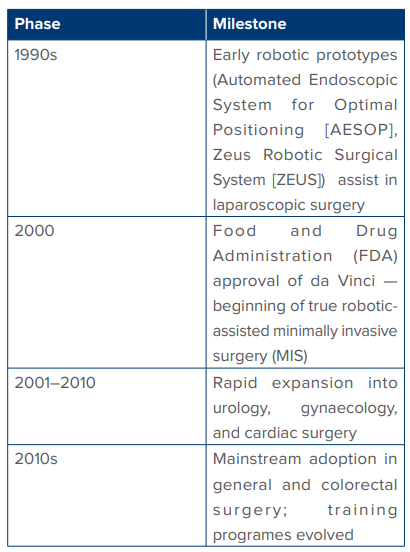
Table 1: Timeline
Cutting edge meets cancer care: What surgical oncology demands?
Surgical oncology demands precision, oncological safety, meticulous dissection, and adherence to sound anatomical and oncological principles such as achieving negative margins, appropriate lymphadenectomy, and minimising tumour handling. These goals often require complex dissections in confined anatomical spaces and careful preservation of critical structures. Robotic surgery, with its enhanced three-dimensional (3D) visualisation, wristed instruments, and improved dexterity, offers a powerful tool to fulfil these demands. It allows the surgeon to perform precise and controlled movements even in anatomically challenging areas, facilitating oncological resections with minimal morbidity. Furthermore, robotic platforms enable better access to deep pelvic, mediastinal, and retroperitoneal regions, making them highly suitable for complex cancer surgeries where conventional open or laparoscopic approaches may fall short. When used judiciously and with proper oncologic intent, robotic surgery serves as a valuable extension of the surgical oncologist’s armamentarium.
Case Report 1: Robotic Oesophagectomy in a Frail Patient
A 53-year-old male with an Eastern Cooperative Oncology Group (ECOG) score of 2, presented with complaints of dysphagia for solid food for 2 months duration associated with significant weight loss. Upon evaluation patient was found to have a circumferential growth in the lower third oesophagus, 34 cm from the incisor teeth. Positron emission tomography–computed tomography (PET–CT) scan revealed bulky lower third oesophageal growth with enlarged subcarinal nodes with no evidence of distant metastasis. There were features of chronic obstructive pulmonary disease (COPD) and aspiration pneumonitis in CT. Patient was advised four cycles of neoadjuvant fluorouracil, leucovorin, oxaliplatin, and docetaxel (FLOT) chemotherapy. After a shared decision in the multidisciplinary tumour board, we decided to go ahead with robotic oesophagostomy. Since the patient was a known case of COPD with a poor ECOG score, we felt that it was prudent to offer a robotic platform for this patient.
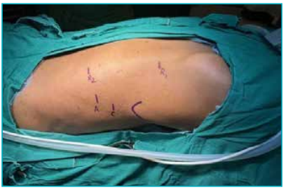
Figure 1.1: Camera port is placed about 2 intercostal spaces (ICS) below the tip of the right scapula. Robotic arm 1 (R1) is taken 7 cm from the midline, about 2–3 ICS above the camera port, lying parallel to the medial border of the scapula. Robotic arm 2 (R2) is taken about 2 ICS below the camera port, about 7 cm from the midline. The assistant port is triangulated between R2 and the camera.
The patient was placed in a prone position. Usually, a single-lung ventilation is initiated using a double-lumen endotracheal tube, but in this patient, we wanted to avoid single-lung ventilation owing to poor pulmonary functions. The ports were placed as described above (Figure 1.1). Circumferential dissection of the oesophagus from surrounding structures was carried out. The azygos vein was clipped and divided to carry out the supra-azygos dissection. Enlarged subcarinal nodes were removed with precision (Figures 1.2A–B). This was followed by gastric conduit creation (Figures 1.3A–B) through mobilising the stomach.
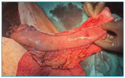
Figure 1.2A: Enlarged subcarinal nodes were removed. The movements made by the robotic arms help in precise dissection at these crucial areas. This would have been difficult with the conventional laparoscopic techniques.
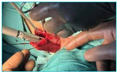
Figure 1.2B: Subcarinal region post-dissection showing complete clearance.
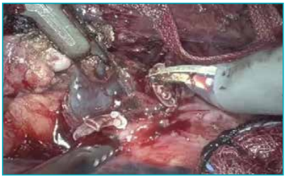
Figure 1.3A: Gastric conduit creation.
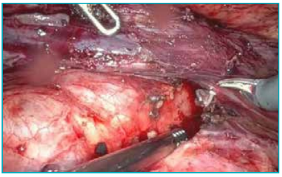
Figure 1.3B: Anastomosis between the gastric conduit and the cervical oesophagus made using the a small neck incision on the left side of neck through ‘Barcelona Technique’ where a side-to-side anastomosis was made using a linear cutter stapler.
The patient was started on a liquid diet on postoperative Day 10 (POD 10) and was subsequently discharged. There were no major post operative events despite the poor general condition of the patient. The final histopathological examination (HPE) report revealed poorly differentiated adenocarcinoma; the resected margins were free. A total of 15 lymph nodes were retrieved. The subcarinal nodes and intrapulmonary nodes showed metastatic deposits. The patient was started on adjuvant chemotherapy after 3 weeks of surgery.
Key considerations:
-
Preoperative optimisation in the form of nutritional support, spirometry, etc., plays a crucial role during early postoperative recovery.
-
In frail patients with poor pulmonary function, it is better to avoid single-lung ventilation; instead, both lungs can be ventilated with low tidal volumes.
-
Identify and preserve the thoracic duct to prevent chyle leak.
-
The conduit viability can be assessed using intraoperative indocyanine green (ICG) dye.
Case Report 2: Pushing the Limits — “Salvage Transoral Robotic Surgery (TORS) After Dual Radiation Exposure”
A 74-year-old male was previously diagnosed in 2007 with squamous cell carcinoma of the right tonsil (cT4cNxMx) and treated with radical radiation therapy. After an 18-year disease-free interval, he developed a second primary cancer in the soft palate, staged as cT1–2N0M0. He was planned for definitive radiation with concurrent chemotherapy, but chemotherapy was not given due to unfitness. He received intensity-modulated radiotherapy (IMRT) to the head and neck region from 21st February 2025 to 9th April 2025, delivered in 33 fractions with a total dose of 60–66 Gy. During follow-up, the patient had a residual lesion in the soft palate. PET–CT scan revealed a low-grade metabolism in the oropharynx. There were no significant fluorodeoxyglucose (FDG) avid neck nodes. A biopsy was done, which reported low-grade squamous cell carcinoma. After discussion in the tumour board, we decided to go ahead with salvage surgery. The lesion involved the soft palate, extending to the uvula medially and posteriorly to the posterior pharyngeal wall (Figures 2.1A–B).
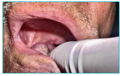
Figure 2.1A: Showing the lesion in the soft palate extending to the uvula medially and posteriorly extending to the posterior pharyngeal wall
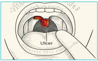
Figure 2.1B: Schematic diagram of the palatal lesion shown
Discussion:
Surgical access to tumours in the oropharynx and base of the tongue has traditionally posed significant challenges due to the anatomic complexity and limited exposure of these regions.
The conventional mandibular swing approach — an invasive technique involving osteotomy of the mandible — has long been used to achieve sufficient visualisation and access for tumour resection. While effective in terms of access, this method is associated with significant morbidity. Patients often endure complications such as malocclusion, non-union or malunion of the mandible, damage to the inferior alveolar nerve resulting in numbness, prolonged hospitalisation, and cosmetic deformity due to surgical scars.3 The conventional mandibulotomy approach and extent of exposure achieved are illustrated in Figures 2.2A–C.
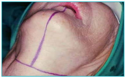
Figure 2.2A: The skin incision being outlined. Source: Jatin Shah textbook of Head and Neck Surgery
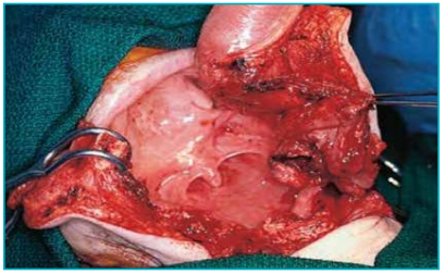
Figure 2.2B: Exposure of tumour through the mandibulotomy approach. Source: Jatin Shah textbook of Head and Neck Surgery.
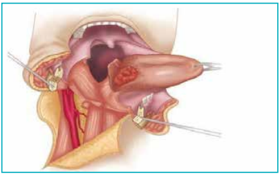
Figure 2.2C: Diagrammatic representation of the exposure obtained through a mandibulotomy. Source: Jatin Shah textbook of Head and Neck Surgery.
TORS represents a paradigm shift in the surgical management of oropharyngeal tumours (Figures 2.3A–C). Speech and swallowing functions are better preserved with TORS, reducing the need for long-term rehabilitation and improving overall quality of life. Patients undergoing TORS typically experience less postoperative pain, shorter hospital stays, and quicker return to oral intake. Tracheostomy and feeding tubes are often avoided or required only for short durations.
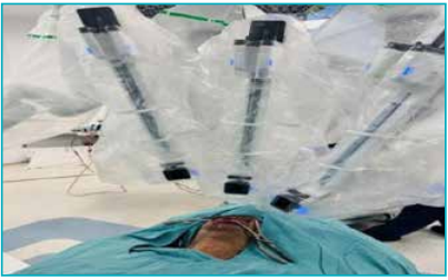
Figure 2.3A: Patient positioning and docking of robotic arms being done.
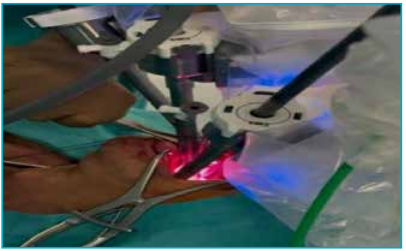
Figure 2.3B: The endoscope and the robotic arms being introduced in the oral cavity.
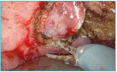
Figure 2.3C: The assistant retracts the tongue with a tongue depressor. Dissection over the posterior pharyngeal wall becomes easy with of monopolar scissors.
Key considerations:
-
Conventional surgery in a previously irradiated neck would add to the morbidity of the patient. TORS would be ideal in these cases.
-
Use an appropriate retractor system (example : Feyh–Kastenbauer [FK] retractor) to prevent pressurerelated injury to the lips, teeth, or tongue.
-
Monitor closely for delayed haemorrhage, which is most common during the first two weeks.
Case Report 3: Thyroid Surgery Re-Imagined — Robotic Techniques for a Scarless Neck
A 35-year-old female working in the information technology (IT) industry presented with complaints of swelling in the front of the neck for 1 month. Upon evaluation, she was found to have a solitary thyroid nodule with fine-needle aspiration cytology (FNAC) showing Bethesda III. She was advised to undergo surgery. She was more concerned about her cosmesis and requested a scarless surgery. Hence, a robotic platform was chosen.
The trans axillary approach to the thyroid surgery was pioneered by the South Korean team from Seoul, led by Chung, in late 2007.4,5 The robotic-assisted transaxillary thyroid surgery (RATS) approach was first described in North America by Kupersmith and Holsinger in 2011.6 Since it was first introduced, more than 3,000 RATS procedures were performed in South Korea, and more than 6,000 worldwide.7 Subsequent advances in robotic retraction and access systems have further improved visualisation and ergonomics for upper aerodigestive and thyroid procedures.⁸ Patient positioning and port placement for the bilateral axillo-breast approach are shown in Figures 3.1 and 3.2.
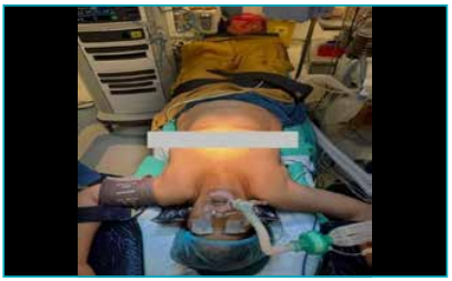
Figure 3.1: This image depicts the patient positioning before docking the robotic arms. The patient lies in supine position with both arms abducted and raised up.
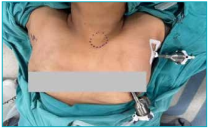
Figure 3.2: Image describes the port positioning. Bilateral axillary and bilateral circumareolar ports are placed.
We have described the steps of the bilateral axillo-breast approach robotic thyroidectomy (BABA-RT). The patient lies in the supine position, with the neck slightly extended. Both arms are raised and fixed to expose the axilla. A 5–12 mm incision is made along the natural skin crease of each axilla. Two circumareolar ports are placed. Blunt dissection is done through the subcutaneous tissues to create a tunnel towards the neck. A working space is developed over the pectoralis major, clavicle, and up to the thyroid cartilage. Retractors or carbon dioxide (CO₂) insufflation may be used to maintain space. The robotic arms of the da Vinci surgical system are docked. Instruments such as the endoscope (camera), force bipolar, monopolar scissors and prograsp forceps are inserted. The strap muscles are retracted laterally to expose the gland. Lateral dissection is carried out until the common carotid artery is visualised. Parathyroid and write it together laryngeal nerve are safeguarded. After ligating the superior pedicle and inferior thyroid vessels, the gland is dissected off from the trachea. Key intraoperative steps, including identification of the external laryngeal nerve, gland dissection, and final specimen retrieval, are illustrated in Figures 3.3A–C.
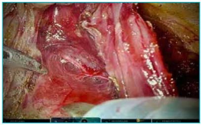
Figure 3.3A: External laryngeal nerve identified and safeguarded before ligating the superior thyroid. The sternothyroid muscle was cut and retracted.
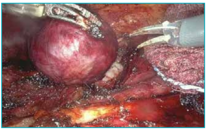
Figure 3.3B: Final dissection where the nodule is being dissected from the tracheal surface.
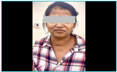
Figure 3.4: Two weeks after surgery, the patient was reviewed in follow-up and expressed satisfaction with the cosmetic result of her robotic thyroidectomy.
Key considerations:
-
Create a working space via subplatysmal flap from the axilla to the thyroid.
-
Use blunt and energy-based dissection carefully to avoid injury to nerves or vessels
-
Use an endo bag through the axillary incision to avoid spillage.
Conclusion
Our case series demonstrates that robotic surgery is a safe and feasible approach in selected patients undergoing procedures in anatomically challenging regions. While the learning curve and cost remain important considerations, our experience highlights that with appropriate patient selection and surgical expertise, roboticassisted techniques can be successfully integrated into head, neck, and upper gastrointestinal oncology practice. Larger prospective studies and longer follow-up will be essential to further validate the oncological safety, functional outcomes, and long-term benefits of this evolving surgical modality
Abhinav Deshpande, Kawadu Jawade, Kapilnath, Nimish Ganvir. Dexterity in Complexity: Case Series of Challenging Cervicothoracic Surgeries via Robotic Platforms. MMJ. 2025, December. Vol 2 (4).
References
-
Szabolcsi R. The Birth of the Term Robot. Advances in Military Technology. 2014;9(1):117–28.
-
Wahlstedt ER, Kronstedt SM, Saffati G, et al. Overview of Robotic Surgery in the U.S. Military: From Conception to Utilization, and Future Applications. Mil Med. 2025;190(3- 4):66–8.
-
Shah JP, Patel SG, Singh B, Wong R. Jatin Shah's head and neck surgery and oncology. 5th Edition. London: Elsevier; 2019.
-
Jackson NR, Yao L, Tufano RP, et al. Safety of robotic thyroidectomy approaches: meta-analysis and systematic review. Head Neck. 2014;36(1):137–43.
-
Lee J, Kang SW, Jung JJ, et al. Multicenter study of robotic thyroidectomy: short-term postoperative outcomes and surgeon ergonomic considerations. Ann Surg Oncol. 2011;18(9):2538–47.
-
Kuppersmith RB, Holsinger FC. Robotic thyroid surgery: an initial experience with North American patients. Laryngoscope. 2011;121(3):521–6.
-
Son H, Park S, Lee CR, et al. Factors contributing to surgical outcomes of transaxillary robotic thyroidectomy for papillary thyroid carcinoma. Surg Endosc. 2014;28(11):3134–42.
-
Rodney JP, Vasan NR. Robotic microlaryngeal surgery: a new retractor that provides improved access to the glottis. SpringerPlus. 2016;5:188.