 WhatsApp Us (24/7)
 +91 926 888 0303 (24/7)


Abstract:
Mayer–Rokitansky–Küster–Hauser (MRKH) syndrome is a rare congenital disorder characterised by vaginal agenesis with normal ovarian function and karyotype. Neovaginoplasty remains the cornerstone of management for patients seeking functional sexual outcomes. While laparoscopic sigmoid vaginoplasty has been widely reported, the robotic approach offers enhanced precision, ergonomics, and three-dimensional (3D) visualisation. We present the case of a 21-year-old female diagnosed with MRKH syndrome who underwent robotic sigmoid neovaginoplasty. The procedure was successfully completed with an operative time of 5 hours and an estimated blood loss of less than 50 mL. Postoperative recovery was uneventful, and the patient was discharged on postoperative Day 7. At 8 weeks of follow-up, the neovagina demonstrated satisfactory length, cosmetic appearance, and functional outcomes. This case highlights that robotic sigmoid neovaginoplasty is a safe and feasible technique for vaginal reconstruction in MRKH syndrome, combining the advantages of minimally invasive surgery with enhanced surgical dexterity.
Key words: Mayer–Rokitansky–Küster–Hauser (MRKH) Syndrome, Robotic Surgery, Sigmoid Neovaginoplasty, Colovaginoplasty.
Introduction
Mayer–Rokitansky–Küster–Hauser (MRKH) syndrome is a rare congenital malformation affecting approximately 1 in 4,500–5,000 female births. It is characterised by congenital absence or hypoplasia of the uterus and upper vagina in phenotypically normal females with a 46,XX karyotype and normal secondary sexual characteristics. The diagnosis is often made during adolescence when patients present with primary amenorrhoea despite normal pubertal development.
Reconstructive options include non-surgical dilation (Frank’s method), McIndoe vaginoplasty, Davydov procedure, and intestinal (sigmoid or ileal) vaginoplasty. Among these, sigmoid vaginoplasty offers several advantages: a well-vascularised mucosa, self-lubrication, and adequate vaginal length with long-term durability
Traditionally performed via open or laparoscopic methods, sigmoid neovaginoplasty has evolved with the advent of robotic-assisted surgery. Robotic technology provides enhanced dexterity, tremor filtration, and superior visualisation in confined pelvic spaces, potentially improving outcomes in such complex reconstructive procedures.
Here, we report a case of robotic sigmoid neovaginoplasty in a patient with MRKH syndrome, highlighting the technical aspects, perioperative outcomes, and clinical implications.
Case Report
A 21-year-old female presented with primary amenorrhoea. She reported normal pubertal development and absence of cyclic abdominal pain. On examination, she was phenotypically normal with normal breast and pubic hair development. Examination of the external genitalia revealed normal labial development with an absent vaginal orifice, as shown in Figure 1.
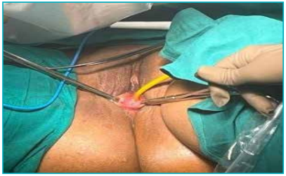
Figure 1: Examination of the external genital showing normal labial folds and vaginal dimple.
Ultrasonography and magnetic resonance imaging (MRI) revealed a non-functional hypoplastic uterus and absent vagina, with normal bilateral ovaries and kidneys, as shown in Figures 2A–C. Karyotyping confirmed 46,XX. A diagnosis of MRKH syndrome with a variation was established.
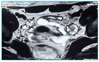
Figure 2A: View of bilateral polycystic ovaries (PCO).
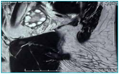
Figure 2B: View of string of pearls — polycystic ovaries (PCO)
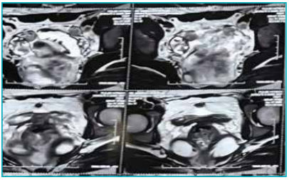
Figure 2C: View of rudimentary bicornuate uterine horns.
The patient was counselled regarding available options for vaginal reconstruction. Considering her preference for surgical creation of a neovagina, a robotic sigmoid neovaginoplasty was planned. Written informed consent was obtained from the patient.
Surgical technique
Under general anaesthesia, the patient was positioned in lithotomy with a steep Trendelenburg tilt. A four-port robotic approach was employed using the da Vinci Xi system.
-
Port placement: An 8-mm camera port was inserted at the umbilicus, with three 8-mm robotic ports and one assistant port placed under direct vision as shown in Figures 3A and 3B
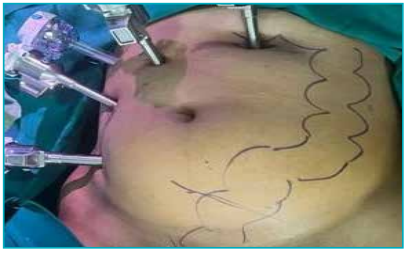
Figure 3A: Placements of the ports.
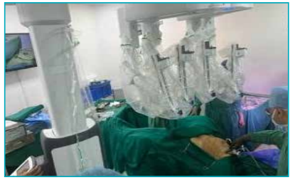
Figure 3B: Left lower docking.
-
Neovaginal space creation: A hysterectomy with bilateral salpingectomy was performed. A perineal incision was made, and blunt dissection was used to create a neovaginal canal between the bladder and rectum up to the peritoneum, as shown in Figure 4.
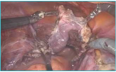
Figure 4: Rudimentary bicornuate hysterectomy specimen.
-
Sigmoid segment mobilisation: A 12–15 cm well-vascularised sigmoid segment, based on its mesenteric blood supply, was mobilised as shown in Figure 5.
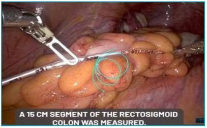
Figure 5: Mobilisation of the sigmoid segment.
-
Anastomosis: The distal end of the sigmoid pedicle was pulled down and sutured to the perineal skin at the neovaginal introitus, while the proximal end was closed using a SureForm stapler. End-to-end anastomosis was performed robotically with the help of a trans-anal circular stapler, allowing precise anastomosis as shown in Figures 6A–C.
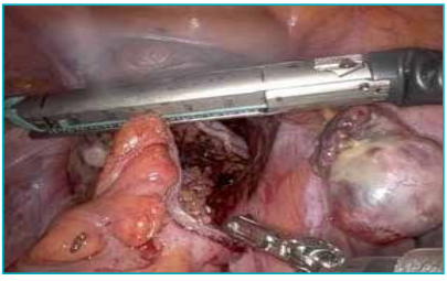
Figure 6A: Application of SureForm stapler.
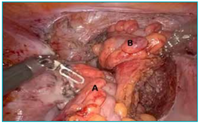
Figure 6B: a. Reanastomosed rectosigmoid, b. Right-sided vascular colo neovaginal pedicle.
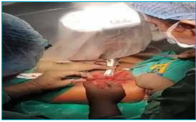
Figure 6C: Colo labial anastomosis.
-
Completion: Haemostasis was secured; anastomosis and bowel integrity were checked using an Asepto syringe, and vascularity was checked with intravenous indocyanine green as shown in Figure 7.
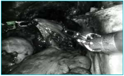
Figure 7: Intraoperative assessment.
The operative time was 5 hours, with an estimated blood loss of less than 50 mL. There were no intraoperative complications.
Postoperative course
The patient recovered uneventfully and was discharged on postoperative Day 7. No vaginal mould was placed intraoperatively. At the 5-week visit, mild stenosis was suspected due to upward traction of the pedicle; dilatation was performed under anaesthesia, and the patient was instructed on a self-dilatation protocol.
At 2-month follow-up, the neovagina was patent with a length of 12 cm. The mucosa appeared healthy with adequate lubrication. The patient reported no complications such as stenosis, fistula, or excessive mucus discharge.
Discussion
The MRKH syndrome presents unique reconstructive challenges, requiring the creation of a functional neovagina that enables satisfactory sexual function and psychosocial well-being. Although non-surgical dilation remains the first-line management, a subset of patients either fail conservative therapy or prefer definitive surgical reconstruction for durable and physiological outcomes.1,2 Among surgical techniques, sigmoid vaginoplasty remains the gold standard due to the segment’s robust vascularity, mucosal lubrication, and tissue similarity to the native vagina. The sigmoid colon provides a wellvascularised, self-lubricating mucosa that minimises postoperative stenosis and ensures long-term patency.3 The advent of minimally invasive techniques — particularly robotic-assisted surgery — has refined this approach, offering enhanced visualisation, precise dissection, and superior instrument control. Robotic-assisted surgery provides substantial ergonomic and technical advantages over laparoscopy, including three-dimensional (3D) magnified vision, tremor filtration, and enhanced dexterity with wristed instruments. These features are particularly valuable during deep pelvic dissection and anastomosis, reducing the risk of injury to adjacent organs and improving operative accuracy.4,5 Multiple reports indicate that robotic sigmoid vaginoplasty achieves comparable or improved perioperative outcomes relative to conventional approaches. Beyond intraoperative precision, the success of neovaginoplasty depends on postoperative functionality. Studies demonstrate that a sigmoid neovagina provides excellent sexual satisfaction, adequate lubrication, and minimal long-term complications such as stenosis or mucous overproduction.3 In our case, the patient achieved adequate vaginal length and calibre with satisfactory cosmetic and functional outcomes within two months postoperatively. The robotic platform likely contributed to reduced intraoperative complications. Psychosexual rehabilitation is integral to recovery and long-term patient satisfaction. Counselling, structured dilation programmes, and gradual resumption of sexual activity are essential to achieving optimal results.1 The minimally invasive and precise nature of robotic surgery may positively influence body image, confidence, and psychological well-being, thereby enhancing overall quality of life in MRKH patients undergoing genital reconstruction. Despite its benefits, robotic sigmoid neovaginoplasty has limitations. The high cost, limited access to robotic platforms, and a significant learning curve remain barriers to widespread adoption. Future multicentric prospective studies with larger sample sizes and long-term follow-ups are warranted to validate the superiority of robotic techniques in terms of anatomical, functional, and psychosocial outcomes.
Conclusion
Robotic sigmoid neovaginoplasty represents a safe, precise, and functionally effective reconstructive technique for patients with MRKH syndrome. It merges the advantages of minimally invasive surgery with improved ergonomics and visualisation, offering excellent anatomical, functional, and psychosocial results when performed in experienced centres.
UddhavRaj Dudhedia, Manmohan Kamat, Anukrati Singh, Shreya Alluri, Akhil Kumar Mishra. Robotic Sigmoid Neovaginoplasty in Mayer–Rokitansky–Küster–Hauser Syndrome: A Case Report of a Feasible and Safe Reconstructive Approach. MMJ. 2025, December. Vol 2 (4).
References
-
Morcel K, Guerrier D, Watrin T, et al. The Mayer-RokitanskyKüster-Hauser (MRKH) syndrome: clinical description and genetics. J Gynecol Obstet Biol Reprod (Paris). 2008;37(6):539–46.
-
Creatsas G, Deligeoroglou E. Vaginal aplasia. Semin Reprod Med. 2001;19(1):89–97.
-
Fedele L, Frontino G, Motta F, et al. Creation of a neovagina with laparoscopic sigmoid vaginoplasty. Int J Gynaecol Obstet. 2010;110(3):249–52.
-
Smith AJ, Vancaillie TG, Abbott J. Robotic-assisted laparoscopic sigmoid vaginoplasty: surgical technique and outcomes. J Minim Invasive Gynecol. 2015;22(7):1125–31.
-
Sinha R, Sanjay M, Rupa B, et al. Robotic surgery in gynecology. J Minim Access Surg. 2015;11(1):50–9.