 WhatsApp Us (24/7)
 +91 926 888 0303 (24/7)


Abstract:
Glioblastoma (GBM) is the most common malignant brain tumour in adults. On radiology, diagnostic dilemmas can arise with other brain pathologies like metastasis, brain abscess, and lymphoma. However, GBM mimicking a meningioma on radiology has been rarely reported. Here, we report a case of GBM with magnetic resonance imaging (MRI) features suggestive of meningioma.
A 46-year-old female presented with a one-month history of headaches. The MRI brain suggested a broad-based extra-axial mass lesion in the left frontal convexity with cerebrospinal fluid (CSF) cleft sign with compression of underlying brain parenchyma. Overall, the MRI brain was suggestive of a cystic meningioma. The patient underwent gross total resection of the tumour under microscopy, and final histopathology confirmed GBM.
Although GBM is usually an intra-axial tumour, it may present as an extra-axial mass lesion with radiological features mimicking meningioma. Therefore, it should be considered a differential diagnosis in patients with an extra-axial tumour displaying atypical malignant features.
Key words: Glioblastoma, Meningioma, Extra-axial, CSF Cleft, Dural Tail
Introduction
Glioblastoma (GBM) is the most common adult brain tumour accounting for 14.6% of all brain tumours.1 The most common sites are the frontal and temporal lobes and it is usually intraaxial.2 On radiology, diagnostic dilemmas can occur with conditions such as metastasis, brain abscess, lymphoma, and arteriovenous malformations.3 However, primary extra-axial GBM mimicking a meningioma on radiology has been rarely reported in literature. Here, we report a case of a middle-aged female with an extra-axial tumour with radiological features suggestive of meningioma but proven to be GBM on histopathology.
Case Discussion
A 46-year-old female presented with complaints of headache of one-month duration. Headache was mild in intensity, holocranial with no diurnal variation. There was no neurological deficit, such as aphasia, motor weakness, or gait abnormalities. Her vitals were stable and laboratory test results were within the normal range.
Magnetic Resonance Imaging (MRI) brain with contrast (Figure 1) was suggestive of a broad-based extra-axial mass lesion measuring 5.8x5cm in the left frontal convexity with cerebrospinal fluid (CSF) cleft and compression of the underlying brain parenchyma with moderate perifocal oedema. A broad dural attachment was seen. The mass was compressing the left lateral ventricle, causing a midline shift of 11.6mm to the right. Overall, the MRI brain findings were suggestive of a cystic meningioma.
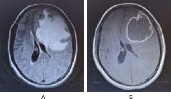
Figure 1: MRI Brain showing left frontal mass with cerebrospinal fluid (CSF) cleft sign [A] with contrast image suggestive of broad dural attachment [B].
The patient was planned for surgery. Preoperative medications such as antiepileptics, steroids and mannitol were started on the morning of surgery. Intraoperatively (Figure 2), the tumour was found to be extra-axial, grey, round, well-defined and soft to firm in consistency.
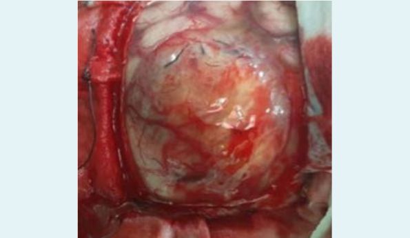
Figure 2: Well defined extra-axial round tumour with feeder vessels is seen. There was no attachment to overlying dura. Tumour is seen compressing the adjacent brain parenchyma.
The tumour had both solid and cystic components, with necrotic tissue, thrombosed vessels, and yellowish serous cystic fluid. There was no dural attachment. The patient underwent gross total resection of tumour under microscopy. The post-operative period was uneventful, and the patient was discharged on the 7th post-operative day. Histopathology (Figure 3) suggested astrocytic proliferation with pleomorphism, nuclear atypia, mitotic activity, an infiltrative appearance, and microvascular proliferation with multinucleate giant cells, consistent with GBM.
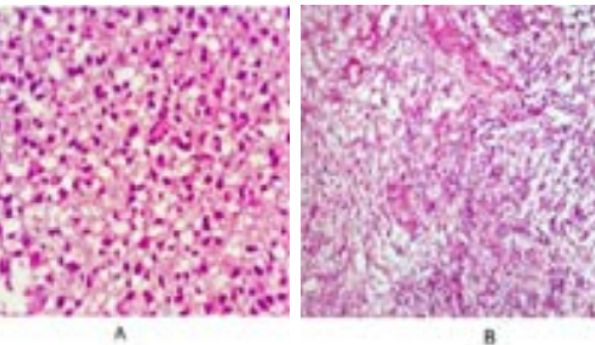
Figure 3: Astrocytic proliferation showing pleomorphism, nuclear atypia, mitotic activity, and infiltrative appearance with multinucleate atypical giant cells; B. Fibrous stroma with extensive microvascular proliferation seen.
Immunohistochemistry (IHC) Panel 3 was performed and was consistent with GBM, wild-type. The tumour cells showed diffuse, strong positivity for glial fibrillary acidic protein (GFAP). The epidermal growth factor receptor (EGFR), and alpha thalassemia/mental retardation syndrome X-linked (ATRX) also showed diffuse positivity with a high Ki67 index. The patient is currently undergoing concurrent radiotherapy and chemotherapy with temezolamide.
Discussion
The WHO Classification of CNS tumours (2021, 5th Edition) define GBM as a diffuse astrocytic tumour in adults that are isocitrate dehydrogenase (IDH) wild-type and is considered a completely separate entity from astrocytoma, IDG mutant, which can be classified as grades 2,3, or 4.4 To make a diagnosis of GBM, the patient must be an adult with diffuse astrocytic, IDH wild-type tumour with at least one of the following features:
- Necrosis
- Microvascular proliferation
- Telomerase reverse transcriptase (TERT) promoter mutation
- EGFR gene amplification
- Combined gain of whole chromosome 7 and loss of chromosome 10 [+7/-10]
IDH-mutant GBMs constitute around 10% of all GBM and develop from grade II or III tumours. They usually occur in persons of less than 55 years of age.5
Radiologically, GBM and meningiomas are usually well differentiated based on their tumour characteristics, location and pattern of enhancement. However, diagnostic dilemmas may occur with other brain pathologies such as abscess, metastases, and lymphomas as radiological features of GBM may resemble these pathologies. The diagnostic dilemma of GBM with meningioma has been rarely reported in literature.
Radiologically, GBM and meningiomas are usually well differentiated based on their tumour characteristics, location and pattern of enhancement. However, diagnostic dilemmas may occur with other brain pathologies such as abscess, metastases, and lymphomas as radiological features of GBM may resemble these pathologies. The diagnostic dilemma of GBM with meningioma has been rarely reported in literature.
Meningiomas usually appear as well defined, lobulated, extraaxial masses with homogenous contrast enhancement. They have a broad dural-based attachment with a dural tail sign. Meningiomas cause compression of adjacent brain parenchyma, resulting in a CSF cleft sign (CSF between the tumour and normal brain parenchyma). They are most commonly located in the cerebral convexities, sphenoid wing, parasagittal, and parafalcine regions. High-grade meningiomas may lack the CSF cleft sign on MRI.
The dural tail sign was initially considered pathognomonic for meningiomas; however, it has also been reported in other brain pathologies like schwannoma, metastasis, GBM, and pituitary adenomas.6 The CSF cleft sign seen in meningiomas has rarely been reported in GBM.
Our patient exhibited MRI features suggestive of meningioma, such as well-defined margins, a broad-based extra-axial mass lesion with a CSF cleft sign, and vascular flow voids, along with compression of adjacent brain parenchyma. Intraoperatively, the tumour was extra-axial, grey, round, well defined, and soft to firm in consistency. The tumour had solid to cystic components with yellowish serous cystic fluid, and there was no dural attachment.
The patient underwent gross total resection of the tumour under the microscope, and histopathology suggested GBM.
There is limited literature on the diagnostic dilemmas between GBMs and meningiomas. Primary extra-axial GBM with a CSF cleft sign has rarely been reported in literature. Patel et al. published a case report regarding two cases of extra-axial GBM with broad dural contact with the first case located in the right temporoparietal region and the second case located in the left parasagittal region. In another case report, Gheyi et al. reported a case of frontoparietal GBM with both intra- and extra-axial components.7 Taghipour Zahir et al. reported a case of frontal calvarial mass proven to be GBM on histopathology.8 Wu et al. reported a primary extra-axial GBM in the cerebellopontine angle region.9 Kartigeyan et al. reported a case of left petroclival extra-axial GBM.10
This case suggests that a GBM can present as an extra-axial tumour on imaging and intra-operatively causing confusion in diagnosis. With limited literature available on extra-axial GBM, further studies are required to explain the nature, course and prognosis of this entity.
Conclusion
Glioblastoma is usually an intra-axial tumour but may present as an extra-axial mass lesion with radiological features mimicking meningioma. So, it should be considered as a differential diagnosis in patients with an extra-axial tumour with atypical malignant features.
Gaurav Sharma, Anu Gupta, Komal Singla. Primary Extra-axial Glioblastoma Mimicking Meningioma:
Case Report and Review of Literature. MMJ. 2024, Dec. Vol 1 (4).
DOI: XXXXX
References
- Gheyi V, Hui FK, Doppenberg EM, et al. Glioblastoma multiforme causing calvarial destruction: an unusual manifestation revisited. American journal of neuroradiology. 2004;25(9):1533-7.
- Karthigeyan M, Ahuja CK, Chatterjee D, et al. Radiologic dilemma in an extra-axial petroclival lesion: low grade versus high grade. World Neurosurgery. 2017;100:713-e1.
- Louis DN, Giannini C, Perry A, et al. Glioblastoma. In: WHO Classification of Tumours Editorial Board. Central nervous system tumours. Lyon (France): International Agency for Research on Cancer; 2021. (WHO classification of tumours series, 5th ed.; vol. 6).
- Ohgaki H, Kleihues P. The definition of primary and secondary glioblastoma. Clinical cancer research. 2013;19(4):764-72.
- Ostrom QT, Cioffi G, Gittleman H, et al. CBTRUS statistical report: primary brain and other central nervous system tumors diagnosed in the United States in 2012–2016. Neuro-oncology. 2019 Oct;21(Supplement_5):v1-00.
- Patel M, Nguyen HS, Doan N, et al. Glioblastoma mimicking meningioma: report of 2 cases. World Neurosurgery. 2016;95:624-e9.
- Shakur SF, Bit-Ivan E, Watkin WG, et al. Multifocal and multicentric glioblastoma with leptomeningeal gliomatosis: a case report and review of the literature. Case reports in Medicine. 2013;2013(1):132679.
- Sotoudeh H, Yazdi HR. A review on dural tail sign. World journal of radiology. 2010;2(5):188.
- Zahir ST, Mortaz M, Yazdi MB, et al. Calvarium mass as the first presentation of glioblastoma multiforme: A very rare manifestation of high-grade glioma. Neurochirurgie. 2018;64(1):76-8.
- Wu B, Liu W, Zhu H, et al. Primary glioblastoma of the cerebellopontine angle in adults: case report. Journal of neurosurgery. 2011;114(5):1288-93.