 WhatsApp Us (24/7)
 +91 926 888 0303 (24/7)


Abstract:
Caecal blow-out is a rare phenomenon that occurs when there is a distal colonic obstruction, leading to the distension of caecum and ascending colon, which in turn thins the walls. If untreated, perforation of the caecum may result. We present here a rare case report of caecal blow-out secondary to distal colonic obstruction due to adenocarcinoma of the descending colon.
Key words: Anastomosis, Caecal Blow-Out, Colon, Obstruction, Perforation, Resection.
Introduction
Caecal blow-out is the perforation of the caecal wall due to increased intraluminal pressure in caecum caused by distal colonic obstruction. It most commonly occurs in elderly individuals due to a malignant colon tumour. Perforation proximal to a colonic mass is termed a diastatic perforation and results from over-distension and blow-out of the caecal wall.1 Thus, in cases of distal large bowel obstruction with a competent ileocaecal valve, the caecum is the most common site of perforation.2,3 The caecum is the first part of the colon and has the largest diameter, becoming distended with minimal intraluminal pressure, which makes it the most vulnerable site for perforation due to obstruction. A competent ileocaecal valve makes caecum excessively dilated with pressure due to colonic obstruction. Caecal perforation is common after radiotherapy and chemotherapy for malignant conditions. Caecal blow-out, also called obstructive perforation of the caecum, occurs when distal colonic obstruction increases intraluminal pressure, thinning the colonic wall and ultimately causing blow-out and perforation. The caecum and ascending colon become a closed-loop pathology in cases of distal colonic obstruction with a patent ileocaecal valve.
Case report
A male patient, Mr. K. H., aged 72 years, was admitted to the hospital with signs of intestinal perforation and generalised peritonitis. There was abdominal distension and generalised guarding and tenderness, and no bowel sounds were noticed.
Per rectal examination revealed an empty rectum. Urgent basic lab tests and an erect abdominal X-ray were performed, showing gas under the diaphragm. The patient presented with low blood pressure and tachycardia, prompting the initiation of intravenous antibiotics and intravenous Ringer’s lactate solution. Due to time constraints, specialised investigations such as an abdominal computed tomography (CT) scan were not performed, and the patient was moved to the operation theatre for exploratory laparotomy. During the operation, a perforation was identified in the anterior wall of the caecum, opposite the mesocolon attachment. Further exploration revealed a tumour in the descending colon obstructing the lumen. A peritoneal washout was conducted with normal saline, followed by a left hemicolectomy and end-to-end anastomosis. A loop ileostomy was not performed, but a cecostomy was created through the caecal perforation. Given the critical condition of the patient, a rapid surgical approach was adopted. The patient later developed wound infection and a burst abdomen, which was repaired as expected. Histopathological examination of the descending colon tumour confirmed adenocarcinoma. The patient gradually improved and was discharged after a 4-week hospital stay
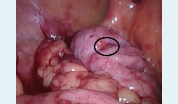
Figure 1: Caecal perforation
In this case, the period between the initial clinical signs and symptoms and definitive diagnosis was approximately 20 hours. The sooner the surgery, the better the outcome. The delay was due to the initial consultation with a general practitioner (GP), who believed it was an intestinal cramp that would improve with some medication. It was only when the pain did not subside and vital signs deteriorated that the GP referred the patient to the hospital for emergency treatment. We did not waste any time even to perform a CT scan or other special laboratory investigations. This was also a factor in saving the life of this gentleman.
Discussion
Although there are numerous cases of caecal perforations, caecal blow-out due to distal obstruction is quite rare. Common causes of spontaneous caecal perforation include trauma, malignant obstruction, ingested foreign bodies, volvulus, and inflammatory conditions such as Crohn’s disease and infections.4 The law of Laplace (Pierre-Simon, Marquis de Laplace, French Scholar (1749-1827), he is referred as French Newton) states, “in a long pliable tube, the site of largest diameter requires the least pressure to distend.” Therefore, in patients with distal large bowel obstruction and a competent ileocaecal valve, the cecum is the most common site of perforation.3 The mortality in between perforation and definitive diagnosis and treatment, as colonic perforation allows bacteria, typically a mixed type, both aerobic and anaerobic type, to rapidly spread within the peritoneal cavity, leading to generalised peritonitis. Toxins released by the bacteria can cause severe complications and may prove fatal. Urgent surgical intervention, as in our case, was crucial for the patient’s survival. The type of surgery in caecal blow-out syndrome with a tumour in distal colon depends upon the site of the tumour.
Hussan et al. treated a similar case of caecal blow-out with a tumour in the descending colon and perforation by performing a left hemicolectomy with end-to-side anastomosis, caecal perforation closure, and loop ileostomy. We also performed a left hemicolectomy with end-to-end anastomosis and a cecostomy through the perforation. Mortality rates associated with caecal perforation are high, ranging from 30% to 72%.5 Early recognition of pathology and prompt intervention are critical for management. A high mortality rate associated with caecal perforation might be related to a delay in diagnosis.6
Declaration of Patient Consent
The authors certify that they have obtained all appropriate patient consent forms. In the form the patient(s) has/have given his/her/their consent for his/her/their images and other clinical information to be reported in the journal. The patient understand that their names and initials will not be published, and due efforts will be made to conceal their identity, but anonymity cannot be guaranteed.
Financial Support and Sponsorship
Nil.
Conflicts of Interest
There are no conflicts of interest
Conclusion
Caecal blow-out is a rare phenomenon with high morbidity and mortality. Early diagnosis and immediate surgical intervention are crucial for improving prognosis. The choice of surgical treatment depends on the general condition of the patient, and site of the distal colonic obstruction. Unnecessary delay in performing extensive investigations can worsen prognosis.
Vinod Kumar Nigam, Siddharth Nigam.Caecal Blow-Out – A Case Report. MMJ. 2024, Dec. Vol 1 (4).
DOI: XXXX
References
- Paramythitis D, Karakatsanis A, Moysides M, et al. Retroperitoneal Caecal Perforation Resulting from Obstruction. Ascending Colon Adenocarcinoma. Case Reports in Surgery. 2020;1-5.
- Hussain MRK, George R. Caecal diastatic perforation due to descending colon adenocarcinoma. Eurorad. 2022, ISSN 1563-5086.
- Slam KD, Calkins S, Cason FD. LaPlace’s law revisited: Caecal perforation as an unusual presentation of pancreatic carcinoma. World J. Surg. Onc. 2017;5:14.
- Yeo R. Spontaneous perforation of the caecum: case reports and a review of the literature. Postgard and J. 1967;43:65-67.
- Laskin MD, Tessler K, Kives S. Caecal perforation due to paralytic ileus following primary caesarean section. J. Obstet. Gynaecol. Can. 2009;31:167-171.
- Balas HA, Balas MA, Wiswasy MA. Idiopathic spontaneous caecal perforation: A rare pathology with high mortality. Ann Med Surg (Lond). 2020;60:518-521.