 WhatsApp Us (24/7)
 +91 926 888 0303 (24/7)


Abstract:
A bicornuate uterus is a Müllerian duct anomaly that develops during embryogenesis. It is usually associated with poor pregnancy outcomes such as preterm birth, recurrent miscarriages and malpresentations. Most cases of uterus bicornuate non-communicant rudimentary horn (UBNCRH) present with endometriosis. The association of unilateral renal agenesis with adenomyosis in the non-communicating rudimentary horn is a rare occurrence and is associated with severe symptoms which was found in our case. Clinical suspicion and diagnosis, along with the removal of adenomyotic horn in a bicornuate uterus, significantly improved the symptoms and led to better obstetrical outcomes during the second pregnancy. This case highlights the diagnostic dilemma and management of such cases, leading to excellent symptomatic improvement and better pregnancy outcomes.
Key words: Bicornuate, Adenomyosis, Rudimentary Horn
Introduction
Müllerian duct anomalies are rare, and the overall incidence of such anomalies in general population is about 0.4%.1 These anomalies are associated with increased perinatal morbidity and mortality, including recurrent miscarriages, preterm births, malpresentations, and disruptions in foetal growth.2
A bicornuate uterus with non-communicant rudimentary horn is usually associated with endometriosis. However, in our case, the findings of unilateral renal agenesis and adenomyosis with severe symptoms were present, which is a rare occurrence and poses a challenge in diagnosis and management. This case report highlights the high index of suspicion, and the surgical excision of the adenomyotic non-communicating horn significantly improved symptoms and resulted in excellent pregnancy outcomes.
Case Report
A 32-year-old Para 1, Living 1, Abortion 0, known case of bicornuate uterus presented with progressive secondary dysmenorrhoea after one year of first preterm birth. Her initial pelvic ultrasound showed bicornuate uterus with normal ovaries, but no obvious cause for her symptoms could be found. She was started on oral analgesics and spasmolytic for almost 6 months, with no response. Due to her progressively severe dysmenorrhoea, she was put on injectable analgesics, which also did not show much improvement.
She gradually developed intractable severe dysmenorrhoea, and since there was no improvement, an injection of gonadotropin releasing hormone (GnRH) was used to induce iatrogenic amenorrhoea for a period of 18 months. She was advised to undergo a hysterectomy as a last resort at another hospital, where she had been receiving treatment, because she did not want to continue GnRH due to its side effects. She then revisited us for further management.
She had previously booked with us at 24 weeks of her first pregnancy. She had a history of spontaneous pregnancy after 1.5 years of marriage. Her previous menstrual, medical, and surgical history was insignificant. She was diagnosed with a bicornuate uterus and an absent right kidney during her booking antenatal scan at 7 weeks of gestation. Cervical cerclage was performed at 20 weeks due to cervical length of less than 2.4cm, and she was advised to take progesterone injections weekly until term.
She was admitted with threatened preterm labour at 33+ weeks of gestation, and despite conservative management, she progressed to active labour and had a vaginal delivery at 34 weeks of a 2.1kg baby, who was active and healthy.
She revisited us for her intractable dysmenorrhoea, and during this visit we found that her pain was mainly toward the right side of the suprapubic area. A 3D ultrasound revealed a bicornuate, non-communicating right horn of the uterus with signs of adenomyosis, but no hematometra. She was counselled and advised to proceed with diagnostic hysteroscopy and laparoscopy to decide on further management.
She underwent surgery at our hospital. Hysteroscopy showed only the left cornua with ostia. Laparoscopy revealed right horn of 5x3.5x2cm, which was almost the same size as the left side (Figure 1).
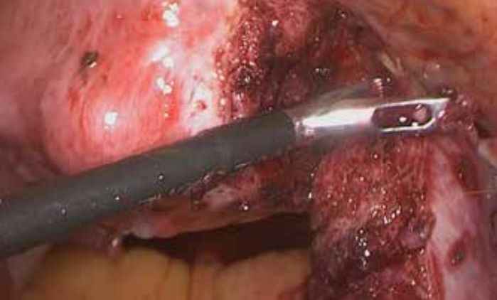
Figure 1: Laparoscopic removal of the rudimentary horn.
The pelvic cavity did not show any endometriotic spots. Both ovaries were normal. Excision of the right horn along with ipsilateral salpingectomy was performed laparoscopically and the specimen was sent for histopathology. The procedure was uneventful, and she was discharged on the second day. Histopathology revealed sections of the endometrium with proliferative phase changes and the myometrium with foci of adenomyosis (Figure 2). The serosa was free of tumours, and fallopian tube was unremarkable.
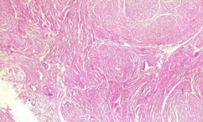
Figure 2: Histopathology showing focal adenomyosis in resected rudimentary horn of the uterus.
Immediately after surgery, her dysmenorrhoea improved and she did not require any analgesics. Figure 3 shows hysteroscopic view of the uterine cavity post-surgery.

Figure 3: Post-procedure hysteroscopic view of the uterine cavity.
She had spontaneous conception one year after surgery, with an uneventful antenatal period. During this pregnancy, she did not require cervical cerclage, as cervical length monitoring showed a long cervix with a closed internal os. Her entire antenatal period was uneventful. She underwent an elective lower segment caesarean section (LSCS) and tubal ligation at 37 weeks, delivering a healthy baby of 3.1kg.
Discussion
The overall incidence of Müllerian anomalies in the general population is around 0.4%. Müllerian anomalies are developmental defects resulting from abnormalities in the fusion or resorption of the paired Müllerian ducts, which are responsible for forming the uterus, cervix, and upper two-thirds of the vagina.
Uterus bicornuate non-communicant rudimentary horn (UBNCRH) is a rare malformation of the uterus. It is classified as a Müllerian duct abnormality Type II b, according to the American Fertility Society classification.3 The anomalies are most often described as de novo congenital occurrences and are often asymptomatic. Some may present with dysmenorrhoea, endometriosis, recurrent miscarriages, and infertility.4
Diagnosis is typically made via 2D or 3D ultrasound, hysteroscopy, hysterosalpingography, or magnetic resonance imaging.5 Renal anomalies are frequently associated with Müllerian anomalies due to the interlinked development of mesonephric and Müllerian ducts, along with the urogenital sinus. The most common defect is renal agenesis, as seen in our case.6
A bicornuate uterus increases the need for surveillance during pregnancy. Although most women with this condition can have healthy pregnancies, there is an increased risk of recurrent miscarriages, preterm birth, disruption of foetal growth, placenta praevia, and retained placenta. These complications arise due to the distortion of the uterus and cervix, leading to cervical insufficiency, reduced uterine capacity, and irregular uterine contractions.
Although endometriosis has been reported in association with UBNCRH, the occurrence of adenomyosis in the rudimentary horn is extremely rare. Adenomyosis can occur in both syndromic and non-syndromic congenital uterine malformations and may arise from several aetiologies.7 One hypothesis is that obstructions in these malformations increase uterine pressure, promoting the development of adenomyosis. This hypothesis remains to be further elucidated, and additional findings may also play a role. The patient’s genetic, epigenetic, and hormonal patterns, as well as normal physiological processes, such as pregnancy, may influence the growth of adenomyosis.
Our case was unique because the patient presented with severe dysmenorrhoea after her first preterm delivery. A thorough clinical history, and examination along with 3D ultrasound confirmed our suspicion of UBNCRH and adenomyosis in one horn without haematometra, along with associated unilateral renal agenesis. Laparoscopic excision of the rudimentary horn, with ipsilateral salpingectomy and histopathological findings of adenomyosis, confirmed our diagnosis.8
This management relieved all her symptoms, and she had an uneventful full-term pregnancy following the surgical removal.
Conclusion:
The management of UBNCRH, renal agenesis, and severe dysmenorrhoea with adenomyosis, through early diagnosis and laparoscopic excision of the rudimentary horn with salpingectomy, leads to significant improvement in symptoms and obstetrical outcome.
Rajni Gupta. Non-Communicant Rudimentary Horn (UBNCRH), Renal Agenesis, and Severe Dysmenorrhoea
with Adenomyosis: A Case Report on Improved Obstetrical Outcomes. MMJ. 2024, Dec. Vol 1 (4).
DOI: XXXXXXXXXXXXXXXXXX
References
- Khati NJ, Frazier AA, Brindel KA. The unicornuate uterus and its variants, clinical presentation, imaging findings and associated complications. J Ultrasound Med. 2012;31(2):319-32.
- Venetis CA, Papadopoulos SP, Campo R, et al. Clinical implications of congenital uterine anomalies: A meta-analysis of comparative studies. Reprod Biomed Online. 2014;29(6):665-83.
- Grimbizis GF, Gordts S, Di Spiezio SA, et al. The ESHRE/ ESGE consensus on the classification of female genital congenital anomalies. Hum Reprod. 2013;28(8):2032-2044.
- Borah T, Das A, Panda S, et al. A case of unilateral dysmenorrhea. J Hum Reprod Sci. 2010;3(3):158-159.
- Deutch TD, Abuhamad AZ. The role of 3-dimensional ultrasonography and magnetic resonance imaging in the diagnosis of müllerian duct anomalies: A review of the literature. J Ultrasound Med. 2008;27(3):413-23.
- Hall-Craggsn MA, Kirkham A, Creighton SM. Renal and urological abnormalities occurring with Mullerian anomalies. J Pediatr Urol. 2013;9(1):27-32.
- Frontino G, Bianchi S, Ciappina N, et al. The unicornuate uterus with an occult adenomyotic rudimentary horn. J Minim Invasive Gynecol. 2009;16(5):622-625.
- Falcone T, Gidwani G, Paraiso M, et al. Anatomical variation in the rudimentary horns of unicornuate uterus, implications for laparoscopy surgery. Human reproduction. 1997;12(2):263-5.