 WhatsApp Us (24/7)
 +91 926 888 0303 (24/7)


Abstract: Steroid-dependent optic neuropathies generally have an immune-mediated origin, with neuromyelitis optica spectrum disorder (NMOSD) and myelin oligodendrocyte glycoprotein antibody-associated disease (MOGAD) remain the most common aetiologies, among other systemic inflammatory disorders. Here, we present a rare case of a young male with human immunodeficiency virus (HIV) and neurosyphilis presenting with steroid-dependent optic neuropathy. To the best of our knowledge, this is the first case report of such a presentation.
Key words: Neurosyphilis, Optic Neuropathy, Neuromyelitis Optica Spectrum Disorder (NMOSD), Myelin Oligodendrocyte Glycoprotein Antibody-Associated Disease (MOGAD)
Case Report
A 42-year-old male was diagnosed as human immunodeficiency virus (HIV)-positive in March 2023 while being evaluated for a three-month history of weight loss and decreased appetite. His CD4 count at diagnosis was 220 cells/mm3 . He was started on highly active antiretroviral therapy (HAART) which included tenofovir, lamivudine, and dolutegravir (TLD regimen). Two months after starting treatment, he presented with recurrent episodes of blurred vision, detailed as follows:
Episode 1: May 2023
He experienced acute onset of blurred vision in the right eye. There was no accompanying headache or eye movement pain, and the blurring progressed over a week to a visual acuity of 3/60 at the peak deficit. There was no history of any preceding fever, vaccination, or travel prior to onset of ocular complaints. He received injectable steroids outside for three days, followed by a tapering course of oral steroids over 4 weeks. His vision improved to 6/12 within three weeks. However, a week after tapering off steroids, he again experienced blurred vision in the same eye.
Episode 2: July 2023
The second episode of visual blurring in right eye occurred a week after tapering steroids. This was again painless, with no complaints of headache or eye pain. The records of visual examination in this event were not available. He received a three-day course of intravenous methylprednisolone at an outside centre, followed by oral steroids. The steroids were maintained at a full dose (i.e., 1 mg/kg) for 4 weeks before gradually tapering them. This time, while on 10 mg of oral steroids, he again started experiencing blurred vision in both eyes.
Episode 3: October 2023
Five days after being started on 10 mg of oral steroids, he developed bilateral blurring of vision, which was associated with bifrontal headaches. He had difficulty perceiving brightness of objects from both eyes, especially the red colour. At peak deficit, vision in the right eye was 3/60, and 6/24 in the left eye. He was brought at our centre during this episode. Evaluation revealed normal optic fundi (Figure 1) with some loss of neuroretinal rim on optical coherence tomography (OCT) in right eye, consistent with a diagnosis of retrobulbar neuritis. Extraocular movements were normal, and there were no signs of rash, lymphadenopathy, or organomegaly. Other motor and sensory examinations were also normal. Clinically there was no evidence of any other opportunistic infection in the body
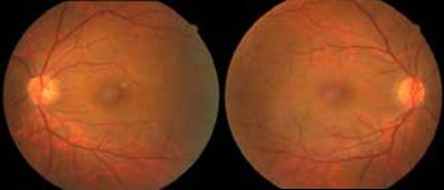
Figure 1: Fundus images of the patient; Bilateral normal fundi.
Evaluation and Management
Routine haematology and biochemistry parameters were normal. Erythrocyte sedimentation rate (ESR) was 12 mm/ hr and antinuclear antibody (ANA) was negative. Tests for cytoplasmic and perinuclear antineutrophilic cytoplasmic antibody (c-ANCA, p-ANCA) and vasculitis panel were noncontributory. Vitamin B12 levels were 828 pg/ml, serum homocysteine levels were 14 mcmol/l and serum folate levels were 10 mg/ml. Serum neuromyelitis optica (NMO) and myelin oligodendrocyte glycoprotein (MOG) antibodies were negative. Serum angiotensin converting enzyme (ACE) levels were normal (24 mcg/l). He underwent lumbar puncture and cerebrospinal fluid (CSF) testing. CSF opening pressure was normal (150 mm CSF). CSF had 25 cells/cumm - all mononuclear, with protein of 45 mg/dl and sugars of 90 mg/dl (corresponding blood sugar levels - 120 mg/ml). CSF NMO and MOG levels were also negative. His magnetic resonance imaging (MRI) brain was normal, while MRI orbits showed bilateral optic atrophy (Figure 2).
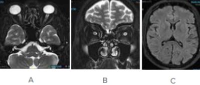
Figure 2: MRI brain and orbit images
(a) Axial and (b) coronal T2 MRI brain image showing bilateral optic nerve atrophy.
(c) Axial Flair MRI brain image showing normal brain parenchyma.
In view of his retropositive status, a serum venereal disease research laboratory (VDRL) test was conducted, which resulted positive (titer - 1:32 dilution). Serum Treponema pallidum haemagglutination assay (TPHA) was also positive. With high clinical suspicion of neurosyphilis, CSF TPHA was tested and found positive, meeting criteria for neurosyphilis due to CSF leukocytosis. He was started on injectable ceftriaxone coupled with injectable steroids. He completed 14 days intravenous course of ceftriaxone. After a 5-day course of intravenous steroids, oral steroids were gradually tapered over a 6-week period. 10 months down the line, he remains relapse free. At last follow-up, his visual activity was 6/12 in the right eye and 6/6 in the left eye.
He has turned seronegative for syphilis at 6-month follow-up. His current CD4 count is 500/mm3 .
Discussion
The case highlights systemic infectious mimics of steroiddependent optic neuropathies. Our patient presented with steroid dependent optic neuritis and was found to have neurosyphilis on evaluation. He was treated for treponemal infection, and did not relapse even after complete cessation of steroids during follow-up. The differentials considered in our case, especially in the background of retropositive status, included inflammatory optic neuritis secondary to seroconversion illness, other inflammatory optic neuritis as myelin oligodendrocyte glycoprotein antibody disease (MOGAD) or aquaporin-4 antibody disease, and other infective/ granulomatous aetiologies.
After ruling out serological immune-mediated entities, neurosyphilis was diagnosed based on a high index of clinical suspicion, along with positive serology and CSF lymphocytosis. Although there is a heterogeneity in the diagnostic criteria for neurosyphilis, the gold standard criterion rests on either a positive CSF-VDRL, or a negative CSF-VDRL but with evidence of CSF biochemical abnormalities (i.e., CSF cells - more than 5 cells per microliter or protein more than 450 mg/l or neurological symptoms without a known cause).1 Optic nerve involvement in syphilis can be seen at any stage, and is sine-que-non with involvement of central nervous system (CNS).2 Syphilis is a great imitator of other, more common conditions presenting with visual dysfunction. In corollary, ocular manifestations are seen in 2-10% of patients with systemic syphilis.3 Secondary stage of syphilis is the stage with a higher proportion of involvement of ocular structures with an incidence of nearly 10%.4
Ocular manifestations of syphilis can be extensive, ranging from anterior segment involvement to pupillomotor pathway affliction. Of all ocular manifestations of neurosyphilis, optic nerve involvement is uncommon and has been reported in nearly 20% of patients with ocular involvement.5 The spectrum of optic nerve involvement is illustrated in table 1. When a young male presents with acute-onset recurrent vision loss in a steroid-dependent fashion, the diagnostic exercise can be challenging. Immune-mediated optic neuropathies are a primary consideration in such cases. There have been reports of concurrent presence of treponemal infection and aquaporin-4 disease in patients presenting with acute myelitis.6,7 These patients had evidence of neurosyphilis along with positive aquaporin-4 antibody in both CSF and serum. They received intravenous steroids along with treatment for syphilis and were not started on long-term immunomodulation. In their reported follow-up periods, they had remained relapse free even without immunomodulation. The theory remains consistent with parainfectious NMO. Similarly, in our patient, we had a presentation akin to an autoinflammatory optic neuropathy, although the serological tests were negative. We believe that treponemal antigen here served as an immune trigger which probably led to an ongoing hyperimmune response, resulting in consistent response to steroids. However, as long as the underlying infection was not treated, he continued to relapse. Hence our case adds to the existing spectrum of syphilitic optic involvement.
| Sr. No. | Manifestations |
|---|---|
| 1. | Inflammatory optic disc oedema |
| 2. | Papilloedema |
| 3. | Gumma of optic disc |
| 4. | Optic atrophy |
| 5. | Optic neuritis |
| 6. | Optic perineuritis |
| 7. | Neuroretinitis |
| 8. | Non-arteritic ischaemic optic neuropathies |
Table 1: Spectrum of optic nerve involvement in neurosyphilis reported in literature.3,8,9
Conclusion:
Optic nerve manifestations of syphilis can be extensive. The presentation of our patient was unique, with a steroiddependent optic nerve syndrome for the past 1.5 years, later achieving complete remission with anti-syphilitic therapy and not requiring long-term immunomodulation. Truly, syphilis is a great imitator, and this entity must be considered when dealing with autoimmune CNS syndromes.
Kamakshi Dhamija, Sitla Prasad Pathak, Namita Kaul1, Sanjay Kumar Saxena, Rinky Anand Gupta,
Ruby Bansal. Neurosyphilis as an Imitator of Autoimmune Optic Neuropathy:
A case report. MMJ. 2024, Dec. Vol 1 (4).(3)
DOI: XXXXXXXXXXXXXXXXXX
References
- Boog GHP, Lopes JVZ, Mahler JV, et al. Diagnostic tools for neurosyphilis: a systematic review. BMC Infect Dis–2021;21:568.
- Smith GT, Goldmeier D, Migdal C. Neurosyphilis with optic neuritis: an update. Postgrad Med J. 2006;82:36–39.
- Klein A, Fischer N, Goldstein M, et al. The great imitator on the rise: ocular and optic nerve manifestations in patients with newly diagnosed syphilis. Acta Ophthalmol (Copenh). 2019;97:e641–e647.
- Tamesis RR, Foster CS. Ocular syphilis. Ophthalmology. 1990;97:1281–1287.
- Tucker JD, Li JZ, Robbins GK, et al. Ocular syphilis among HIV-infected patients: a systematic analysis of the literature. Sex Transm Infect. 2011;87:4–8.
- Chen H-Q, Zhang Y, Wang S-B, et al. Concurrent aquaporin-4- positive NMOSD and neurosyphilis: A case report. Mult Scler Relat Disord. 2019;34:137–140.
- Youming Long FS. Neuromyelitis Optica Spectrum Disorder and Neurosyphilis Coexist in A Chinese Woman. J Mol Biomark Diagn; 05. Epub ahead of print 2014.
- AlKhayal FA, Albusair MK. Secondary syphilis presenting with non-arteritic anterior ischemic optic neuropathy (NAION). Oxf Med Case Rep. 2024; omae012.
- Alqurashi MM, Badr M, Bukhari A, et al. Ocular Syphilis Presenting As Non-arteritic Anterior Ischemic Optic Neuropathy. Cureus; 13. Epub ahead of print 28 July 2021.