 WhatsApp Us (24/7)
 +91 926 888 0303 (24/7)


Abstract:
Squamous cell carcinoma (SCC) of the oral cavity is a significant global health concern, accounting for approximately 90% of all oral malignancies. This case report describes a 45-year-old male diagnosed with SCC of the right alveolus sulcus extending to the buccal mucosa. The treatment involved a multi-disciplinary team employing advanced planning and surgical techniques, including 3D Computer-Aided Design (CAD) and Computer-Aided Manufacturing (CAM) technology, for precise preoperative planning of the composite resection and reconstruction. This approach facilitated accurate mandibular resection, fibula flap harvest, and immediate dental implant placement, paving the way for future prosthetic rehabilitation. This report highlights the importance of comprehensive treatment planning and the integration of cutting-edge technologies in managing oral SCC. The outcome was a scarless, contoured face with excellent anatomical and functional outcomes.
Key words: Free Fibula Flap, Osseointegrated Implants, CAD-CAM, SCC, Oncologic Rehabilitation
Introduction
Oral squamous cell carcinoma (SCC) is the sixth most common cancer globally, with an estimated annual incidence of 275,000 cases.1,2 The alveolar ridge and buccal mucosa are frequently affected sites, accounting for approximately 10% and 10%-15% of all oral cavity cancers, respectively.3 These lesions often present unique challenges due to their proximity to vital structures and their impact on oral function and aesthetics.
This report describes a case of alveolar sulcus carcinoma extending to the buccal mucosa, emphasising the surgical approach, reconstruction techniques, and the use of advanced technologies in treatment planning and execution.
Case report
A 45-year-old man, presented to the Max Hospital, Patparganj, with a chief complaint of a non-healing ulcer on the right side of his cheek and near his mandibular teeth (Figure 1), which had persisted for four months. The patient had initially sought ayurvedic treatment but was given a poor prognosis and was later referred to another hospital for further evaluation.
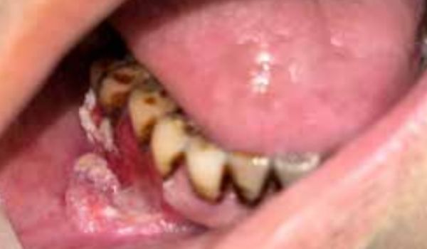
Figure 1: Intraoral lesion
Medical History: The patient had a history of myocardial infarction in 2022, for which he had undergone angioplasty. He was a former tobacco chewer but had quit following his cardiac event. This history of tobacco use was noted as a significant risk factor for his current condition.
Clinical examination
Extraoral examination revealed:
- No facial asymmetry
- Mouth opening of 3 fingers
- No palpable neck nodes
- No skin involvement
Intraoral examination showed:
- A 2x0.5cm ulceroproliferative lesion on the right mandibular buccal mucosa, extending from the canine to the second molar
- The retromolar trigone (RMT) was not involved.
An incisional biopsy confirmed the diagnosis of SCC
Imaging findings:
Positron Emission Tomography-Computed Tomography (PET -CT) scan (Figure 2): A 2X0.8 cm lesion on the right buccal mucosa involving the lower gingivobuccal sulcus, cortical erosion of the right hemimandible, and subcentrimetric lymph nodes bilaterally at levels IB and II. The RMT was free from tumour involvement.
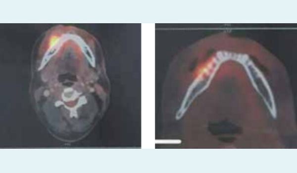
Figure 2: Positron Emission Tomography (PET) Scan
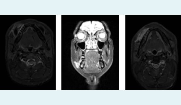
Figure 3,4,5: Showing MRI face and neck in coronal and axial section T1 and T2 weighted images
Treatment plan
Based on the clinical and radiological findings, the oncologic & plastic surgery team recommended the following treatment plan:
- Composite resection
- Marginal mandibulectomy
- Bilateral neck dissection (levels I-IV)
- Free fibula flap reconstruction
- Immediate dental implant placement for future dental rehabilitation.
The treatment approach involved a multidisciplinary team utilising advanced planning and surgical techniques, including 3D Computer-Aided Design (CAD) and Computer-Aided Manufacturing (CAM) technology for precise preoperative planning of the composite resection and reconstruction (Figures 6,7,8). This approach facilitated accurate mandibular resection, fibula flap harvest, and immediate dental implant placement, ensuring optimal functional and aesthetic outcomes. Immediate dental implants were placed to facilitate future prosthetic rehabilitation. A 3D-printed surgical guide was fabricated to ensure accurate bone resection and reconstruction.
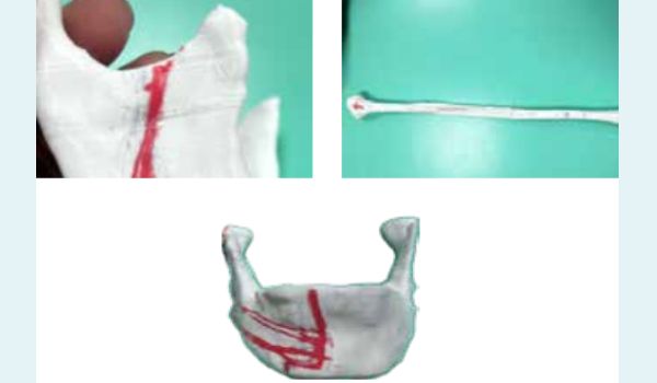
Figure 6,7,8: Showing preoperative planning of the composite resection and reconstruction
The surgical procedure began with composite resection, including the tumour with adequate margins, followed by marginal mandibulectomy (Figure 9). The resected mandible specimen was placed along the bone cutting zig , which guided the precise amount of bone to be replaced. The reconstruction phase was performed by the plastic and reconstructive surgery team. The free fibula flap was harvested and shaped. After securing the flap to the residual mandible with titanium plates, immediate dental implants were placed in the fibula graft to support future prosthetic rehabilitation. (Figure 10)
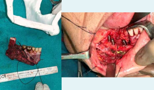
Figure 9,10: Images showing marking and placement of dental implants for prosthesis
Figure 6,7,8: Post operative picture of patient showing minimal facial discrepancies
The patient was scheduled for follow-up visits (Figure 12) to monitor healing and plan for implant-supported denture rehabilitation, which would restore normal masticatory function.
Figure 9,10: Patient on the 10th day of subsequent follow-up. The patient was well and comfortable and greeted the entire team and showed his gratitude
Results
The post-operative recovery was uneventful. By day three, the patient was stable and able to take oral fluids (Figure 11). Facial aesthetics were well-preserved, with no visible external scarring, as the entire procedure was performed intraorally. The patient expressed high satisfaction with the outcome, particularly noting the absence of visible scars. The histopathological examination confirmed clear surgical margins and the absence of nodal metastases.
Surgical technique
The procedure was performed under general anaesthesia (GA) in the right lateral decubitus position with a supine tilt. Pneumoperitoneum was achieved using a Veress needle at a point 4cm above and to the left of the umbilicus. A 10-mm optical trocar was introduced at that point. Three additional 5-mm ports were placed: two in the lower quadrants and one at Palmer’s point. Laparoscopic examination revealed a large left AIH with displacement of diaphragm (Figure 2), along with the spleen and splenic flexure of colon herniating through the intercostal space, with absence of any diaphragmatic defect. The hernia contents were reduced by releasing the lateral attachments, and the intercostal defect was closed with 1-0 non-absorbable barbed suture in a continuous uninterrupted manner after releasing the edges (Figures 3 and 4). A composite mesh (20x25cm, Parietex®, Covidien, USA.) was positioned to completely cover the defect. The mesh was secured with absorbable tacks (Absorbable Tack®, Covidien) to the abdominal wall with care taken to avoid the chest wall (Figure 5) The operative time was 110 minutes, and the postoperative course was uneventful, with mild postoperative pain (VAS 1-2). The patient was discharged on post-operative day 2 and followed up a week later, with no noteworthy events thereafter.
Discussion
Mandibular reconstruction following major resective surgery remains complex. Vascularised free tissue transfer with hard tissue, especially using a vascularised fibula free flap, provides one of the best possibilities for full functional mandibular rehabilitation. Dental implants placed in reconstructed areas have demonstrated normal integration with high success and survival rates when compared to those implants placed in native bone. The use of computer-assisted surgery and navigation technology in head and neck oncology was described in the early 1990’s by A. Wagner. In recent years, the concept of “precision medicine” has become part of standard hospital practice, allowing different products to be adapted to each patient in a specific way. Recent technological advances such as virtual surgical planning (VSP), CAD-CAM design and modelling, and intraoperative navigation techniques have contributed to simplify and improve the accuracy of surgeries. These techniques allow the pre-planning of the oncological resection, the dimensions of the neomandible and the precise location of the osteotomies in bone flaps. The CAD-CAM cutting guides help the surgical team to faithfully carry out the treatments, improving the precision, accuracy and reliability of the results in oncological resections and reconstructions. Intraoperative dynamic navigation systems allow immediate insertion of osseointegrated implants, contributing to faster dental rehabilitation. The fibula width is sufficient for the placement of commonly used dental implants, facilitating optimal oral rehabilitation after mandibular reconstruction.
Conclusion
This case report highlights the successful management of alveolar sulcus carcinoma extending to the buccal mucosa using a multidisciplinary approach. The integration of advanced surgical techniques, 3D planning, and immediate dental rehabilitation demonstrates the potential for improved outcomes in complex oral cancer cases. Further research is needed to establish long-term outcomes and refine patient selection criteria for these advanced interventions.
Pawan Gupta. Primary Osseointegrated Free Flap in a Middle-Aged Oral Cancer Patient with Excellent
Functional and Anatomical Outcomes: A Case Report. MMJ. 2024, Dec. Vol 1 (4).
DOI: XXXX
References
- Bray F, Ferlay J, Soerjomataram I, et al. Global cancer statistics 2018: GLOBOCAN estimates of incidence and mortality worldwide for 36 cancers in 185 countries. CA Cancer J Clin. 2018;68(6):394-424.
- Warnakulasuriya S. Global epidemiology of oral and oropharyngeal cancer. Oral Oncol. 2009;45(4-5):309-16.
- Shah JP, Gil Z. Current concepts in management of oral cancer–surgery. Oral Oncol. 2009;45(4-5):394-401.