 WhatsApp Us (24/7)
 +91 926 888 0303 (24/7)


Abstract:
Inflammatory disorders of pituitary are rare, and often present as space occupying lesions arising from it. A preoperative diagnosis of hypophysitis is a challenge due to the absence of any specific clinical signs other than those of a pituitary mass, and the magnetic resonance imaging (MRI) showing a pituitary mass lesion. Our patient, a female in her thirties, presented with headache, galactorrhoea and amenorrhoea. She underwent transsphenoidal endoscopic excision of a fluid-filled tumour arising from the pituitary. Histobiopsy and immunohistochemistry revealed the tumour to be granulomatous hypophysitis (GHy). In view of granulomas, she was put on antitubercular chemotherapy for twelve months. A follow up MRI showed no residual lesion.
Key words: Brain Tumour; Granulomatous; Hypophysitis; Pituitary; Tuberculosis.
Introduction
A patient presenting with headache and visual field defects raises a suspicion of a pituitary tumour. Appropriate neuroimaging (Gadolinium enhanced MRI) provides rapid confirmation and enables treatment planning. The atypical appearances of pituitary lesions can be seen regularly in high-volume neurosurgical centres, many of whom turn out to be inflammatory hypophysitis. There are only small series or case reports in literature that discuss this pathology. It is a rare pathology that causes tissue destruction and endocrine dysfunction. Granulomatous hypophysitis (GHy) is a common subtype featuring discrete granulomas of multinucleated giant cells, histiocytes and plasma cells. Surgery is essential to confirm the diagnosis and to relieve the symptoms of chiasmatic compression and headache.
Case Report
A 28-year-old female patient presented with a six-month history of intermittent frontal headache without accompanying vomiting or visual disturbances. Her menarche was at 12 years of age, and her menstrual cycles were normal. However, for about ten months prior to presentation she was having scanty periods, each lasting barely a day. In addition, she had noticed galactorrhoea. On examination, she was normotensive, with normal facies. Galactorrhoea was confirmed on clinical examination. Visual fields charting (Goldman) was normal. Gadolinium-enhanced MRI of brain revealed a peripherally enhancing sellar mass extending into the suprasellar space
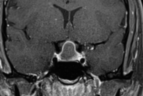
Figure 1: Contrast enhanced T1-weighted coronal MR image showing pituitary lesion with peripheral enhancement
(Figures 1 and 2). Her endocrine profile revealed mildly elevated prolactin levels (150 µg/L). The growth hormone (GH), thyroidstimulating hormone (TSH), cortisol, and adrenocorticotropic hormone (ACTH) levels were normal.
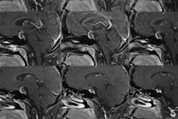
Figure 2: Contrast-enhanced T1-weighted sagittal MR image showing pituitary lesion with peripheral enhancement
The tumour was exposed by an endoscopic transsphenoidal approach. A dural incision in the sellar floor revealed a pale tumour with gel-like consistency. The tumour was curetted out and the operative corridor was sealed after placing a small pad of adipose tissue and application of biological sealant. Her postoperative period was uneventful, and she was discharged after 72 hours of surgery. She was free from headaches at the time of discharge. Her oligomenorrhoea however persisted during her follow-up period.
Histopathology (H & E stain) showed destruction and infiltration of pituitary parenchyma by lymphocytes, non-caseating granulomas with Langhans giant cells (Figures 3 & 4). Staining for acid fast bacilli and fungi was negative. She was put on antitubercular chemotherapy (rifampicin, isoniazid, ethambutol, pyrazinamide) for nine months. Further evaluation for cutaneous, skeletal and visceral survey for granulomatous lesions was negative.
A post-operative MRI six months later did not reveal any residual or recurrent lesion (Figure 5).
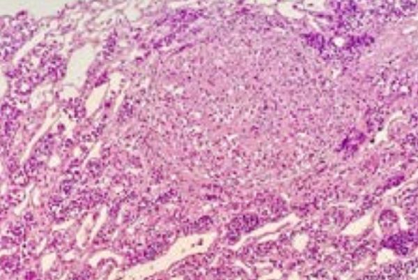
Figure 3: H & E section showing epithelioid cell granuloma in the background of native pituitary parenchyma. (presence of pituitary parenchyma confirmed by synaptophysin stain)
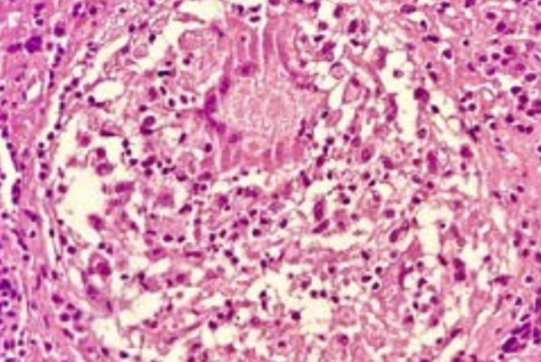
Figure 4: High power view of H & E section showing epithelioid cell granuloma, Langhans giant cell and foci of necrosis
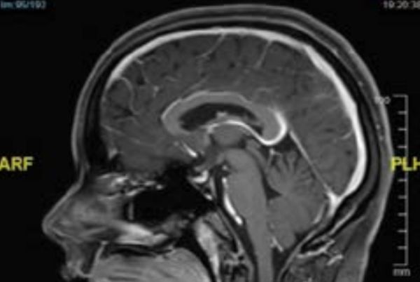
Figure 5: Postoperative contrast-enhanced T1-weighted sagittal MR image showing no residual pituitary lesion
Discussion
GHy is one of the five types of inflammatory hypophysitis (lymphomatous, granulomatous, xanthomatous, xanthogranulomatous and necrotizing). GHy is a rare lesion, seen more often in females.1 The aetiology of GHy may be idiopathic, tuberculous, fungal, systemic inflammatory disorders (Crohn’s disease, Sarcoidosis, Takayasu’s arteritis, Wegener’s granulomatosis) and foreign body reactions (rupture of Rathke’s cleft cyst, mucocele).2 Presentation is usually in the form of an expanding pituitary mass with or without diabetes insipidus. While headache is non-specific, visual field defects may be in the form of bitemporal hemianopia. Gross endocrinological derangements are unusual, while few patients have shown visual field defects. Our patient had galactorrhoea and scanty menstruation on presentation. Acute presentation may include extraocular paralysis, fever, and meningeal signs.3,4 Neuroimaging (contrast MRI) shows a pituitary mass expanding cranially without lobulations or lateral spread. Although the lesion can be suspected when the appearance of the space occupying lesion is atypical, firm diagnosis is achieved only when the tissue is submitted after surgery. Preoperative appearances on MRI are those of a uniformly enhancing space occupying lesion; a fluidfilled lesion with enhancing walls can be mistaken for a pituitary abscess or a cystic tumour. There may be dural enhancement (‘dural tail’) and pituitary stalk thickening.5 Almost half the patients with inflammatory hypophysitis are preoperatively diagnosed on MRI as pituitary adenoma.6
Surgery has diagnostic and therapeutic utility and provides rapid decompression of the optic apparatus and relief from headache. Surgery is by transsphenoidal approach. Postoperatively, the patient may require hormonal replacement.
Conclusion:
GHy is a rare disease of varied aetiology, of which tuberculosis is important in our country. The outcome of surgery is favorable, although hormone replacement is frequently required and long-term follow-up is important.
Harjinder S Bhatoe, Anjali Bhutani, Anup Roy. Granulomatous Hypophysitis: A Case
Report. MMJ. 2024, December. Vol 1 (4).
DOI: XXXXXXXX
References
- Hunn BH, Martin WG, Simpson S Jr, et al. Idiopathic granulomatous hypophysitis: a systematic review of 82 cases in the literature. Pituitary. 2014;17:357-365.
- Imber BS, Lee HS, Kunwar Set al. Hypophysitis. A single center case series. Pituitary. 2015;18:630-641.
- Bhansali A, Velayutham P, Radotra BD, et al. Idiopathic granulomatous hyphysitis presenting as non-functional pituitary adenoma: description of six cases and review of literature. Br J Neurosurg. 2004;18:489-494.
- Yoshioka M, Yamakawa N, Saito H, et al. Granulomatous hypophysitis with meningitis and hypopituitarism. Intern Med. 1992;31:1147-1150.
- Su SB, Zhang DJ, Yue SY, Zhang JN. Primary granulomatous hypophysitis: a case report and literature review. Endocr J . 2011;58:467-473.
- Leung GK, Lopes MB, Thorner MO, Vance ML, Laws ER, Jr. Primary hypophysitis: a single center experience of 16 cases. J Neurosurg. 2004;101:262-271.