 WhatsApp Us (24/7)
 +91 926 888 0303 (24/7)


Background:
Rotational atherectomy (RA) has been studied in instent restenosis (ISR) and underexpanded, undilatable stents implanted in calcified lesions, but the data is limited. Further, ostial side branch (SB) lesions (Medina 0,0,1) are a difficult subset to treat and need an individualised approach. Our case report shows how RA helped in transverse partial stent ablation of ostial SB stent with significant main vessel protrusion.
Case Summary:
An 81-year-old female presented to our emergency room (ER) with Wellens syndrome. She had a history of stenting to the diagonal (D1) artery 2 years back. Echocardiography showed a left ventricular ejection fraction (LVEF) of 50% with no wall motion abnormalities. Coronary angiography showed a significant proximal left anterior descending artery (LAD) disease involving D1 origin that had 50% ISR. Our plan was to perform provisional left main (LM)-LAD stenting followed by kissing balloon inflation of LAD-D1. However, we could not cross the smallest available (1x5mm semi-compliant [SC] balloon) beyond the origin of D1. Hence, RA was performed, ablating the D1 stent struts that were protruding into the LAD, following which percutaneous coronary intervention (PCI) was successfully completed.
Discussion:
Stent ablation with RA for the management of ISR and underexpanded or undilatable stents has been documented, though its use in ablating ostial SB stents is rare. Our case highlights the potential use of RA in transverse partial stent ablation of ostial SB stents protruding into the main vessel.
Key words: LAD, RCA, Diagonal (D1), Instent restenosis (ISR), Rotational atherectomy (RA)
Case presentation
An 81-year-old female presented to our emergency room (ER) with chest pain for 4 hours. There was history of percutaneous coronary intervention (PCI) with drug-eluting stents (DES) to the diagonal (D1) artery 2 years back. Her general physical and systemic examination was normal. Baseline investigations revealed anaemia with a haemoglobin (Hb) level of 10.7g/dl, total leukocyte count (TLC) of 8.6x10⁹/L, mean corpuscular volume (MCV) of 83.2fL, mean corpuscular haemoglobin (MCH) of 26.3pg, and platelet count of 207x109/L. Her renal function test revealed urea of 29.8mg/dL, and creatinine of 0.95mg/dL. Highsensitive troponin I was 0.02ng/ml. The electrolytes included Na 134mmol/L, K 4.3mmol/L, Cl 98mmol/L, HCO3 17.5mmol/L, and Ca 9.11mg/dL. Her liver function test was normal. Her electrocardiogram (ECG) showed a Wellens pattern (Figure 1).
Case Discussion
A 46-year-old female presented with complaints of headache of one-month duration. Headache was mild in intensity, holocranial with no diurnal variation. There was no neurological deficit, such as aphasia, motor weakness, or gait abnormalities. Her vitals were stable and laboratory test results were within the normal range.
Magnetic Resonance Imaging (MRI) brain with contrast (Figure 1) was suggestive of a broad-based extra-axial mass lesion measuring 5.8x5cm in the left frontal convexity with cerebrospinal fluid (CSF) cleft and compression of the underlying brain parenchyma with moderate perifocal oedema. A broad dural attachment was seen. The mass was compressing the left lateral ventricle, causing a midline shift of 11.6mm to the right. Overall, the MRI brain findings were suggestive of a cystic meningioma.
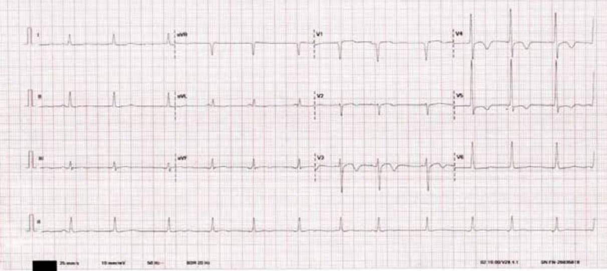
Figure 1: The patient’s ECG shows sinus rhythm, with heart rate of around 100/min. T-wave inversions noted in the precordial leads, most prominently in V3-V5. Notably, no significant ST elevation & no precordial Q waves. This T-wave abnormality pattern is typical of Wellens syndrome
Echocardiography showed concentric left ventricular hypertrophy (LVH), and Grade I diastolic dysfunction with left ventricular ejection fraction (LVEF) of 50%. No wall motion abnormalities were observed. The patient was diagnosed with Wellens syndrome and taken for coronary angiography. The coronary angiography showed significant disease in the proximal left anterior descending (LAD) artery as well as the ostial and proximal right coronary artery (RCA) (Figures 2a, 2b).
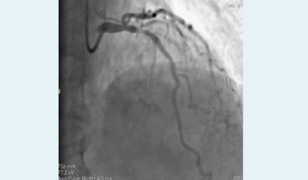
Figure 2a:
Figure 2a & 2b: Right anterior oblique (RAO) cranial & left anterior oblique (LAO) caudal views show distal left main disease with 70%-80% hazy lesion in the proximal left anterior descending (LAD) artery at the diagonal (D1) bifurcation (Medina 1,1,1). There is focal in-stent restenosis (ISR) near the proximal diagonal (D1) stent edge causing around 50% luminal narrowing in the ostioproximal D1. The right coronary artery (RCA) is the dominant vessel showing around 80% stenosis at the ostium followed by a 60%-70% eccentric lesion in the proximal RCA. The distal RCA has mild plaquing. The posterior descending artery (PDA) and posterolateral ventricular (PLV) branches are normal.
Our plan was to perform provisional left main (LM)-LAD stenting from the mid-LM up to the first major septal branch, followed by kissing balloon inflation of LAD-D1. We started with 6F extra backup (EBU) guide catheter via the right radial route and wired the LAD with a Runthrough NS wire. However, we were unable to cross the smallest available balloon [1x5mm semi-compliant (SC)] beyond the origin of D1. We realized that the old D1 stent was protruding well into the LAD. We then switched to the femoral route with a 7F EBU to increase guide support, rewired multiple times with multiple wires, used buddy wire and anchoring balloon in D1; however, we were still not able to cross. Hence, we decided to ablate the D1 stent struts protruding into LAD with rotational atherectomy (RA). We parked a Finecross microcatheter proximal to D1 as we could not cross it, and then wired the LAD directly with a Rotafloppy wire. Using a 1.25mm burr at 180,000 rpm, we performed RA for a duration of 2.5minutes, which allowed us to cross successfully. Next, we completed the PCI with 4x28mm DES, achieving a good final result (Figure 3a-3d & 4a-4c). The patient remained asymptomatic at her six-month follow-u
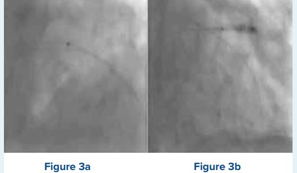
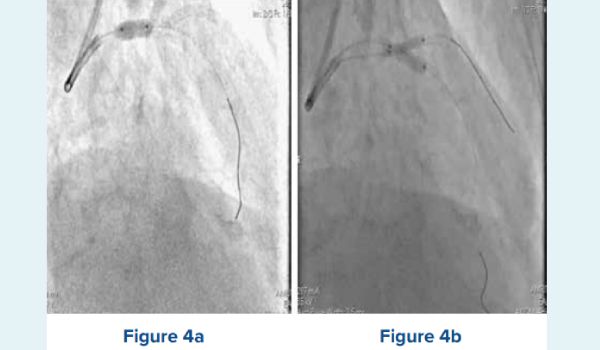
Figure 3a-3d: Figure 3a-3d: Showing lesion preparation & stent implantation from the left main (LM) to the left anterior descending (LAD) artery. Figure 3a shows a buddy wire being used to negotiate a 1x5mm semi-compliant (SC) balloon across, which was unsuccessful. Figure 3b shows a 2.5x12mm non- compliant (NC) balloon anchored in Diagonal (D1), with attempt to cross the 1x5mm SC balloon, which was also unsuccessful. Figure 3c shows the 1.25mm burr successfully crossing the origin of D1. Figure 3d shows the 4x28mm drug-eluting stent (DES) being deployed from the LM to the LAD.
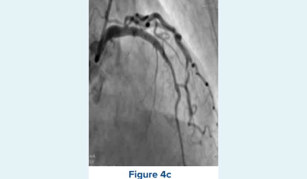
Figure 4a-4c: Showing the optimisation of the deployed stent and the final angiographic result. Figure 4a shows post dilation (POT) being done in the left main (LM) with a 5x8mm noncompliant (NC) balloon. Figure 4b shows simultaneous kissing balloon inflation being done using 4x10mm and 2.75x10mm NC balloons. Figure 4c shows a satisfactory final angiographic result.
Discussion
Our case highlights two key points; first, that ostial side branch (SB) lesions are a challenging subset requiring an individualised management approach, and second, the potential role of RA in transverse partial stent ablation of ostial SB stents with significant main vessel protrusion. Figure 5 shows a proposed approach to non-left main ostial SB (Medina 0,0,1) lesions.
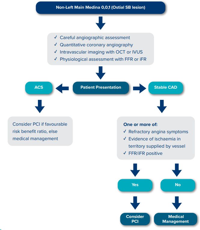
Figure 5: Shows an approach to non-left main Medina 0,0,1 [ostial side branch (SB)] lesions. Lesion definition using intravascular imaging & physiological assessment is paramount. Moreover, the clinical presentation of the patient will dictate the best management strategy. Hence this lesion subset deserves an individualised approach to management.
OCT: Optical Coherence Tomography; IVUS: Intravascular Ultrasound; FFR: Fractional Flow Reserve; iFR: Instantaneous WaveFree Ratio; SB: Side Branch; CAD: Coronary Artery Disease; PCI: Percutaneous Coronary Intervention.
There are some technical considerations in the interventional management of Medina 0,0,1 lesions as it is not possible from a geometric standpoint to place a stent right at the ostium of a SB except with a bifurcation angle of 90°.
There will inevitably be some ostial miss or main vessel protrusion if we try to place a stent right at the ostium of SB in bifurcation angles <90°.1 Our case demonstrates how main vessel protrusion can cause marked difficulty in performing main vessel interventions if needed in the future. Several techniques have been proposed for ostial SB stenting, including the Stent Drawback technique, Szabo technique, Inverted Provisional Stenting/Crossover Technique, Crush technique without Main Vessel Stenting & Modified Flower Petal technique. Observational studies on drug-eluting balloons (DEB) in Medina 0,0,1 lesions showed that DEBs are safe and technically feasible therapeutic options.2 Studies of RA in ISR have yielded conflicting results.3,4 Small case series on the use of RA for treating undilatable, underexpanded stents implanted in calcific lesions have shown acceptable results, given the complexity of these lesions and limited intervention options.5,6 Transverse partial stent ablation of stent struts jailing the left circumflex (LCX) ostium with RA for suboptimal culotte technique in LM bifurcation has been reported.7
Conclusion
Ostial SB lesions are a challenging subset of coronary lesions that need an individualised approach. Studies on longitudinal stent ablation in ISR have yielded conflicting results. However, stent ablation with RA for managing underexpanded and undilatable coronary stents has been shown to be a safe and effective procedure with acceptable outcomes. Our case highlights the potential use of RA in transverse partial stent ablation of ostial SB stents that protrude into the main vessel.
Ishtiyaq Masood, Haider Rashid, Bupinder Brar, Neeraj Singla. An Unusual Balloon Uncrossable Lesion.
MMJ. 2024, Dec. Vol 1 (4)
References
- Suleiman S, Coughlan JJ, Touma G, et al. Contemporary management of isolated ostial side branch disease: an evidence-based approach to medina 001 bifurcations. Interventional Cardiology Review. 2021 Apr;16.
- Vaquerizo B, Fernández‐Nofreiras E, Oategui I, et al. Second‐ generation drug‐eluting balloon for ostial side branch lesions (001‐bifurcations): mid‐term clinical and angiographic results. Journal of Interventional Cardiology. 2016;29(3):285-92.
- Sharma SK, Kini A, Mehran R, et al. Randomized trial of rotational atherectomy versus balloon angioplasty for diffuse in-stent restenosis (ROSTER). American heart journal. 2004;147(1):16-22.
- vom Dahl J, Dietz U, Haager PK, et al. Rotational atherectomy does not reduce recurrent in-stent restenosis: results of the angioplasty versus rotational atherectomy for treatment of diffuse in-stent restenosis trial (ARTIST). Circulation. 2002;105(5):583-8.
- Ferri LA, Jabbour RJ, Giannini F, et al. Safety and efficacy of rotational atherectomy for the treatment of undilatable underexpanded stents implanted in calcific lesions. Catheterization and Cardiovascular Interventions. 2017;90(2):E19-24.
- Whiteside HL, Nagabandi A, Kapoor D. Stentablation with rotational atherectomy for the management of underexpanded and undilatable coronary stents. Cardiovascular Revascularization Medicine. 2019;20(12):1203-8.
- De Maria GL, Kharbanda R, Banning AP. Transverse partial stent ablation with rotational atherectomy for suboptimal culotte technique in left main stem bifurcation. Catheterization and Cardiovascular Interventions. 2018;91(6):1074-8.