 WhatsApp Us (24/7)
 +91 926 888 0303 (24/7)


Abstract:
Acquired abdominal intercostal hernias (AIH) are rare and represent an uncommon presentation of herniation of abdominal contents through an acquired weakness in the musculature, secondary to trauma or other connective tissue disorder. We discuss the case of a 66-year-old gentleman who presented with a left lateral abdominal swelling and a history of trauma one year prior. Intercostal hernias should be suspected following high-impact thoracic wall injuries. A computed tomography (CT) scan is the investigation of choice. Surgical repair of the defect, including the placement of a prosthetic mesh, ensures a favourable outcome.
Key words: Acquired Abdominal Intercostal Hernia (AIH), Costal Margin Rupture (CMR), Diaphragmatic Rupture (DR), Diaphragmatic Collapse.ine (T-DM1)
Introduction
Intercostal hernias are a rare and exceptional clinical condition that involve the protrusion of an organ or part of an organ through a defect in the intercostal muscles or ribs in the chest wall.1 This phenomenon has been referred to by various terms, including "intercostal pleuroperitoneal hernia" and "transdiaphragmatic intercostal hernia". The first cases were reported during the 1960s. Acquired abdominal intercostal hernias (AIH) typically stem from trauma or previous surgical procedures, with the preferred management approach being surgical repair.2 Insufficient experience and incorrect techniques frequently result in missed diagnosis and suboptimal surgical interventions, leading to high recurrence rates.
Case report
A 66-year-old male presented to our hospital with an abdominal swelling below the left lower chest wall, which he first perceived around 8 months ago and which had gradually increased in size. He experienced mild pain but no obstructive symptoms. The patient also reported a history of a road traffic accident one year earlier. The swelling increased while coughing and decreased when in the right lateral decubitus position. He had no other significant medical history or surgical risk factors. Physical examination demonstrated a parietal swelling in the left lateral aspect of the left lower abdomen and inferior costal margin. A reducible swelling was noted between the left 9th and 10th ribs, extending into the left hypochondrium. Blood investigations were unremarkable. Contrast-enhanced computed tomography (CECT) of the abdomen revealed a lateral hernia bulge in the left hypochondrium extending into the left 9th and 10th intercostal spaces, with inferior displacement of the lower ribs and fractures involving the left 9th and 10th ribs (Figure 1). The spleen and the splenic flexure of the colon were identified as the contents of the hernia sac. A diagnosis of left AIH with costal margin rupture (CMR) was made and an elective laparoscopic repair was planned.
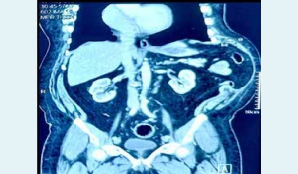
Figure 1: Computed Tomography (CT) scan shows acquired abdominal intercostal hernia (AIH) with colon and spleen prolapsing into the hernia.
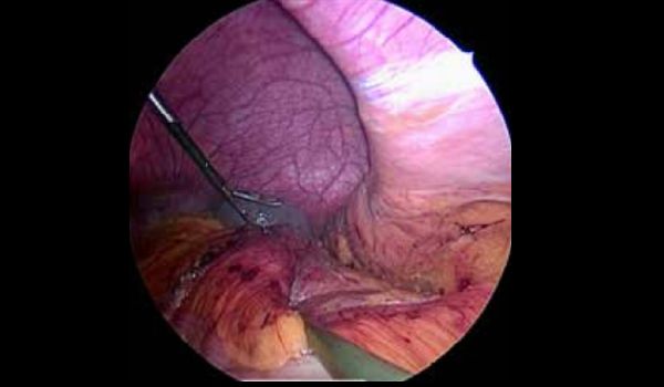
Figure 2: Large intercostal hernia with contents
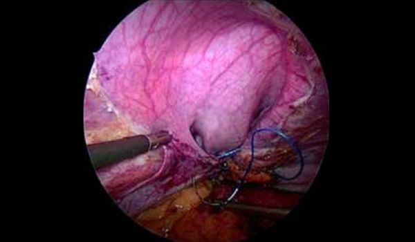
Figure 3: Suture repair of hernia from lateral to medial with diaphragmatic collapse into hernia noted
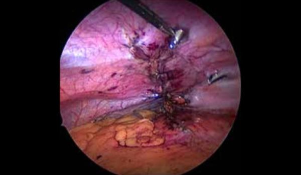
Figure 4: Complete anatomical approximation with sutures
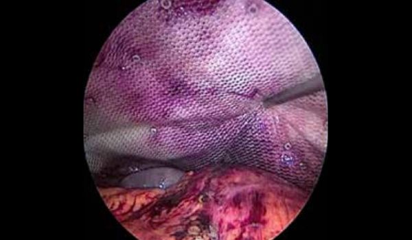
Figure 5: Mesh placement and fixation.
Surgical technique
The procedure was performed under general anaesthesia (GA) in the right lateral decubitus position with a supine tilt. Pneumoperitoneum was achieved using a Veress needle at a point 4cm above and to the left of the umbilicus. A 10-mm optical trocar was introduced at that point. Three additional 5-mm ports were placed: two in the lower quadrants and one at Palmer’s point. Laparoscopic examination revealed a large left AIH with displacement of diaphragm (Figure 2), along with the spleen and splenic flexure of colon herniating through the intercostal space, with absence of any diaphragmatic defect. The hernia contents were reduced by releasing the lateral attachments, and the intercostal defect was closed with 1-0 non-absorbable barbed suture in a continuous uninterrupted manner after releasing the edges (Figures 3 and 4). A composite mesh (20x25cm, Parietex®, Covidien, USA.) was positioned to completely cover the defect. The mesh was secured with absorbable tacks (Absorbable Tack®, Covidien) to the abdominal wall with care taken to avoid the chest wall (Figure 5) The operative time was 110 minutes, and the postoperative course was uneventful, with mild postoperative pain (VAS 1-2). The patient was discharged on post-operative day 2 and followed up a week later, with no noteworthy events thereafter.
Discussion
Acquired intercostal hernias were first discussed by Precht Z in 1959, marking the beginning of their formal description.3 According to the Sheffield classification of injuries involving the costal margin, these hernias are classified into multiple types, mainly traumatic diaphragmatic and intercostal hernia (TDIH), CMR with diaphragmatic rupture (CMR+DR), CMR with intercostal hernia (CMR+IH), CMR, traumatic diaphragmatic and intercostal hernia without CMR (TDIH w/o CMR), intercostal hernia (IH) and diaphragmatic rupture (DR).4 The term AIH remains undefined, with no clear distinction between instances where viscera reach the intercostal space directly from the peritoneal cavity or through a transdiaphragmatic defect.4 Any chest swelling post-trauma raise concern for acquired abdominal costal hernia triggering an urgent need for a computed tomography (CT) scan to rule out diaphragmatic or abdominal injury.4,5 These hernias are most commonly found below the 9th rib, without significant differences between sides. The primary symptoms are chest swelling (85%), often associated with discomfort or pain (76%). Acute complications such as incarceration and strangulation, are rare.5 A recent review of the literature revealed that intercostal hernias typically occur below the 9th rib, are a rare form of hernia, and are characterised by protrusions through the spaces between the ribs.5 They can be either congenital or acquired, with the acquired form usually stemming from trauma, surgery, or chronic coughing. The lower placement of these hernias is thought to be due to increased weakness in the lower intercostal muscles and decreased support from surrounding tissues. Diagnosis can be challenging due to their rarity and atypical presentation, often leading to delays in treatment. Traditionally, these hernias were treated by thoracoabdominal incisions.6,7 Our method of repair aimed to provide suture closure of the defect with intraabdominal reinforcement with a large size mesh, leveraging the physiologic benefits of a minimally invasive procedure. The literature offers limited insights into laparoscopic repair methods, except for a few case studies exploring robot-assisted repairs.8-11 It is advisable to prioritise tension-free prosthetic repair over rib approximation. When rib fractures are detected and there is no urgency for surgery, delaying hernia repair until complete bone healing has occurred may be more beneficial.12
Conclusion
A bulge in the costal region should invite a high index of suspicion following major or minor trauma. Imaging by CT plays a crucial role in the diagnosis and planning management, guiding treatment strategies and surgical interventions when necessary. Intercostal defects can be effectively repaired through surgical interventions. The laparoscopic approach offers a minimally invasive method for repairing intercostal defects, reducing postoperative pain, shortening recovery times, and delivering excellent cosmetic outcomes compared to traditional open surgeries.
Parikshith Mahesh Chandra, Vandana Soni, Anil Sharma, Manish Baijal, Rajesh Khullar, Pradeep Chowbey.
Exploring the Uncommon: Intercostal Herniation with Costal Margin Rupture – A Case Report. MMJ. 2024,
Dec. Vol 1 (4).
DOI: XXXX
References
- Abunnaja S, Chysna K, Shaikh I, et al. Acquired Abdominal Intercostal Hernia: A Case Report and Literature Review. Case Rep Surg. 2014;2014:1-5.
- Sharma A, Mehare S, Rakesh CR. Traumatic abdominal intercostal hernia: A rare experience. Med J Armed Forces India. 2023;79(1):101-4.
- PRECHT Z. Intercostal diaphragmatico-abdominal hernia. Pol Przegl Chir. 1959;31:697-9.
- Gooseman MR, Rawashdeh M, Mattam K, et al. Unifying classification for transdiaphragmatic intercostal hernia and other costal margin injuries. Eur J Cardiothorac Surg. 2019;56(1):150-8.
- Erdas E, Licheri S, Calò PG, et al. Acquired abdominal intercostal hernia: case report and systematic review of the literature. Hernia. 2014 Oct 13;18(5):607-15.
- Szentkereszty Z, Boros M, Sápy P, et al. Surgical treatment of intercostal hernia with implantation of polypropylene mesh. Hernia. 2006;10(4):354-6.
- Takeuchi Y, Kurashima Y, Nakanishi Y, et al. Mesh trimming and suture reconstruction for wound dehiscence after huge abdominal intercostal hernia repair: A case report. Int J Surg Case Rep. 2018;53:381-5.
- Kurer MA, Bradford IMJ. Laparoscopic Repair of Abdominal Intercostal Hernia: A Case Report and Review of the Literature. Surg Laparosc Endosc Percutan Tech. 2006;16(4):270-1.
- Dan D, Ramraj P, Solomon V, et al. Laparoscopic repair of a rare acquired abdominal intercostal hernia. Int J Surg Case Rep. 2014;5(12):1041-3.
- Luqman MQ, Mughal A, Waldron R, et al. Laparoscopic IPOM repair of an acquired abdominal intercostal hernia. BMJ Case Rep. 2018; bcr-2018-227158.
- Drexel S, Watkins J, Tseng D. Robotic thoracoabdominal hernia repair: a novel approach. Hernia. 2023;28(1):249-54.
- Visagan R, McCormack DJ, Shipolini AR, et al. Are intracostal sutures better than pericostal sutures for closing a thoracotomy? Interact Cardiovasc Thorac Surg. 2012;14(6):807-15.