 WhatsApp Us (24/7)
 +91 926 888 0303 (24/7)


Introduction
The wide availability and recent improvement in technology coupled with portability, low cost and safety makes ultrasonography (USG) the first-choice imaging investigation for the evaluation of musculoskeletal (MSK) diseases. Diagnostic use of USG findings is greatly enhanced by knowledge of the clinical presentation. Conversely, USG skills with its prerequisite anatomical knowledge make the clinical diagnosis more precise and reduce uncertainty in the choice of therapy. USG examination provides an excellent opportunity for patient education and to explain the rationale for therapy. This review summarises the indications for MSK USG and describes its role in diagnosis, monitoring and prognosis.
USG is the most practical and rapid method of obtaining images of the MSK system. It can be performed readily in the clinic, with assessment of multiple joints at the same appointment, providing a ‘one stop’ answer to many MSK problems. This relatively inexpensive technology with the benefits of portability and real-time dynamic examination has made it possible to provide a diagnostics service in the community or even on the sports field.
USG technology offers several inherent advantages. Being noninvasive, with a quick scan time and without radiation makes it well accepted by patients. There are several advantages from the clinician’s point of view. It allows contralateral examination and does not pose limitations due to metal artefacts, which can be problematic in magnetic resonance imaging (MRI). The ability to visualise needles and target structures in real time makes it an ideal tool for the guidance procedures used in diagnosis and management.[1]
There are several applications of real-time dynamic USG examination in the MSK system. USG can show tendon instability such as anterior dislocation of the extensor carpi ulnaris (ECU)[2]. It plays an important role in the diagnosis of impingement of the shoulder by showing which structure is being impinged and reveals potential intrinsic and extrinsic causes[3].
Despite these advantages, there are some limitations of this technology. USG is considered to be an operator-dependent technology with poor repeatability. However, it is reassuring to see that recent studies have established moderate to good interobserver reliability.[4,5,7]
Even with advances in the resolution of the transducers, deeper structures are difficult to visualise as the higher-frequency transducers have lower tissue penetration capability. MRI has clear advantages over USG in the imaging of deeper structures. MRI scans can also examine a larger area. Another limitation is the restricted access to certain joints which are difficult to image with an US probe.
Acquisition of US skills takes time depending on trainee’s hand–eye coordination skills. A long training period may be an important limiting factor in its popular use. In addition, examination of multiples joints in the clinical setting can be time consuming. Evidence is accumulating for focused examination with concentration on a small number of active joints to reduce examination time.[3]
Applications of ultrasonography in musculoskeletal disorders
Tendon abnormalities: The superficial tendons are well assessed on USG. Common pathologies include tendinosis (inflammation of the tendon), calcific tendinitis and tendon tear. (Fig 1-9)
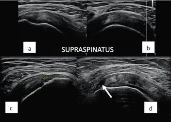
Figure 1: Supraspinatus tendon- Longitudinal and short axis (a and b) images showing heterogenous and thick tendon, suggesting tendinosis. (c) Complete tear of the tendon with retraction of fibers as marked by cursors. (d) Partial tear of the footprint of the tendon (arrow)
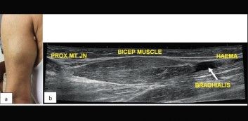
Figure 2: Biceps tendon- (a) Clinical picture of swelling in the mid arm. (b) There is a tear of the distal biceps tendon (arrow) with the formation of a small hematoma (haema)
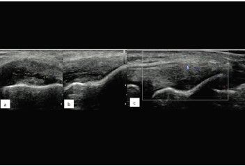
Figure 3: The power doppler signal indicates active inflammation in the tendon, which is indicated by cursors
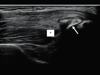
Figure 4: Triceps tendon – Longitudinal images show bony avulsion (arrow) of the deeper muscular attachment of triceps tendon (asterisk)
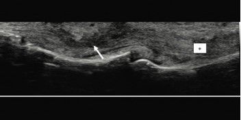
Figure 5: Flexor tendon of index finger- There is partial tear of the distal segment as seen by decreased echogenicity and loss of normal orientation of the fibers (arrow). Asterisk shows the normal proximal tendon
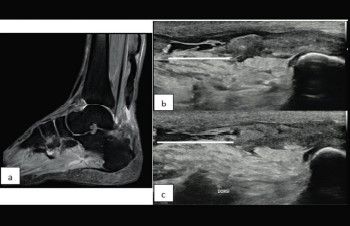
Figure 6: Achilles tendon – (a) Sagittal MRI image shows complete tear of the tendon with retraction of fibers (arrow). Dynamic USG in plantar flexion (b) and dorsifexion (c) redemonstrate the tear (white line) with increased tear gap in dorsiflexion
Tenosynovitis, trigger finger and volar plate injury are some other common abnormalities seen in relation to the tendon which can be well assessed on USG.
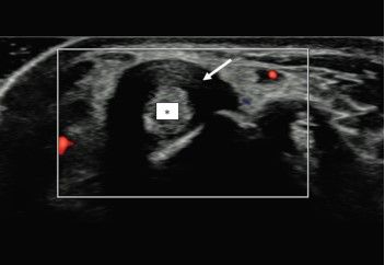
Figure 7: De Quervain’s tenosynovitis - Diffuse thickening of the sheath (arrow) of 1st compartment extensor tendons of the wrist (asterisk). This can be a part of the inflammatory arthritis or may be mechanical in nature
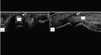
Figure 8: Trigger Finger – Short axis and longitudinal view (a and b) of the flexor tendon (asterisk) of the ring finger showing thickening of the A1 pulley (arrows) leading to trigger finger
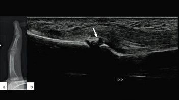
Figure 9: Volar plate avulsion – (a) Lateral radiograph of the index finger shows hyperextension at distal interphalangeal joint due to an avulsion injury of the volar plate from the base of the distal phalanx (arrow)
Ligament injury Superficial ligaments of the small joints of the hand and foot are easily assessed by high frequency linear probes. The common abnormalities of the ligament include sprain and tear. (Fig 10-11)
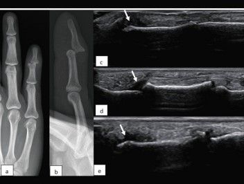
Figure 10: Frontal and lateral (a and b) radiographs of the little finger shows dorsal and medial subluxation of the distal interphalangeal (DIP) joint due to complete tear of the terminal extensor tendon at distal phalangeal attachment (arrow in c) and ulnar collateral ligament at DIP joint (arrow in e). There is also injury to the volar plate at DIP (arrow in d)
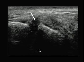
Figure 11: Longitudinal image of the medial aspect of carpometacarpal joint of thumb shows complete tear of the ulnar collateral ligament (arrow) which was post-traumatic in nature
Cystic lesion: USG is the first modality of choice for palpable surface swellings. Cystic lesions are one of the commonest lesions in clinical practice and ganglion cyst is one such commonly encountered lesion that can be diagnosed on USG. (Fig.12)
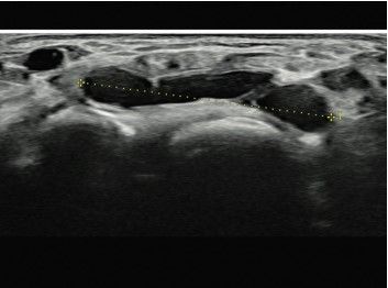
Figure 12: Dorsal wrist ganglion cyst - USG of the wrist shows a well-defined lobulated and septate cystic lesion in the dorsal aspect overlying the intercarpal joints
Nerve abnormalities: Superficial nerves of the upper and lower limbs are well assessed by USG, including the median, ulnar and radial nerves in upper limb and sciatic, posterior tibial and common peroneal nerves in lower limb. Nerve entrapment, nerve injury and masses of the nerve can be diagnosed with confidence on USG. (Fig.13-14)
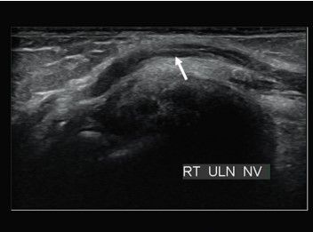
Figure 13: Cubital tunnel syndrome – Longitudinal image at the cubital tunnel shows long segment thickening of the ulnar nerve (arrow) suggesting entrapment of the nerve within the tunnel
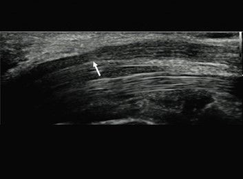
Figure 14: Carpal tunnel syndrome - Longitudinal image of the carpal tunnel shows compression of the median nerve (arrow) within the tunnel and thickening of the nerve proximal to it
Bony injuries: Cortical breach in some of the superficially placed bones can be detected on USG without the need of radiographs or CT scans which involve radiation. Some examples are avulsion fractures, stress fractures of the metatarsals and patella fracture. (Fig.15)
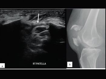
Figure 15: (a) Long axis USG image shows displaced fracture of the mid part of patella with fracture gap (arrow) which is redemonstrated on radiograph (b)
Arthritis: USG is the modality of choice in clinical suspicion of inflammatory arthritis of the hand and foot. Based on the pattern of distribution and radiological features like synovitis, tenosynovitis and bone erosion, differentiation between various types of inflammatory arthritis can be suggested. (Fig.16-20)
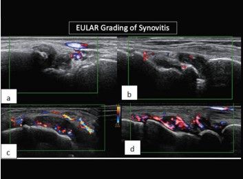
Figure 16: Grading of synovitis – USG of the wrist joint of different patients show grade-I (a), grade-II (b), grade-III (c) and grade-IV (d) synovitis as indicated by area of involvement by power doppler signal
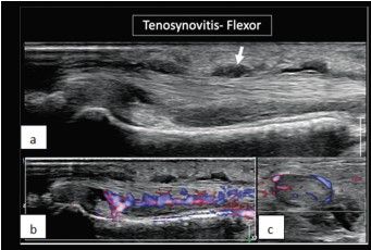
Figure 17: Tenosynovitis – Longitudinal grey scale image (a) shows thickening of the flexor tendon sheath of middle finger (arrow) which shows a power doppler signal in longitudinal (b) and short axis (c) views, suggesting inflammation
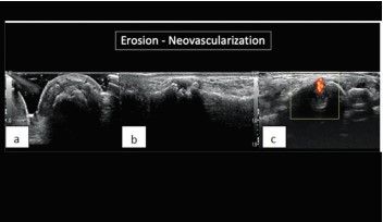
Figure 18: Bone erosion – Short axis (a) and longitudinal (b) image of the carpal bone shows erosion, which has a power doppler signal (c) suggesting active erosion with neovascularisation
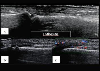
Figure 19: Enthesitis - is inflammation at the site of attachment of the tendon or ligament to the bone. It is usually seen in seronegative arthritis. (a,b) There is thickening of the extensor tendon at attachment to the base of the middle and distal phalanges of fingers which shows a power doppler signal (c)
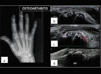
Figure 20: Osteoarthritis – (a) A radiograph of the hand shows degenerative changes at the 1st carpometacarpal and distal interphalangeal (DIP) joints. USG demonstrates osteophytes (b) and synovitis(c) at 1st carpometacarpal joint, along with osteophytes at one of the DIP joints(d)
Crystal deposition disease: Gout and calcium pyrophosphate deposition disease (CPPD) are two common crystal deposition diseases causing arthropathy. The crystals can be seen as echogenic deposits and there can be erosions in the bones. (Fig.21-22)
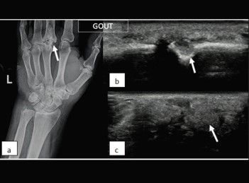
Figure 21: Gout - (a) radiograph shows punched out erosions in the head of the 3rd metacarpal (arrow). The erosions can be well appreciated on USG (b) with overlying large echogenic tophus (arrow in c)
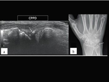
Figure 22: CPPD - Longitudinal USG image (a) shows calcification in the region of triangular fibrocartilage complex (arrow) which is redemonstrated on radiograph (arrow in b)
Conclusion CUSG provides a safe, cost-effective and rapid means of assessing MSK abnormalities. This review has emphasised the role of USG examination as the primary imaging investigation in initial evaluation of MSK diseases. In most aspects of assessment of MSK diseases, USG is comparable to or even better than the expensive imaging techniques such as MRI. The combination of high-frequency probes and improved power doppler technology provides a great opportunity to study image aspects of inflammatory conditions such as tenosynovitis and enthesitis that were traditionally considered difficult to image. Recent advances in technology such as small footprint high frequency probes, high image resolution, contrast USG and elastography have made USG a final diagnostic tool rather than just a screening modality. The long learning curve remains an important limiting factor in the widespread use of USG in routine clinical practice.
References
- Del Cura J.L. (2008) Ultrasound-guided therapeutic procedures in the musculoskeletal system. Curr Probl Diagn Radiol 37: 203–218 [PubMed] [Google Scholar]
- Bianchi S., Martinoli C., Sureda D., Rizzatto G. (2001) Ultrasound of the hand. Eur J Ultrasound 14: 29–34 [PubMed] [Google Scholar]
- Bureau N.J., Beauchamp M., Cardinal E., Brassard P. (2006) Dynamic sonography evaluation of shoulder impingement syndrome. Am J Roentgenol 187: 216–220 [PubMed] [Google Scholar]
- Scheel A.K., Hermann K.G., Kahler E., Pasewaldt D., Fritz J., Hamm B., et al. (2005a) A Novel ultrasonographic synovitis scoring system suitable for analyzing finger joint inflammation in rheumatoid arthritis. Arthritis Rheum 52: 733–743 [PubMed] [Google Scholar]
- Scheel A.K., Schmidt W.A., Hermann K.G., Bruyn G.A., D’agostino M.A., Grassi W., et al. (2005b) Interobserver reliability of rheumatologists performing musculoskeletal ultrasonography: results from a EULAR “Train the Trainers” course. Ann Rheum Dis 64: 1043–1049 [PMC free article] [PubMed] [Google Scholar]
- Schmidt W.A., Blockmans D. (2005) Use of ultrasonography and positron emission tomography in the diagnosis and assessment of large-vessel vasculitis. Curr Opin Rheumatol 17: 9–15 [PubMed] [Google Scholar]
- Naredo E., Moller I., Moragues C., De Agustin J.J., Scheel A.K., Grassi W., et al. (2006) Interobserver reliability in musculoskeletal ultrasonography: results from a “teach the teachers” rheumatologist course. Ann Rheum Dis 65: 14–19 [PMC free article] [PubMed] [Google Scholar]