 WhatsApp Us (24/7)
 +91 926 888 0303 (24/7)


Abstract: The goal of pregnancy ultrasound scans during the first trimester is to provide accurate information for optimised antenatal care, ensuring the best outcomes for both mother and foetus. Divided into early and late stages, early scans between 6 to 8 weeks focus on determining gestational sac implantation, viability and identifying multiple pregnancies. First-trimester screening, conducted at 11 + 0 to 13 + 6 weeks, includes biometry, anomaly scan, chromosomal abnormality assesment and screening for pre-eclampsia through maternal serum biochemistry. This pictorial essay emphasises the importance of a systematic examination of foetal anatomy during first-trimester screening, aiding in the early detection of structural anomalies. The protocol-based approach ensures reliability in detecting or ruling out abnormalities, allowing for timely counselling and further testing if necessary. While both transabdominal and transvaginal approaches may be utilised, the latter may offer better resolution, especially in certain patient populations. Despite limitations, a comprehensive first-trimester anomaly scan serves as a crucial tool in detecting major foetal abnormalities and providing reassurance to expecting mothers.
Key words: Ultrasound scan, pregnancy, first-trimester pregnancy
Introduction
The goal of a pregnancy ultrasound scan is to provide accurate information which will facilitate optimised antenatal care and ensure the best possible outcomes both for the mother and foetus. The first trimester is divided into early (6-8 weeks) and late first (11 + 0 to 13 + 6 weeks) trimester. During an early first trimester scan, which is performed typically between 6 to 8 weeks of gestation, the site of implantation of the gestational sac, viability along with the number of foetuses and in the presence of multiple pregnancy chorionicity and amnionicity, must be determined. First-trimester screening is performed at 11 + 0 to 13 + 6 weeks’ gestation with the crown rump measurement between 45mm and 84mm.
First trimester screening includes:
- Biometry (BPD, HC, AC, FL and CRL)
- Anomaly scan
- Assessment for chromosomal abnormalities (NT, NB, DV, TVD)
- Screening for Pre-Eclampsia (Mean UAD PI)
- Maternal serum biochemistry (First trimester quadruple test/NIPT)
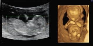
Figure 1: First trimester 2D and 3D image
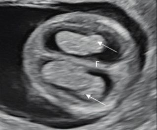
Figure 2: Head and Brain Axial View [(Cranial calcification, two brain halves separated by interhemispheric falx(F), Choroid plexus-Butterfly sign (Arrows)]
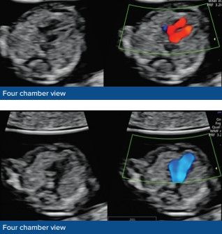
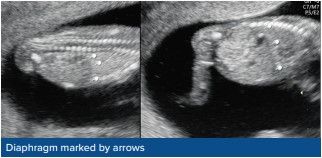
Figure 3: Cardia - Axial View (Four-chamber view with two distinct ventricles on grayscale and colour doppler in diastole, Three Vessel view -Y sign, Thorax - axial and sagittal view, symmetry with cardia occupying 1/3rd of the chest, Diaphragmatic continuity)
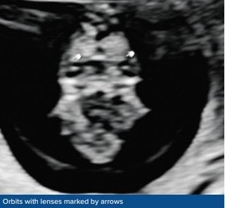
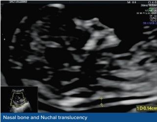
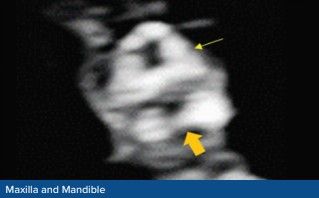
Figure 4: Face and neck (Bilateral orbits, Nasal bone, Nuchal translucency thicknes, maxilla, mandible)
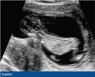
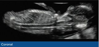
Figure 5: Spine-Sagittal and coronal view [Regular shape and continuity of spine].
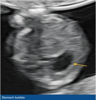
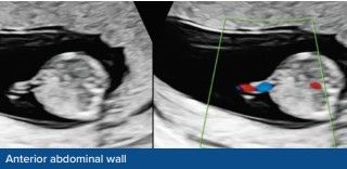
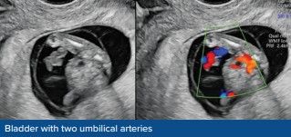
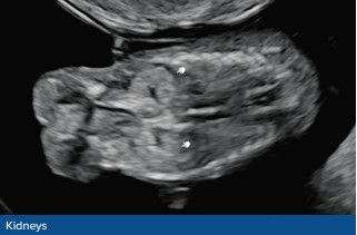
Figure 6: Abdomen- Axial View [Stomach bubble, intact anterior abdominal wall, foetal bladder (< 7mm longest diameter) with 3 vessel cords, Kidneys]
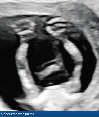
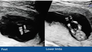
Figure 7: Extremities (Upper limbs with three segments, Lower limbs with three segments)
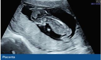
Figure 8: Placenta [Size and texture normal, without cystic appearance]
Conclusion The first-trimester anomaly scan provides an opportunity to assess foetal structural abnormalities and their presence or absence should be assessed as a minimum standard in all patients presenting for an 11 + 0 to 14 + 0-week scan, providing early reassurance. Early detection allows ample time for counselling and further testing if needed.
Both transabdominal and transvaginal approaches may be required to complete a systematic evaluation. A Transvaginal approach may provide better image resolution for the assessment of foetal anatomy, especially in women with high body mass index, uterine fibroids or retroverted uterus
Many major malformations may develop later in pregnancy or may not be detected even with appropriate equipment and in the most experienced of hands. However, a protocol-based, detailed first-trimester anomaly scan allows detection of major foetal abnormalities and reassurance to the expecting mother.
References
- ISUOG Practice Guidelines (updated): performance of 11–14- week ultrasound scan. Ultrasound Obstet Gynecol 2023
- Snijders RJ, Johnson S, Sebire NJ, Noble PL, Nicolaides KH. First-trimester ultrasound screening for chromosomal defects. Ultrasound Obstet Gynecol. 1996;7(3):216-226. doi:10.1046/j.1469-0705.1996.07030216.x
- Sonek J. First trimester ultrasonography in screening and detection of fetal anomalies. Am J Med Genet C Semin Med Genet. 2007;145C(1):45-61. doi:10.1002/ajmg.c.30120
- Bromley B, Henningsen C, Jones D, Timor-Tritsch I, Simpson L, Thiel L. AIUM practice parameter for the performance of detailed diagnostic obstetric ultrasound examinations between 12 weeks 0 days and 13 weeks 6 days. J Ultrasound Med. 2021;40(5):E1-E16. doi:10.1002/jum.15477
- Karim JN, Roberts NW, Salomon LJ, Papageorghiou AT. Systematic review of first trimester ultrasound screening in detecting fetal structural anomalies and factors affecting screening performance. Ultrasound Obstet Gynecol 2017;50:429–441