 WhatsApp Us (24/7)
 +91 926 888 0303 (24/7)


Abstract: Donath Landsteiner (DL) antibodies are biphasic IgG autoantibodies formed in response to a viral or bacterial infection and can induce paroxysmal cold hemoglobinuria (PCH), an autoimmune hemolytic anaemia. It is more frequently seen in children post-infective episodes and is transient and self-limiting in nature. We report a case of a 4.5-year-old male who presented with sudden intravascular hemolysis and AKI with a 2-weekold history of fever and acute viral hepatitis. AKI in PCH is a rarity, and the diagnoses of PCH could only be made correctly with the help of a DL antibody test, which helped with subsequent patient management and avoiding unnecessary treatment escalation.
Key words: AIHA, donath–landsteiner test, paroxysmal cold hemoglobinuria, acute kidney injury
Introduction
Paroxysmal cold hemoglobinuria (PCH) is an acquired complement-mediated autoimmune hemolytic process associated with polyclonal biphasic IgG autoantibodies directed against the P antigen on human red blood cells (RBCs). Molecular mimicry stimulates the production of these antibodies as some microbial antigens share structural similarities to the P antigen on RBCs.[1] These antibodies are biphasic and bind at cold temperatures, causing activation of the complement cascade and intravascular hemolysis at 37°C. Most commonly seen in children < 5 years old with acute hemolysis, 1 - 3 weeks following a viral or bacterial infection or after immunisation.[2] Generally, the disease is transient and self-limiting. Diagnosis is based on high clinical suspicion of PCH, confirmed by a positive Donath Landsteiner (DL) test in which biphasic haemolysins are detected. However, in a real clinical setting, the serological diagnosis of PCH is not always easy. The Direct antiglobulin test (DAT) is usually positive for C3 only and can be wrongly diagnosed as a cold autoimmune hemolytic anaemia (AIHA). Here, we report a rare case of AIHA with AKI confirmed as PCH by the DL antibody test.
Case A 4.5-year-old male was admitted to an outside hospital with hemoglobinuria and a history of fever with the passage of high-coloured urine and yellowish discolouration of eyes for two days and presenting complaints of decreased urine output. The child had previously received treatment for acute viral hepatitis at another hospital. On investigation, the complete blood count (CBC) showed haemoglobin (Hb) – 2.6 gm/dL, total leucocyte count (TLC) - 15.0 x 10⁹/L and platelet count- 2.8 x 10⁹/L. Peripheral smear showed normocytic normochromic red cells with some anisocytosis and spherocytes; however, no schistocytes were seen. Total Bilirubin was 3.66 mg/dl; direct bilirubin was 0.8 mg/dl, ALT- 13 IU/dl, LDH- 2897 mg/ dl, S.Creatinine- 3.9 mg/dl, corrected reticulocyte count (CRC) was 1.09% and serum complement level (C3) was low (40 mg/ dl). ANA, C-ANCA, P-ANCA, Immunoglobulin level, Hepatitis B, Hepatitis C, and HIV were negative. Based on this, the possibility of HUS/ AIHA was considered. On admission, the child received two units of leukodepleted packed red blood cell (PRBC) and one methylprednisolone dose. Urine microscopy showed 2+ proteinuria and no RBC.
Differential diagnosis and laboratory workup
An initial clinical diagnosis of HUS/AIHA was made, and the child was started on IVIG, Intravenous (IV) methylprednisolone and IV antibiotics. Hemoglobinuria improved, but S. creatinine kept rising. In view of rising S. Creatinine (4.36 mg/dl) and blood urea nitrogen (132.5 mg/dl) along with altered sensorium and decreased urine output, the child started on hemodialysis on day 3 of admission. Subsequently, the child was intubated and put on ventilatory support. Amlodipine was used to control hypertension. Despite the above therapy, the child had worsening renal function. The prompt onset of severe hemolysis triggered suspicion of PCH; therefore, the patient’s samples were sent to our hospital blood centre for investigations for PCH.
Confirmation of the diagnosis
Direct antiglobulin test (DAT), indirect antiglobulin test (IAT), and test for Donath Landsteiner antibodies were performed. While IAT was negative, Polyspecific DAT was positive. Additional tests with monospecific antiglobulin reagent exhibited antiC3d antibody
As the clinical presentation and acute onset of hemolysis favoured the possibility of PCH, DL testing was performed. A fresh clotted blood sample collected and transported at 37°C was sent to our laboratory. The clotted sample was centrifuged at 37°C, and the serum was separated from the red blood cells. Freshly collected pooled normal sera were used as a complement source. Three sets of three tubes labelled as: A1, A2, A3; B1, B2, B3; and C1, C2, C3. Ten volumes of the patient’s serum were added to tubes 1 and 2 of each set, and 10 volumes of fresh normal serum were added to tubes 2 and 3 of each set. One volume of the 50% suspension of washed P-positive red cells was added to all tubes and mixed well. Three “A” tubes are placed in a bath of melting ice for 30 min and then at 37°C for 1 hour. Three “B” tubes were placed in a bath of melting ice and kept in melting ice for 90 min. Three “C” tubes were placed at 37°C and kept at 37°C for 90 min. All tubes were gently mixed and centrifuged, and the supernatant fluid was examined for hemolysis. DL test was positive, confirming PCH (Figure 1).
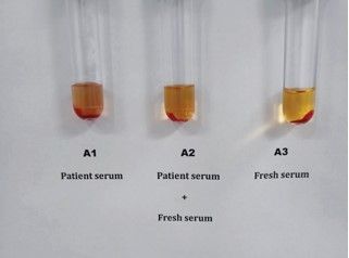
Figure 1: DL antibody detection
Management and follow-up
Hb stabilised on 4 days of hospitalisation after three units of PRBC transfusion. Hemoglobinuria resolved, and steroids rapidly tapered off and subsequently stopped. He received a total of 10 cycles of hemodialysis. His sensorium improved gradually, and urine output increased. His s.creatinine and LDH normalised (259 mg/dl at discharge). He was on ventilatory support for 3 days with 12 days of PICU stay
Discussion
There are limited population-level data on the prevalence of PCH from India. Still, most cases are seen in children before their fifth birthday who present with a postinfectious acute hemolytic episode that is self-limiting.
In the study, we report a case of PCH presenting with severe acute hemolysis with acute AKI and diagnoses clichéd by DL test. The case presented with oliguria, highly coloured urine, pallor and icterus, and a history of fever preceding the onset of AIHA, although no causative organism could be identified. Hb at presentation was < 3 g/L and showed normal reticulocyte count. Reticulocytopenia at presentation likely occurs due to both physiologic lag in reticulocyte generation as well as direct destruction of reticulocytes expressing the P antigen.[4] Polyspecific DAT was positive (3+), monospecific DAT was positive for C3d. High LDH, approximately 3000mg/dl, indicative of intravascular hemolysis. We noticed a low complement level in this case, which presented as AKI with intravascular hemolysis, which may correlate with its severe presentation, although no existing literature has shown such a correlation. Further data is needed to confirm the association of complement level with severity.
The case showed no difficulty in compatibility testing at the blood centre, probably due to the presence of cold reactive IgG antibodies, which do not agglutinate at room temperature. This case did not require prolonged transfusion support, reinforcing the transient nature of PCH despite an acute AKI in the backdrop. As hemoglobinuria stopped, the diagnosis of PCH was reenforced, corticosteroids were rapidly tapered, and the patient showed no recurrence on follow-up.
AKI in PCH is a rarity Das et al., in their report, have described a two-year-old boy who developed AKI secondary to PCH. The boy required peritoneal dialysis and plasma exchange.[6] Gunawardena et al. described a three-year-old girl with PCH and AKI who did not require renal replacement therapy.[7] Jessica et al. described a 4-year-old girl child with severe hemolytic anaemia and renal failure requiring two weeks of renal replacement therapy due to PCH. They also gave a trial of Eculizumab in this case. The 8 DL test is a specific and key test for establishing the diagnosis of PCH, and keeping the sample at 37°C during transportation to the laboratory helps maximise the number of antibodies in the serum and avoid a false negative test result. Coordination between the treating physician and laboratory is important for diagnosing and managing such patients. This case report highlights how a positive DL test helped in the timely diagnosis of PCH. Helped with subsequent management, avoiding unnecessary treatment escalation.
Learning points
Donath Lansteiner Hemolytic anaemia, or PCH, is a rare form of cold AIHA.
Sudden onset severe intravascular hemolysis following an infective episode with other investigations not correlating; a positive DL test is required for diagnosis.
Patients require predominantly only supportive care:
- Warm ambient temperature
- Blood transfusions as necessary are the mainstay of treatment
- Hemoglobin frequently falls below 6 g/dL during active hemolysis
From a transfusion medicine perspective, keeping the sample at 37°C helps maximize the quantity of antibodies in the serum and enables performing a DL test correctly
Patient consent
Obtained.
Financial support and sponsorship
Nil.
References
- Tiwari AK, Aggarwal G, Mitra S, et al. Applying DonathLandsteiner test for the diagnosis of paroxysmal cold hemoglobinuria. Asian J Transfus Sci. 2020;14(1):57-59. doi:10.4103/ajts.AJTS_132_17
- Cooling LL. Kids, colds, and complement: paroxysmal cold hemoglobinuria. Transfusion. 2017;57(6):1332-1335. doi:10.1111/trf.14128
- Sokol RJ, Hewitt S, Stamps BK, Hitchen PA. Autoimmune haemolysis in childhood and adolescence. Acta Haematol. 1984;72(4):245-257. doi:10.1159/000206397
- Gertz MA. Management of cold haemolytic syndrome. Br J Haematol. 2007;138(4):422-429. doi:10.1111/j.1365- 2141.2007.06664.x
- Das K, Hughes D. Paroxysmal cold haemoglobinuria leading to acute renal failure: A case report. Scottish Medical Journal. 2008;53(1):1-5. doi:10.1258/RSMSMJ.53.1.60A
- Gunawardena D, Velu M, Senaviratne SN. Case report on a child with paroxysmal cold haemoglobinuria. Indian J Hematol Blood Transfus. 2012;28(2):112-113. doi:10.1007/ s12288-011-0094-y
- Daniel M, Adamowicz-Salach A, Szymanik-Grzelak H. Diagnostic difficulties in a patient with paroxysmal cold haemoglobinuria and acute kidney injury. Cent Eur J Immunol. 2017;42(4):404-406. doi:10.5114/ceji.2017.72816
- Pelletier J, Ward C, Borloz M, Ickes A, Guelich S, Edwards E Jr. A Case of Childhood Severe Paroxysmal Cold Hemoglobinuria with Acute Renal Failure Successfully Treated with Plasma Exchange and Eculizumab. Case Rep Pediatr. 2022;2022:3267189. Published 2022 Apr 22. doi:10.1155/2022/3267189