 WhatsApp Us (24/7)
 +91 926 888 0303 (24/7)


Abstract: A 20-year-old girl presented to us with a history of septic arthritis of the right hip in childhood, with complaints of painful limp and shortening. Examination revealed restricted hip ROM and an ipsilateral hypoplastic limb. Radiographs revealed a high overriding trochanter with destruction of the head and neck of the femur. CT scan revealed a dysplastic acetabulum, giving a dysplastic hip picture. After thorough pre-operative evaluation and ruling out of quiescent infection, we performed total hip replacement with Subtrochanteric osteotomy followed by a limb lengthening procedure. Union at the osteotomy site was achieved at four months follow-up. The shortening was managed with a limb lengthening procedure using a limb reconstruction system on the tibia. On subsequent follow-up, the patient did well with a painless stable hip and elimination of limp.
Conclusion: Meticulous pre-operative planning and diverse intra-operative options are essential to approach a difficult case like postseptic sequelae of the hip. Total hip arthroplasty provides a stable, mobile hip and good patient satisfaction. S-ROM stem is a good option for a stem to be used in cases requiring subtrochantric shortening osteotomy
Case presentation
A 20-year-old girl, a medical student by profession, presented with complaints of a painful limp and shortening of her right lower limb since childhood. The patient had a history of septic arthritis of the hip in childhood. On examination, the patient had a short limb trendelenberg gait, puckered scar over the anterolateral aspect of the thigh, wasting of thigh and calf muscles, restriction of hip movements, limb shortening of 7 cm, ipsilateral hypoplastic limb and intact neurovascular status. The ipsilateral knee range of movement was normal. As the patient approached marriageable age, patient demands were that of a painless, stable hip with equal leg lengths.
The pre-operative Modified Harris hip score was 32
Methods
Radiological evaluation
Anteroposterior view of bilateral hip through X-ray with pelvis proximal 2/3rd of femur revealed a markedly proximally migrated greater trochanter and almost destroyed head and neck of femur. The acetabulum showed dysplastic changes. The ipsilateral hemipelvis was hypoplastic as compared to the opposite side. The affected femur showed thinned-out cortices and a narrow medullary canal, as shown in (Figure 1A). Lateral view of bilateral hips with proximal femora showed an absence of a head of the right femur, hypoplastic right femur dysplastic changes in the right acetabulum as shown in (Figure 1B). CT of the pelvis and proximal femur was done to create a 3D morphology for preoperative planning (Figure 2B).
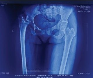
Figure 1A: Anteroposterior view of X-ray bilateral hip with pelvis proximal 2/3rd of femur revealing a markedly proximally migrated greater trochanter and almost destroyed head and neck of femur. The acetabulum showed dysplastic changes. The ipsilateral anterior the pelvis was hypoplastic as compared to the opposite side. The femur showed thinned-out cortices and a narrow medullary canal
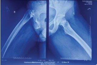
Figure 2B: Lateral view of bilateral hips with proximal femora showing an absence of head of right femur, hypoplastic right femur dysplastic changes in the right acetabulum
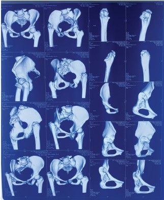
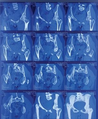
Figures 2A and 2B: CT of the pelvis and proximal femur was done to create a 3D morphology for preoperative planning
Laboratory evaluation
Markers of active infection i.e. total leukocyte count was 4800 WBCs/ml, ESR was 9 mm/hr and CRP was 0.3mg/dl. Routine preoperative blood examinations were done and found to be normal.
Challenges
As a post-septic sequela, the case posed the issue of marked fibrosis of the tissues, loss of tissue planes and difficult identification of the sciatic nerve. The radiological evaluation revealed no femoral offset, so restoration of the same posed a difficult task. It was hard to find the proper version. Moreover, there was no bone to augment the acetabulum. Marked overriding and shortening posed the threat of tractional injury to the sciatic nerve during correction. As the whole limb was found to be hypoplastic, the correction of deformity and shortening, as well as hardware placement was going to be difficult.
Treatment
After meticulous pre-operative planning, we subjected the patient to Primary Uncemented Right Total hip replacement with subtrochanteric shortening osteotomy on 13th November 2018. The procedure was undertaken under C-arm cover. A standard lateral approach to the hip was taken. Extensive fibrosis and loss of tissue planes were noticed. Following meticulous dissection, iliopsoas was found to be tight and was carefully released. The acetabulum was covered with dense fibrous tissue which was gradually and carefully cleared. Intraoperative tissue samples were sent for culture and sensitivity. Acetabular socket preparation was done. We didn’t perform a cotyloplasty as we got sufficient depth of the Acetabular cup. Using a 38mm shell, the cup was placed in desired inclination and anteversion and secured with one screw. (Figure 3A) Intraoperatively, the femur was found to be anteroposteriorly flattened. The greater trochanter was distalised by 2 cm. (Figure 3B) S-ROM stem preparation was done. Prophylactic circulate wiring was done. A subtrochanteric shortening osteotomy was performed and 1 cm of bone was removed. (Figure 3C) Clinical trialing of the stem was done and checked for rotation and stability. SROM stem of size 14x9x130mm was used with precise offset and version. On the C-arm image, the stem closure of distal flutes indicated well-fitting stem. (Figure 3D) Osteotomy was found to be stable. (Figure 3E). The final C-arm image confirmed adequate offset and version. (Figure 3F)
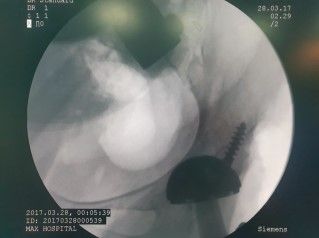
Figure 3A: The cup was placed in desired inclination and anteversion and secured with 1 screw
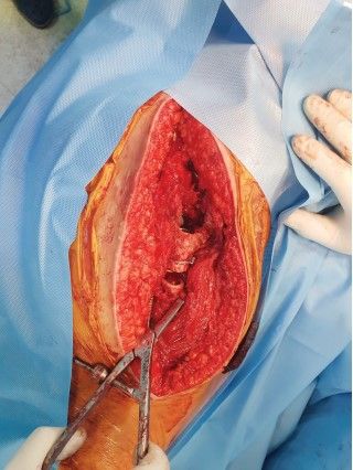
Figure 3B: Greater trochanter destabilised by 2 cm
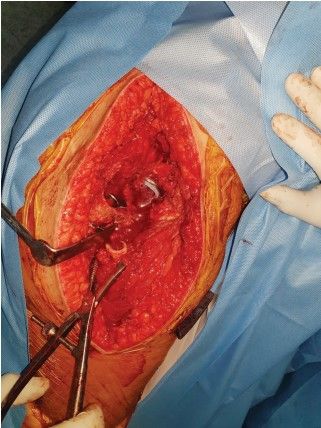
Figure 3C: Subtrochanteric shortening osteotomy with 1 cm of bone removed
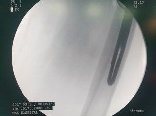
Figure 3D: Stem closure of distal flutes indicates well-fitting stems.
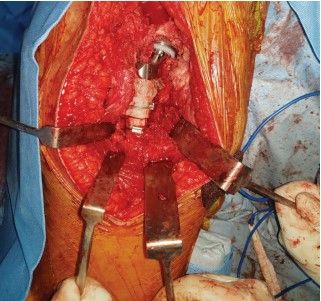
Figure 3E: Osteotomy was found to be stable. The final constitution was satisfactory
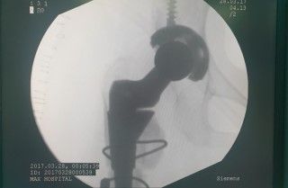
Figure 3F: Final C arm image confirmed adequate offset and version
Follow up
The patient was kept on toe-touch weight-bearing mobilisation for the first 6 weeks and gradually progressed to full weightbearing as the osteotomy site united
Follow up
The surgical scar healed well with no signs of complications. Intra-operative samples were culture-negative. Union at the osteotomy site was achieved at the 4th-month follow-up (Figure 4A)
In the next stage, the patient also demanded equalisation of limb length as the limb was hypoplastic. The residual shortening was noted to be 5 cm clinically and radiologically. The patient was further planned for a limb lengthening procedure using the Limb Reconstruction System. In July 2019, the patient underwent Right proximal tibia corticotomy and application of a Limb Reconstruction System (Rail Road Fixator) right tibia. (Figure 4B) Gradual distraction of 1 mm per day was given for 3 months, followed by 3 months of static fixation, after which the external fixator was removed.
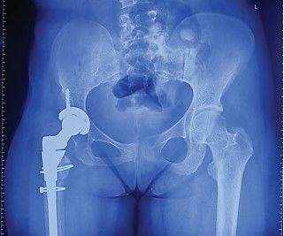
Figure 4A: Post-operative X-ray of the pelvis with both hips and proximal femora showing excellent offset, stem placement and overall constitution after the procedure
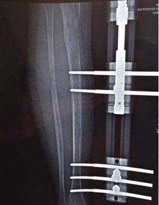
Figure 4B: LRS external fixator was used for the limb lengthening procedure
At 1 year follow-up, the patient was walking independently, with no pain or shortening. The range of motion at the hip was painless and with no impingement. (Figure 4C). The Modified Harris hip score at 1-year follow-up was 67.
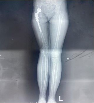
Figure 4C: On 1-year follow-up, the patient showed equal limb lengths, with a stable and painless limp
Discussion
Sequelae of Septic arthritis of the hip joint in the paediatric population may result in an array of clinical presentations ranging from ankylosis to pathological dislocation. The pathophysiology consists of hematogenous or local spread of pyogenic bacteria. The majority of cases present with pain, gait disturbance or both.[1]
Our case presented us with a completely absent head and neck of the femur with a dysplastic acetabulum. The demands that our patient had were that of a painless stable hip with no shortening. The challenges were the absence of offset, a dysplastic shallow acetabulum, presence of extensive fibrosis with a previous scar.
The patient was given an option of pelvic support osteotomy which could provide her with a painless and stable hip, with a compromise in range of movement and a possibility of loss of correction and need for re-operation in the future. The patient opted for total hip arthroplasty as it granted her a painless, stable hip with a good range of movement.[2
We decided to tackle the problems of the hip joint first and work on the limb length in the second stage. Our goal was to provide a painless stable hip with a good range of movement. Bochatey E et al reported significant improvement in hip scores of patients undergoing the first stage total hip replacement with quiescent septic arthritis with a period gap ranging between 5 to 46 years. They were also advised to go for the procedure after obtaining negative ESR and CRP values.[2]
In our case, the episode of septic arthritis was more than 10 years old and we obtained negative values of ESR and CRP. We took the Lateral approach for the procedure for the reason of the presence of substantial fibrous and contractured tissue both anterior and posterior to the joint. The lateral approach gave us good access to both these areas and allowed adequate soft tissue release. Intra-operatively iliopsoas was found to be tight and released carefully. In the setting of obscure tissue planes and fibrotic tissue, the lateral approach also had the advantage of being distant from the region of traversing the Sciatic nerve. On the Acetabular aspect, we found dense fibrous tissue which was gradually and carefully released. We did not perform a cotyloplasty as we procured sufficient depth. The hip center was restored.
The next challenge that was posed to us was the high-riding trochanter. The longevity of hip arthroplasty in high hip dislocation patients is improved by restoring the anatomic hip centre, which decreases hip joint reaction forces and creates an improved lever arm for the abductor musculature.
Restoration of the hip centre in our case would have resulted in excess lengthening which would increase the chances of neuralgic injury. Subtrochantric osteotomy, first described by Klisic and Jancovic, has been used for high dislocation of the hip with a good rate of success. Hence, we decided to incorporate the STSO in our procedure.
We kept all implants ready for any intra-operative difficulty. Our stems included CDH Cemented stem, Wagner stem and S-ROM stem. The modular S-ROM prosthesis is unique. The independent neck and sleeve can accommodate a proximaldistal mismatch while providing ample opportunity for leg length adjustment. The S-ROM stem also allows placement of the version to maximise the range of motion and resulting hip stability. These features combined with the availability of highoffset stems achieve restoration of patient biomechanics. Yang et al concluded the cementless S-ROM modular femoral stem used with subtrochanteric transverse shortening osteotomy is safe and effective for high hip dislocation secondary to quiescent pyogenic arthritis and provides satisfactory midterm results. They observed significant improvements in clinical function were observed, as were high rates of stable fixation of the cementless implant, restoration of more normal limb lengths, and a low incidence of complications.[7] Multiple studies report the effectiveness of S-ROM stems from conditions like developmental dysplasia of hip and revision surgeries.[8][9][10] With this literature support, we chose the SROM stem for our procedure. Intra-operative samples were culture negative.
We preferred the tibia for the lengthening procedure as we did not want to risk the chances of pin-track infection at the femoral aspect. Proximal tibial corticotomy and LRS is a proven method of limb lengthening we adopted. In summary, this case was a highly technically challenging one that dealt with careful pre-operative planning and execution intra-operatively with preparedness to handle any intraoperative surprises
Conclusion: Meticulous pre-operative planning and diverse intraoperative options are essential to approach a difficult case like post-septic sequelae of the hip. Total hip arthroplasty provides a stable, mobile hip and good patient satisfaction. S-ROM stem is a good option for a stem to be used in cases requiring subtrochantric shortening osteotomy. With all the above considerations, our treatment led to significant improvement in the patient’s Modified Harris hip score of 80.
References
- Spiegel DA, Penny JN, Banskota AK, Shrestha OP. Sequelae of septic arthritis of the hip. Seattle: Global-HELP publications. 2007
- Gursu S, Demir B, Yildirim T, Er T, Bursali A, Sahin V. An effective treatment for hip instabilities: pelvic support osteotomy and femoral lengthening. Acta Orthopaedica et Traumatologica Turcica. 2012;45(6):437-45
- Bochatey E, Del Sel H, Tillet F, Lopreite F. Arthroplasty as a treatment for acute and quiescent septic arthritis in native hips. Acta ortopédica mexicana. 2021 Feb;35(1):11-6
- Morag G, Zalzal P, Liberman B, et al. Outcome of revision hip arthroplasty in patients with a previous total hip replacement for developmental dysplasia of the hip. J Bone Joint Surg Br 2005;87-B:1068
- Pagnano MW, Hanssen AD, Lewallen DG, et al. The effect of superior placement of the acetabular component on the rate of loosening after total hip arthroplasty: long term results of patients who have Crowe type II congenital dysplasia of the hip. J Bone Joint Surg Am 1996;78:1004
- Kelly SS. High hip center in revision arthroplasty. J Arthroplasty 1994;9:503
- Yoder SA, Brand RA, Pederson DR, et al. Total hip acetabular component position affects acetabular loosening rates. Clin Orthop 1988;228:79
- Yang Y, Yu QP, Wang SL, Zhang SL, Li J, Zhou Y, Fan HQ, Zhang X, Zhou Y, Zhou M, Zhou MQ. Outcomes after Total hip arthroplasty using a Cementless S-ROM modular stem for patients with high hip dislocation secondary to hip pyogenic arthritis. Orthopaedic Surgery. 2019 Jun;11(3):460-6
- Tamegai H, Otani T, Fujii H, Kawaguchi Y, Hayama T, Marumo K. A modified S-ROM stem in primary total hip arthroplasty for developmental dysplasia of the hip. The Journal of Arthroplasty. 2013 Dec 1;28(10):1741-5
- Moreta J, Uriarte I, Foruria X, Loroño A, Agirre U, Jáuregui I, Martínez-de Los Mozos JL. Medium-term outcomes of the S-ROM modular femoral stem in revision hip replacement. European Journal of Orthopaedic Surgery & Traumatology. 2018 Oct;28:1327-34
- Li L, Yu M, Yang C, Gu G. Total hip arthroplasty (S-ROM stem) and subtrochanteric osteotomy for Crowe type IV developmental dysplasia of the hip. Indian Journal of Orthopaedics. 2016 Apr;50:195-200