 WhatsApp Us (24/7)
 +91 926 888 0303 (24/7)


Abstract: IgG4-related disease (IgG4-RD) is a complex systemic condition that can affect virtually every body organ. It has highly variable clinical presentation, indistinguishable from many inflammatory and neoplastic diseases. There are overlapping biochemical and imaging features making prospective diagnosis difficult without a high index of suspicion. One of the varied presentations of IgG4-RD is the formation of pseudotumours, which are benign masses composed of inflammatory cell infiltrates and fibrous components. It is relatively straightforward to suspect the occurrence of such pseudotumours in patients with known IgG4-RD disease or with multiorgan or systemic involvement. However, if present as an isolated initial manifestation of this disease, they may be underdiagnosed in the preoperative period, as biopsy may be complicated in every patient with a resectable tumour before surgery with imaging features consistent with a typical malignant mass for that particular organ. The present case describes the occurrence of IgG4-related inflammatory pseudotumours (IPT) in a patient who was diagnosed postoperatively due to the absence of any other manifestation of this disease elsewhere in the body.
Key Words: IgG4 related disease, hepatic pseudotumor
Background
After its description in 2003 by Kamisawa, IgG4-related disease has increasingly been recognised as a severe immune-mediated condition, closely simulating a variety of neoplastic or nonneoplastic diseases.[1] It may present as mass, inflammation or fibrosis involving multiple body organs at different locations. The common sites of involvement include the pancreas, biliary system, salivary glands, orbit, kidneys, liver, lungs, retro peritoneum and lymph nodes. The disease is characterised by infiltration with IgG4-positive plasma cells, causing inflammation and eventually leading to fibrosis. Elevated serum IgG4 level may serve as an adjunct marker, but it may be normal in up to 40% of cases and falsely elevated in other cases with no definite IgG4-RD.[2] The disease usually responds well to steroids and immunosuppressive agents like rituximab in a 2-4 week duration.[2] If left untreated, it can lead to systemic inflammation and may eventually progress to sclerosing disease. Here, we present a rare case of this disease, closely simulating intrahepatic cholangiocarcinoma in the liver, but subsequently diagnosed as IgG4-related inflammatory pseudotumor on histopathological examination.
Case details
A 65-year-old male patient presented to the outpatient department of our institution with a complaint of fatigue and abdominal pain for three weeks. There was no pyrexia or jaundice. Abdominal sonography revealed a well-circumscribed 5 cm hypoechoic mass in the left lobe of the liver. On lab evaluation, liver function tests were within normal limits with mild elevation of serum carbohydrate antigen 19-9 levels- 56U/ml (3– 34U/ml). There was no elevation of C-reactive or alpha protein levels with negative viral serology. Further investigation with contrast-enhanced multiphasic CT scan demonstrated an irregular 5 cm mass in the left lobe of the liver at segment IV A/II junction, showing inhomogeneous enhancement in the arterial phase with progressive centripetal enhancement in subsequent equilibrium phases and mild peripheral washout in delayed venous phases (Figure 1 A-D). An irregular necrotic hypo enhancing area in the centre of the lesion gave rise to a targetoid appearance with mild peripheral biliary dilatation in the left lateral segments of the liver. No vascular thrombosis was seen in portal or hepatic veins. The imaging findings closely resembled intrahepatic cholangiocarcinoma and the patient underwent left hepatectomy with regional lymph node dissection. The post-operative histological examination of the resected tumour revealed the presence of lymphocytic infiltration, mainly by plasma cells and proliferation into the surrounding connective tissue with fibrosis and obliterative phlebitis, suggesting the diagnosis of IgG4-related hepatic inflammatory pseudotumor (IPT) (Figure 2 A-B).
This case highlights the difficulties in differentiation between intrahepatic cholangiocarcinoma and hepatic IPT, based on imaging findings alone.
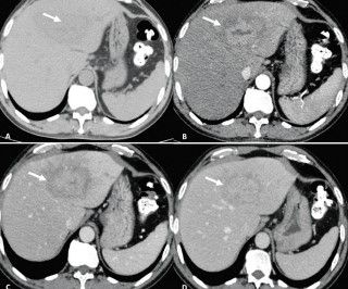
Figure 1 (A-D): Contrast-Enhanced Multiphasic CT scan shows an irregular mildly hypodense mass (Arrow) in the left lobe of the liver at segment IVA/ II junction on non-contrast image (A) demonstrating inhomogeneous enhancement in arterial phase (B), progressing in portal venous phase (C) and in equilibrium phase (D) with mild peripheral hypodensity. The mass shows a targetoid appearance on postcontrast images
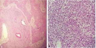
Figure 2 (A, B): Low (A) and High magnification images of the Haematoxylin Eosin-stained slide demonstrates the presence of lymphocytic infiltration, mainly by plasma cells and proliferation into the surrounding connective tissue with fibrosis
Discussion
IgG4-RD can present as a tumefactive lesion of nearly any organ, mainly containing glandular tissues, which may be indistinguishable from malignancy clinically or radiologically. The median age of diagnosis is 60-70 years, with an M: F ratio of 2:1.[2] An international set of classification criteria termed ‘Revised Comprehensive Diagnostic (RCD) criteria for IgG4-RD’ was developed by the American College of Rheumatology/ European League Against Rheumatism (ACR-EULAR) to help diagnose mimickers of IgG4-RD by considering a set of clinical, radiological, serological and pathological criteria, summarised in Table 1.[3]
Hepatic inflammatory pseudotumours (IPT) are relatively rare lesions and may present as well-circumscribed parenchymal masses, closely mimicking Intrahepatic cholangiocarcinoma. Hepatic IPT has been reported to account for 0.4% of all focal liver lesions treated by hepatectomy.[4] IgG4-related hepatic IPT usually occur over a background of IgG4-related sclerosing cholangitis. Histopathologically, it may be classified into fibro histiocytic type and lymphoplasmacytic type. The fibrohistiocytic type is characterised by xanthogranulomatous inflammation, multinucleated giant cells and neutrophilic infiltration, while the lymphoplasmacytic type shows infiltration by lymphocytes and IgG4-positive plasma cells.[5] Our case belonged to the lymphoplasmacytic variety as there was infiltration by dense lymphoplasmacytic infiltrate with storiform fibrosis and obliterative phlebitis. Chougule et al. did a systemic review of hepatic IPT and found a higher predilection of IgG4-related hepatic IPT in older males with a mean age of 60.8 years, like our case.[6] There are no specific radiologic findings in hepatic IPT, with most cases presenting as solitary or multiple nodular lesions in the liver. The post-contrast enhancement of hepatic IPT may vary from a hyper-vascular lesion showing early hyperenhancement to a rather hypo-enhancing mass. The lymphoplasmacytic IPTs usually present as infiltrative tumours, developing in the hepatic hilum and extending along the hilar bile ducts. Few cases of hepatic IPT may undergo regression of the mass and reduction of symptoms after medical treatment; however, in a few other instances, IPT may have a recurrence and persistent local growth necessitating resection.
Learning Objectives: IgG4-RD is usually underdiagnosed due to its polymorphic presentation. However, radiologists must be familiar with this disorder because patients generally respond well to corticosteroid therapy and timely recognition may help avoid unwanted procedures.
| Clinical and radiological features | One or more organs show diffuse or localised swelling or a mass or nodule characteristic of IgG4-RD. In single-organ involvement, lymph node swelling is omitted |
| Serological diagnosis | Serum IgG4 levels greater than 135mg/dl |
| Pathological diagnosis | Positive for two of the following three criteria
|
| Diagnosis: |
|
Table 1.The 2020 revised comprehensive diagnostic (RCD) criteria for IgG4-RD
CONCLUSION:
IgG4-RD is a complex condition with a highly variable clinical appearance that can mimic various inflammatory and neoplastic diseases. It should be considered in the differential diagnosis of hepatic mass-forming lesions, especially when multisystem involvement is present. If clinically suspected, further workup with serum protein electrophoresis, IgG subclass measurement and tissue biopsy will help make a preprocedural diagnosis.
Acknowledgement
We acknowledge the contribution of Dr Anjali Gupta, Department of Pathology, Max Super Speciality Hospital, Saket, New Delhi in proving the histopathological images and guiding the preparation of the manuscript.o
References
- Kamisawa T, Funata N, Hayashi Y, Eishi Y, Koike M, Tsuruta K, et al. A new clinicopathological entity of IgG4-related autoimmune disease. J Gastroenterol. 2003;38(10):982–4
- Deshpande V, Zen Y, Chan JK, Yi EE, Sato Y, Yoshino T, et al. Consensus statement on the pathology of IgG4-related disease. Mod Pathol. 2012 Sep;25(9):1181–92
- Wallace ZS, Naden RP, Chari S, Choi H, Della-Torre E, Dicaire JF, et al. The 2019 American College of Rheumatology/ European League against rheumatism classification criteria for IgG4-related disease. Arthritis Rheum. 2020;72(1):7–19
- Torzilli G, Inoue K, Midorikawa Y, Hui AM, Takayama T, Makuuchi M. Inflammatory pseudotumors of the liver: prevalence and clinical impact in surgical patients. Hepatogastro- enterology 2001;48:1118–23
- Zen Y, Fujii T, Sato Y, Masuda S, Nakanuma Y. Pathological classification of hepatic inflammatory pseudotumor with respect to IgG4-related disease. Mod Pathol. 2007 Aug;20(8):884–94
- Chougule A, Bal A. IgG4-related inflammatory pseudotumor: A systematic review of histopathological features of reported cases. Mod Rheumatol. 2017 Mar;27(2):320–5