 WhatsApp Us (24/7)
 +91 926 888 0303 (24/7)


Abstract: Chylothorax is a rare and serious complication of thoracic surgeries. Although most cases occur immediately post-operatively, late-onset chylothorax is rare and there is a lack of definitive consensus on its management. In this report, we describe a case of chylothorax that occurred several years after thoracic surgery which was treated successfully with a combination of conservative measures, including octreotide and lymphangiography and review current management strategies.
Keywords: Chylothorax, octreotide, lymphangiography
Introduction
Chylothorax is defined as pathological accumulation of chyle in pleural space following traumatic or spontaneous disruption of thoracic duct. Traumatic chylothoraxes are more common than spontaneous. Iatrogenic chylothorax, especially postoesophageal surgeries, are the leading cause of chylothorax accounting for 4% of cases. Although rare, chylothorax is a serious complication of thoracic surgery. Given the rarity of this complication, there is no definitive consensus regarding its management.[1,2,3]
Delayed chylous leak is uncommon since most cases of postoperative chylothorax are diagnosed within 3 days. There have been few case reports describing late onset chylothorax after a few months in post-operative patients.[4,5] In this case report, we describe a late-onset chylothorax that occurred many years after thoracic surgery, which was successfully treated with a combination of conservative management and lymphangiography
Case An 87-year-old male presented with complaints of sudden onset breathlessness with orthopnoea for 2 days and drowsiness in the past one day. There was no history of fever, cough or chest pain. He was a non-smoker with no history of chronic obstructive pulmonary disease. His past history included a coronary artery bypass surgery (CABG) in 2014, esophagectomy in 2016 for carcinoma oesophagus, pacemaker implantation in 2020 for complete heart block.
On examination, the patient was drowsy, responding to commands with tachypnoea and tachycardia. He had reduced breath sounds bilaterally in the infra-scapular region on auscultation. His arterial blood gases (ABG) on admission showed acute respiratory acidaemia with- pH- 7.11, PCO2- 75, HCO3- 26. The patient was admitted in the intensive care unit (ICU) and started on non-invasive ventilation (NIV). His chest x-ray showed a left moderate effusion with right costophrenic angle blunting (Figure.1). An ultrasonography (USG) of chest showed around 2 litres of fluid on the left and 750 ml on the right. Laboratory tests and echocardiography was normal with an ejection fraction of 55- 60%.
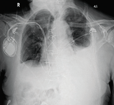
Figure 1: Chest x-ray on presentation showing left moderate effusion with right costophrenic angle blunting
A left pigtail catheter was inserted under USG guidance, 1500 ml of whitish pink fluid was drained initially. The pleural fluid analysis confirmed a chylous effusion with pleural fluid triglyceride of 587 mg/dl (normal range <50), pleural fluid protein of 4.2 g/ dl, total cell count (TLC) of 3100 per mm3 and cytology was negative for malignant cells.
The patient was kept on a fat free diet, injection of octreotide 100 microgram (mcg) subcutaneously three times a day was started on the third day post pigtail catheter insertion and continued for a week. A whole-body PET/CT scan did not show any evidence of tumour recurrence. The patient was kept on intermittent NIV support and had a drainage of around 500 ml daily. In view of persistent drainage despite conservative measures, lipiodol lymphangiography (LG) was performed on 7th day in an attempt to occlude the chyle leak.
Pleural fluid drainage reduced significantly and became more haemorrhagic in appearance. NIV support was gradually tapered off and he required minimal supplemental oxygen. A repeat pleural fluid analysis showed resolution of chylothorax with pleural fluid protein of 3.3 g/dl, TLC of 1080 per mm3 and triglycerides of 58 mg/dl. Figure 2 demonstrates a progressive reduction in pleural output post octreotide and LG.
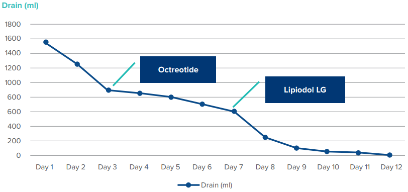
Figure 2: Chest x-ray on presentation showing left moderate effusion with right costophrenic angle blunting
Pigtail catheter was removed on 12th day. The patient was discharged with supplemental oxygen. After one month of follow-up, he is off oxygen support with no recurrence of pleural effusion on chest x-ray (Figure 3)
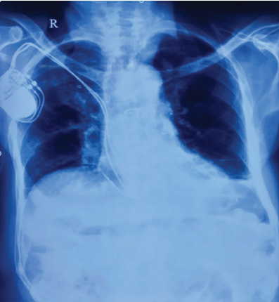
Figure 3: Chest x-ray after one-month of follow-up shows complete resolution of effusion
Discussion
The leading cause of chylothorax is iatrogenic, especially after thoracic surgeries. Most cases of chylothorax occur within 3 days post-surgery. There have been few case reports occurring within weeks to months after thoracic surgery.[4] To the best of our knowledge, only one case report describing chylothorax that occurred 20 years after blunt chest trauma that was successfully treated with video-assisted thoracoscopic surgery (VATS) and pleurodesis was reported.[6] Another case report describing the occurrence of chylothorax 4 months post heart and lung transplant surgery, which was managed through conservative measures..5 In our case, we attributed the cause of chylothorax to the past history of CABG or esophagectomy in 2014 and 2016, respectively.
Chylothorax is diagnosed based on pleural fluid analysis. The milky appearance of Chyle occurs in less than half of cases, others may demonstrate different appearance including serous, serosanguinous, or haemorrhagic. Chylothorax is found in 99% of cases when the triglyceride level exceeds 110 mg/ dL and the cholesterol level is below 200 mg/dL, whereas a triglyceride concentration below 50 mg/dL nearly eliminates the possibility of chylothorax. When triglyceride levels are between 50-110mg/dl, lipoprotein electrophoresis should be performed for demonstration of chylomicrons which are characteristic of chyle leak.[7,8]
Conservative treatment of chylothorax involves total elimination of oral or enteral fat intake, which reduces chyle flow 10-fold. Pleural cavity drainage should be initiated at diagnosis.[8,9] Octreotide resembles natural somatostatin pharmacologically. It causes mild splanchnic vasoconstriction, which leads to reduction of gastric, pancreatic, intestinal secretions and intestinal absorption, thereby reducing the flow of chyle.[7,8] It can be initiated at a dose of 50 mcg per day to a maximum dose of 600 mcg per day. Adverse effects include nausea, abdominal pain, diarrhoea. Rarely, it can cause hypothyroidism, cholelithiasis, bradycardia, chest pain; hence, it should be used cautiously in patients with pre-existing heart and liver diseases.[10] Bryant et al. observed a 90% success rate with octreotide in 41 patients with chylothorax, following pulmonary resection. Barbetakis et al. observed 80% reduction in drain output and drain removal by day 8 in a case of chylothorax following CABG.[11] There is no definite consensus regarding the optimal duration of therapy but it is usually administered for 1-2 weeks before stopping in non-responders.[3] Fujita et al. observed that a high-output chylothorax (>1 L/day), persisting for 48 hours after treatment with octreotide is predictive of treatment failure.[11]
Lymphangiography (LG) and thoracic duct embolisation (TDE) are minimally invasive interventions alternative to surgical ligation. TDE involves percutaneous image-guided occlusion of the thoracic duct (TD). It is a 3-step process that consists of lymphangiography followed by percutaneous transabdominal catheterisation of the cisterna chyli (CC) and embolisation of the TD proximal to the leak or occlusion. Embolisation is usually achieved with a combination of coils and n-butyl cyanoacrylate (n-BCA). It can be accessed via conventional pedal route or an updated inguinal intranodal lymphangiography which has a shorter duration. The procedure is technically challenging with a successful catheterisation rate of 67%. When successful catheterisation is achieved, the chylothorax resolution rate is 90%.[12,13]
LG is invaluable in detecting different types of lymphatic leakages, with a detection rate of 64% to 78%. LG has both diagnostic and therapeutic value. Lipiodol, an ethiodised oil contrast agent infused during LG to identify the lymphatics and chylous leak can induce a granulomatous and inflammatory reaction that seals the leak. It also has embolic properties as it accumulates at points of leakage outside and within lymphatics. A successful therapeutic outcome of 56% to 86% has been reported in patients with chylothorax and chylous ascites. Serious complications include pulmonary embolism, allergic reactions and lipiodol extravasation in soft tissues, which are frequently related to the volume of contrast used, which should be restricted to 10 ml. Contraindications for LG include pulmonary insufficiency, and right to left cardiac shunt.[12] Our patient successfully underwent chylous leak occlusion with LG and lipiodol contrast injection.
Surgical interventions should be kept in mind in post-operative chylothorax with high volume leakage, greater than 1000 mL/ day for more than 5 days, persistent leak for more than 2 weeks despite conservative management, or development of insurmountable nutritional or metabolic complications.[9,12]
CONCLUSION:
Chylothorax is a rare and serious complication of oesophageal and thoracic surgeries that can occur either immediately post-operatively or late after many years of surgery. Lipiodol lymphangiography is an effective and minimally invasive procedure that should be considered early in patients who are unlikely to respond to conservative treatment.
References
- Rieu MC, Mabrut J. Management of postoperative chylothorax. J Visc Surg. 2011;148(5):346–52
- Miao L, Zhang Y, Hu H, Ma L, Shun Y, Xiang J. Incidence and management of chylothorax after esophagectomy. Thorac cancer. 2015;6(3):354–8
- Rehman KU, Sivakumar P. Non-traumatic chylothorax: diagnostic and therapeutic strategies. Breathe. 2022;18(2):1–15
- Zhang C, Zhang R, Pan Y, Wu W, Zhang M. Late-onset chylothorax during chemotherapy after lobectomy for lung cancer. Med. 2019;98(22):1–10
- Shitrit David IG. The Annals of Thoracic Surgery Late-onset chylothorax after heart – lung transplantation Comment. Ann Thorac Surg. 2003;75(1):285–6
- Kamiyoshihara M, Ibe T. Late-Onset Chylothorax After Blunt Chest Trauma at an Interval of 20 Years : Report of a Case. Surg Today. 2008;36(56–58):1–7
- Ag J, Ag J. Chylothorax after off-pump coronary artery bypass graft surgery: Management strategy. Ann Card Anesth. 2018;21(3):300–3
- Mcgrath EE, Blades Z, Anderson PB. Chylothorax : Aetiology, diagnosis and therapeutic options. Respir Med. 2010;104(1):1–8
- Talwar A, Lee HJ. A contemporary review of chylothorax. Indian J Chest Dis Allied Sci. 2008;50(4):343–51
- Swanson MS, Hudson RL, Bhandari N, Sinha UK, Maceri DR, Kokot N. Use of octreotide for the management of chyle fistula following neck dissection. JAMA Otolaryngol - Head Neck Surg. 2015;141(8):723–7
- ESME H. The use of octreotide in the treatment of chylothorax. J Contemp Med. 2019;9(4):432–5
- Lee EW, Shin JH. Lymphangiography to Treat Postoperative Lymphatic Leakage : A Technical Review. Korean J Radiol. 2014;15(6):724–32
- Mikhail C.S.S. Higgins, Auh Whan Park, John F. Angle: Chylothorax: Percutaneous Embolization of the Thoracic Duct. Operative Techniques in Thoracic and Cardiovasculary Surgery. 2016, 20:402-412