 WhatsApp Us (24/7)
 +91 926 888 0303 (24/7)


Here, we present a case of a 5-month-old developmentally normal female presented with loose stools, vomiting, fever and poor oral intake for one day.
The child was dull-looking, irritable and dehydrated. She had tachycardia and tachypnoea but no chest findings. The cardiac exam was normal. Initial diagnosis of Acute Gastroenteritis with some dehydration was made. Relevant investigations were sent, which showed high CRP, hence, Inj. Ceftriaxone were added and dehydration was corrected along with supportive measures. In view of irritability and high CRP, Lumbar Puncture was advised, but the parents refused. wStool analysis was normal and negative for reducing sugar and rotavirus antigen. With the above measures, the child initially showed improvement in lab parameters. Blood culture reported sterile, but fever spikes persisted and again on day 6, increased CRP, with a persistent dull appearance; antibiotics were upgraded to Inj. Meropenem and Amikacin. After 48 hours of upgradation of antibiotics, fever and irritability were persistent. A Neurology opinion was taken, an MRI of the Brain and EEG and a CSF examination was done, which showed no sequelae of Meningoencephalitis. As fever spikes continued, autoimmune causes of fever were considered and inflammatory markers (LDH and Ferritin), within normal range, were sent. 2D ECHO (Figure-1) was done on day 8 of illness, which showed dilated coronaries suggestive of Kawasaki Disease Z scores LM- 5.5, LAD- 6 RCA-5.2. IVIG 2gm/kg over 24 hrs along with Aspirin as per the dose recommended by the Paediatric Cardiologist was given. As the Fever persisted, despite IVIG infusion at 48 hrs, repeat CBC and CRP were done, which showed reduced CRP and improved TLC. 2D ECHO was repeated on day 10 of illness, which showed a further increase in Z-score in dilatation of coronaries (Z scores- LM +6.46. LAD- +10, RCA-+9.1). So, a repeat dose of IVIG along with IV steroids and LMWH was given and the child was shifted to a dedicated cardiac monitoring centre under Paediatric Rheumatologist. Gradually, fever spikes settled on repeat 2D ECHO, Z-score was further increased LM- 5.28, LAD-11.98, RCA-9.2, so started on cyclosporine and steroid continued. On follow-up, a week later, a gradual decrease in LMCA 2.5, LAD- 3.2 and RCA 3.9 in coronary dilatation was seen.
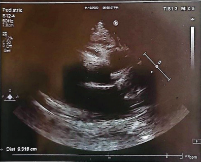
Figure 1: 2D Echo depicting dilated coronaries suggestive of Kawasaki Disease
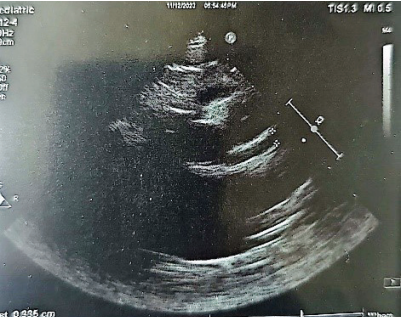
Figure 2: 2D Echo depicting dilated coronaries suggestive of Kawasaki Disease
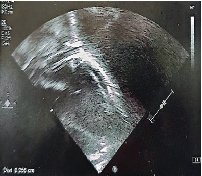
Figure 3: 2D Echo depicting dilated coronaries suggestive of Kawasaki Disease
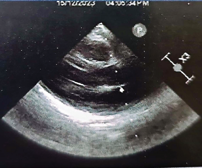
Figure 4: 2D Echo depicting dilated coronaries suggestive of Kawasaki Disease
This case report highlights the need for getting a 2d echo in any case of fever without a focus beyond 7 days in children; cardiac pathology should be ruled out, especially Kawasaki Disease, as the development of coronary artery aneurysms is associated with the need for long-term monitoring and antiplatelet therapy.
Our case is noteworthy because of the young age of the patient, the atypical clinical presentation with the development of coronary artery aneurysms in the absence of other clinical and lab findings and the unresponsiveness to the standard line of management.
Kawasaki disease is an acute necrotising vasculitis of unknown aetiology, with multisystem involvement and inflammation of small to medium-sized vessels, with resulting aneurysm formation in infants and children under 5 years of age. It was named after Tomisaku Kawasaki, a Japanese paediatrician who described this febrile vasculitis for the first time in 1967. KD most commonly occurs in males and in children less than 5 years of age, with a peak between 2 and 3 years; it is rare in children over and above 7 years. It is now the most common cause of acquired heart disease.[1]
Criteria for diagnosis of Kawasaki disease
Fever of >5-day duration associated with at least four of the following five changes:
- Bilateral nonsuppurative conjunctivitis
- One or more changes of the mucous membranes of the upper respiratory tract, including pharyngeal injection, dry fissured lips, injected lips, and “strawberry” tongue
- One or more changes of the extremities, including peripheral erythema, peripheral edema, periungual desquamation, and generalised desquamation
- Polymorphous rash, primarily truncal
- Posterior cervical lymphadenopathy >1.5cm in diameter
- Disease cannot be explained by some other known disease process
*A diagnosis of Kawasaki disease can be made if fever and only three changes are present in conjunction with coronary artery disease documented by two- dimensional echocardiography or coronary angiography
Figure 5: Criteria for diagnosis of Kawasaki disease
Incomplete KD: Fever with less than four of the five principal clinical criteria with compatible laboratory or echocardiography findings suggests incomplete KD. Often seen in infants 6 years of age, the incomplete clinical picture delays the diagnosis. Be aware of KD in children with unexplained fever for >5 days.
Atypical KD: Patients who, along with the usual clinical features of KD, also have a few unusual clinical manifestations, such as pulmonary involvement and renal impairment, are diagnosed to have atypical KD. (Figure-5)
Irritability (aseptic meningitis), arthritis (at onset or delayed), sterile pyuria (urethritis), gastroenteritis (abdominal pain, vomiting, and diarrhoea), Bacillus Calmette–Guérin (BCG) reactivation, uveitis, perianal and periungual desquamation, gallbladder hydrops, and jaundice.[2]
Infants less than six months represent a special age group, as they are more likely to lack classical manifestations and have a higher risk for coronary artery involvement. Currently, the chief uncertainties for clinicians are how to perform a timely diagnosis, how to prevent cardiovascular complications and how to treat intractable forms. Refractory forms have been increasing remarkably and both the young age of the patient and a delay in initiating the treatment seem to be the most important risk factors.[2]
At present
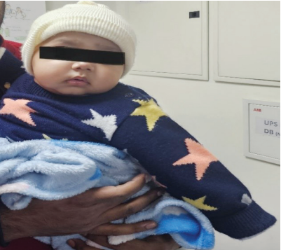
Figure 5: Picture of the child after treatment
References
- Lloyd, A. J., Walker, C., & Wilkinso, M. (2001). Kawasaki disease: is it caused by an infectious agent?. British journal of biomedical science, 58(2), 122–128
- Leonardi, S., Barone, P., Gravina, G. et al. (2013). Severe Kawasaki disease in a 3-month-old patient: a case report. BMC Res Notes 6, 500 https://doi.org/10.1186/1756-0500- 6-500